Treated with TRAIL, and Does So by NF-
k
B Dependent
Induction of cFLIPL
Josep Maria Lluis1, Ulrich Nachbur1, Wendy Diane Cook1, Ian Edward Gentle1, Donia Moujalled1, Maryline Moulin1, Wendy Wei-Lynn Wong1, Nufail Khan1, Diep Chau1, Bernard Andrew Callus2, James Edward Vince3, John Silke1, David Lawrence Vaux1*
1Deparment of Biochemistry, La Trobe University, Bundoora, Australia,2School of Biomedical, Biomolecular and Chemical Sciences, University of Western Australia, Crawley, Australia,3Department of Biochemistry, University of Lausanne, Epalinges, Switzerland
Abstract
Tumor necrosis factor (TNF)-related apoptosis-inducing ligand (TRAIL) is known as a ‘‘death ligand’’—a member of the TNF superfamily that binds to receptors bearing death domains. As well as causing apoptosis of certain types of tumor cells, TRAIL can activate both NF-kB and JNK signalling pathways. To determine the role of TGF-b-Activated Kinase-1 (TAK1) in TRAIL signalling, we analyzed the effects of adding TRAIL to mouse embryonic fibroblasts (MEFs) derived from TAK1 conditional knockout mice. TAK12/2MEFs were significantly more sensitive to killing by TRAIL than wild-type MEFs, and failed to activate NF-kB or JNK. Overexpression of IKK2-EE, a constitutive activator of NF-kB, protected TAK12/2MEFs against TRAIL killing, suggesting that TAK1 activation of NF-kB is critical for the viability of cells treated with TRAIL. Consistent with this model, TRAIL failed to induce the survival genes cIAP2 and cFlipL in the absence of TAK1, whereas activation of NF-kB by IKK2-EE restored the levels of both proteins. Moreover, ectopic expression of cFlipL, but not cIAP2, in TAK12/2MEFs strongly inhibited TRAIL-induced cell death. These results indicate that cells that survive TRAIL treatment may do so by activation of a TAK1–NF-kB pathway that drives expression of cFlipL, and suggest that TAK1 may be a good target for overcoming TRAIL resistance.
Citation:Lluis JM, Nachbur U, Cook WD, Gentle IE, Moujalled D, et al. (2010) TAK1 Is Required for Survival of Mouse Fibroblasts Treated with TRAIL, and Does So by NF-kB Dependent Induction of cFLIPL. PLoS ONE 5(1): e8620. doi:10.1371/journal.pone.0008620
Editor:Andreas Bergmann, University of Texas MD Anderson Cancer Center, United States of America
ReceivedNovember 30, 2009;AcceptedDecember 6, 2009;PublishedJanuary 8, 2010
Copyright:ß2010 Lluis et al. This is an open-access article distributed under the terms of the Creative Commons Attribution License, which permits unrestricted use, distribution, and reproduction in any medium, provided the original author and source are credited.
Funding:This work has been funded by the Leukemia and Lymphoma Society, and National Health and Medical Research Council grant#461221. The funders had no role in study design, data collection and analysis, decision to publish, or preparation of the manuscript.
Competing Interests:The authors have declared that no competing interests exist.
* E-mail: [email protected]
Introduction
TRAIL is a member of the tumor necrosis factor superfamily that selectively induces apoptosis in a wide variety of cancer cells, while sparing normal cells, highlighting its potential as an agent for cancer therapy[1]. So far, the mechanism for differential TRAIL sensitivity has not been established.
Murine TRAIL is known to bind to three different receptors: mTRAIL-R which contains a death domain (DD) in the intracellular portion, and mDcTRAIL-R1 and mDcTRAIL-R2, which are decoy receptors that regulate the binding of TRAIL to mTRAIL-R[2].
TRAIL triggers apoptosis by binding to mTRAIL-R, which leads to the recruitment of Fas associated death domain (FADD) through its DD. The adaptor protein FADD also contains a death effector domain (DED) that allows the binding of inactive procaspase 8 and cellular FLICE-inhibitory protein (cFlip). Once this death-inducing signalling complex (DISC) has been assem-bled, self-cleaved caspase 8 will lead to the activation of effector caspases 3 and 7 resulting in apoptotic cell death.
cFlip is the only protein present in the mTRAIL-R DISC that is capable of blocking death receptor-mediated apoptosis. In mouse cells, cFlip exists mainly in three forms: cFlipL and cFlipR that
arise from mRNA splicing, and the cleaved form, Flipp43 [3,4]. All these variants of cFlip bear two DED domains but only cFlipL possesses a caspase-like domain, which lacks catalytic activity. Therefore, all cFlip forms are potentially able to compete with procaspase 8 for binding to the DED of FADD, preventing its full activation and, thereby, cell death. Interestingly, elevated levels of cFlip protein have been reported in different types of cancer [5,6,7,8], and cFlip gene silencing can sensitize tumor cells to TRAIL induced cell death in many cases[9,10,11,12,13].
While apoptosis is the major outcome for many types of cancer cells exposed to TRAIL, there is accumulating evidence that
TRAIL can also activate NF-kB and c-Jun N-terminal kinase
(JNK) pathways [14,15,16]. The effects of NF-kB and JNK on
TRAIL signalling are controversial, with some reports showing that their activation protects cells from TRAIL induced apoptosis [17] and others suggesting the opposite effect [18]. Activation of
NF-kB by TRAIL is of particular interest, because of its ability to
induce anti-apoptotic genes such as cFlip, cIAPs, A20, and Mcl-1[19,20].
death domain (TRADD), receptor interacting protein (RIP1),
TNF receptor associated factor 2 (TRAF2) as well as IKKc, and
this is crucial for NF-kB and JNK activation by TRAIL [15,21].
On the other hand, TAK1, a member of the MAP3K family,
was originally identified as a kinase involved in TGF-bsignalling.
TAK1 is activated by a wide range of cytokines such as TLR, IL-1 and TNF [22]. Activated TAK1 then is able to phosphorylate
IKK and MKK, leading to the activation of NF-kB and JNK [23].
Recently, TAK1 has been shown to be involved in survival of cells treated with TRAIL [24,25,26] but there are discrepancies between the cellular mechanisms postulated to explain how TAK1 determines TRAIL sensitivity.
Here, we show that the kinase activity of TAK1 is required for transformed mouse fibroblasts to survive treatment with TRAIL.
Although TRAIL induced JNK and NF-kB activation was
abolished in the absence of TAK1, only NF-kB appears to play
a key role in allowing survival of TRAIL treated cells.
Interestingly, NF-kB dependent induction of cFlipL in TAK1
knockout MEFs was able to inhibit TRAIL killing. Thus, we propose that TRAIL induced cFlip upregulation, signalled
through TAK1 and NF-kB, is essential for transformed mouse
fibroblasts to survive in the presence of TRAIL.
Materials and Methods
Ethics Statement
All mouse work was done according to the requirements of La Trobe University Animal Ethics Committees with ethics approval number: AEC 09-01-B.
Constructs, Antibodies, and Reagents
Cre-recombinase and SV40 Large T antigen were cloned into lentiviral vector pFU. In the tamoxifen inducible lentiviral system,
the inducible transcriptional activator Gal4 1-147 ERT2 VP16
(GEV16) was cloned into pFU PGK Hygro, and the genes TAK1, TAK1(K63W), IKK2EE, cFlipL, cFlipp43, cFlipR were cloned into pF 5x UAS SV40 Puro vector. Murine cIAP2 was cloned in doxycycline regulated Tet-Off lentiviral vector, pF 7x tOp MCS RS PGK Hygro TetR VP16. Antibodies directed against TAK1 (4505),
caspase 8 (4927), caspase 3 (9661), PARP (9542), p-p65 (3033), IkBa
(9242), p-cjun (9261), c-jun (9165), jnk (9252), p-jnk (9255), IKK2 (2684), p38 (9212), p-p38 (4511) all from Cell Signaling Technology,
b-actin (Sigma: A-1978), p65 (SC-372), p-b-jun (SC-101724), b-jun
(SC-46) from Santa Cruz, cFlip Dave-2 (ProScience: XA-1008), cIAP1 (Enzo Life Sciences: ALX-803-335-C100) and cIAP2 (homemade). We used in this study murine isoleucine zipper TRAIL where oligomerization of the ligand was promoted and stabilized by fusion of a isoleucine zipper motif to its amino terminus [27]. The irreversible and highly selective TAK1 inhibitor, 5Z-7-oxozeaenol was purchased from Bioaustralis Fine Chemicals. Human shRNA target for TAK1 was from Open Biosystems.
Cell Culture, Lentiviral Infections, and Cell Treatments
HuH7, HT29, 293T cells were purchased from American Type
Culture Collection and were maintained at 37uC, 10% CO2in
DMEM supplemented with 8% FBS, 2 mM L-glutamine, and penicillin/streptomycin.
To generate lentiviral particles, 293T cells were transfected using
effectene (QIAGEN) with packaging constructs pCMV dR 8:2,
VSVg, and the relevant lentiviral plasmid in the ratio 1:0.4:0.6. After 24–48 h, the virus-containing supernatants were harvested and
filtered. 12mg/ml Polybrene was added and target cells were infected
with virus supernatant 24–48 h. Selection was performed with 100–
300mg/ml hygromycin B and 2–5mg/ml puromycin [28].
Generation of TAK12/2 MEFs
MEFs were generated from E14 embryos from conditional knockout TAK1 mice[29] using standard procedures and infected
with SV40 Large T antigen-expressing lentivirus (TAK1flox/flox
MEFs). To remove TAK1, the transformed TAK1flox/floxMEFs were
infected with a Cre-expressing lentivirus (pFU cre SV40 puro) and
deletion was confirmed by PCR and Western Blot (TAK12/2MEFs).
Cell Death Assays
Cells were seeded at 40% confluency and were allowed to adhere overnight. To evaluate survival if TRAIL-treated cells after inducing
TAK1, TAK1 K63W, IKK2, FlipL, Flipp43, FlipR in TAK12/2
MEFs with the inducible lentiviral system, we preincubated cells with 10 nM 4-hydroxytamoxifen (4HT) for 16 h and then added TRAIL
(1mg/ml) to cells for 24–48 h. Cell death was measured by
propidium iodide staining and flow cytometry. MTT cell viability assays were performed in parallel with PI exclusion experiments.
Western Blotting
Cells were washed in PBS and lysed in DISC buffer (1% Triton X-100, 10% glycerol, 150 mM NaCl, 20 mM Tris, pH 7.5, 2 mM EDTA and complete protease inhibitor cocktail (Roche) for
45 min at 4uC. Cell lysate was centrifugated at 14,000 g for
10 min and after addition of loading buffer to the supernatant (1%
SDS and b-mercaptoethanol), boiled for 5 min. Samples were
electrophoresed on 4–12% polyacrylamide gels (Invitrogen) and transferred to nitrocellulose membranes for antibody detection. The blots were stained with Ponceau S to confirm the uniformity of protein loading in each lane. All membrane-blocking steps and antibody dilutions were performed with 5% skin milk in PBST (PBS containing 0.1% Tween 20), and washing steps were performed with PBST. Proteins were visualized by ECL (GE Healthcare) after incubation of membranes with the correspond-ing HRP-coupled secondary antibody.
Preparation of cDNA and Quantitative PCR
Total RNA isolation from MEFs was performed using Trizol Reagent (Invitrogen). Complementary DNA (cDNA) was
synthe-sized from 4mg of total RNA using Superscript III reverse
transcriptase (Invitrogen) as per the manufacturer’s protocol. Quantitative PCR was performed in a Light Cycler 480 instrument using the FastStart Universal SYBR Green Master (Roche). Each reaction was run in triplicate and the threshold (C
p) values for each mRNA were subtracted from that of 18S ribosome mRNA, averaged, and converted from log-linear to linear term. PCR
primers for each target gene: cIAP2 Forward 59-
GGAAATT-GACCCTGCGTTATACAGA -39, Reverse 59
-TCTCGGTCCA-TACACACTTTACACATT-39, A20 Forward 59
-ACTTGGT-CAAAATGGCATCA-39, Reverse 59
-GCAGATAAATCCCAC-CCACT-39, cFlipL Forward 59
-CCTCCAGCTCATCCTCTG-TG-39, Reverse 59-TTTGTCCATGAGTTCAACGTG-39and as
housekeeping gene: 18S Ribosome Forward 59
-TTGGAGGGCAA-GTCTGGTG-39, Reverse 59-CCGCTCCCAAGATCCAACTA-39.
PCR products amplified from RNA were analyzed by agarose gel electrophoresis to confirm that the PCR products were the correct length (data not shown).
Results
The Kinase Activity of TAK1 Is Required for MEFs to Survive Treatment with TRAIL
MEFs from mice in which exon 2 of the TAK1 gene is flanked
immortal-ized with SV40T. Subsequently, we used a lentiviral vector
encoding Cre recombinase to generate TAK12/2cells in vitro
(Fig. 1A). Primary TAK1flox/floxMEFs were also infected with Cre
recombinase protein but TAK1 deleted primary MEFs failed to grow.
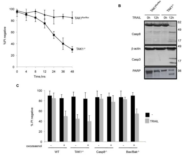
Whereas TAK1flox/flox MEFs survived 48 h treatment with
increasing concentrations of TRAIL, dose-dependent death was
observed in TAK12/2 MEFs (Fig. 1B). To confirm that the
sensitivity of TAK12/2MEFs to TRAIL killing was due the loss of
TAK1 alone, we reintroduced TAK1, or the kinase inactive mutant TAK1-K63W[30], into TAK1 knock out MEFs using a tamoxifen-inducible lentiviral system. As shown in Fig. 1C, the levels of TAK1 and TAK1-K63W increased following induction
with tamoxifen. Complementation of TAK1 in TAK12/2MEFs
effectively protected against TRAIL killing, whereas expression of TAK1-K63W failed to block TRAIL induced death as measured by PI exclusion (Fig. 1D) and MTT cell viability assay (Fig. S1).
In order to determine whether TAK1 deletion could induce TRAIL sensitivity in other cell types, two human cancer cell lines, HT29 and HuH7, were stably infected with lentivirus encoding shRNA against TAK1. The depletion of TAK1 (Fig. S1), sensitized HT29 cells to a greater extent than HuH7 cells to TRAIL (Fig. S1). Lower efficiency of shRNA mediated TAK1 knockdown in HuH7 cells may explain this lower sensitization to TRAIL.
In contrast to TRAIL, TAK1 deletion did not alter sensitivity to killing by FasL (Fig. S1).
TRAIL-Induced Death of TAK1 Deleted Cells Depends on Caspase 8
TAK12/2 MEFs began to die 12–14 h after TRAIL addition
(Fig. 2A), and this was preceded by cleavage of caspase 8, 3 and PARP (Fig. 2B).
To assess whether a Bax/Bak or caspase 8 dependent apoptotic
pathway was required for death of TAK12/2 MEFs caused by
TRAIL, we used 5Z-7-oxozeanol, a selective inhibitor of TAK1[31,32], in caspase 8[33] and Bax/Bak knockout MEFs.
Because TAK1flox/flox cells treated with 5z-7-oxozeanol plus
TRAIL succumbed at a similar rate to TAK12/2MEFs treated
with TRAIL, and the addition of 5z-7-oxozeanol to TAK12/2
MEFs did not cause an increase in cell death beyond that caused by TRAIL alone, at the concentrations used, 5z-7-oxozeanol does not appear to have any off-target effects (Fig. 2C).
Caspase 82/2MEFs were completely resistant to the treatment
with 5z-7-oxozeanol plus TRAIL whereas Bax/Bak double knockout MEFs were efficiently killed by this combination. These results showed that the caspase 8 dependent apoptotic path-way, and not the Bax/Bak pathpath-way, is responsible for death of
TAK12/2MEFs caused by TRAIL.
Figure 1. The kinase activity of TAK1 is required for TRAIL survival in MEFs.(A) Immunoblot analysis of TAK1 levels before and after TAK1flox/floxMEFs were infected with lentivirus expressing Cre protein. (B) Polyclonal populations of wild-type (TAK1flox/flox) and TAK1 knock out (TAK12/2) MEFs were treated with different concentrations of TRAIL for 48 h. (C,D). WT TAK1 but not mutant TAK1 (K63W, kinase null) complementation of TAK12/2 MEFs protects against TRAIL sensitivity. (C) Time course of TAK1 levels after induction with 10 nM of 4-hydroxytamoxifen (4HT). (D) Cells were treated with TRAIL 1mg/ml for 48 h and viability was assessed by PI staining and flow cytometry. The mean
TAK1 Is Required for Activation of NF-kB and JNK Signalling Pathways by TRAIL
TAK1 is essential for NF-kB and JNK activation when cells are
treated with TNF-a, TGF-b, TLR and IL-1[22,23,34]. As there
have been several reports of NF-kB, JNK and p38 activation by
TRAIL [1], we decided to study the role of TAK1 in the activation of these signalling pathways after TRAIL treatment.
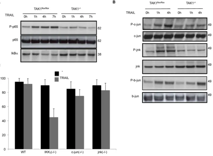
TRAIL induced an increase in the phosphorylation of p65/
RelA NF-kB that peaked at 7 h, and was accompanied by the
degradation of IkBa in TAK1flox/floxMEFs (Fig. 3A). However,
phosphorylation of p65 and IkBa degradation were greatly
impaired in TAK1 knockout MEFs (Fig. 3A).
TRAIL also caused JNK activation, as indicated by an increase in the phosphorylation of both JNK and c-jun in WT MEFs, whereas in the absence of TAK1 this was abolished (Fig. 3B). Moreover, b-jun, a member of the JUN family whose activation is
not regulated by JNK, was phosphorylated in both TAK1flox/flox
and TAK12/2MEFs in response to TRAIL (Fig. 3B), indicating
that TAK1 is not required for b-jun activation.
We also evaluated the kinetics of p38 MAPK activation in response to TRAIL by immunoblot. However, neither short nor
long-term exposure of TAK1flox/floxMEFs to TRAIL was able to
activate p38 pathway (data not shown).
In order to distinguish the relative importance of NF-kB versus
JNK activation in cell survival after TRAIL treatment, we tested the
sensitivity of IKKc, c-jun and jnk knockout MEFs to TRAIL (Fig. 3C).
Interestingly, only IKKc2/2 MEFs were sensitized to
TRAIL-induced cytotoxicity, indicating that NF-kB, but not JNK pathway,
plays a key role in protecting MEFs against killing by TRAIL.
Activation of NF-kB in TAK12/2MEFs Protects Against Killing by TRAIL
To test wether activation of NF-kB was sufficient to protect
TAK12/2MEFs against TRAIL induced cell death, we infected
TAK12/2 MEFs with IKK2EE tamoxifen-inducible lentivirus
(TAK12/2uasIKK2EE). IKK2EE is an active mutant of IKK2
(S177E, S181E) that displays constitutive IKBakinase activity and
activates NF-kB[35].
Figure 2. TRAIL induced death of TAK12/2MEFs is mediated by caspase 8.(A) Time course of viability of TAK1flox/flox
and TAK12/2MEFs exposed to TRAIL 1mg/ml. (B) Cleavage of caspase 8, 3 and PARP in TRAIL treated WT and TAK12/2MEFs. (C) Wild-type, TAK12/2, caspase 8 knock
out (Casp82/2), Bax and Bak double knock out (Bax2/2/Bak2/2) MEFs were coincubated with 30 nM 5Z-7-oxozeanol (an inhibitor of TAK1’s catalytic activity) and TRAIL 1mg/ml for 24 h. The mean and SEM of three independent experiments is shown.
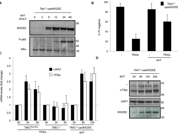
Expression of IKK2EE in TAK12/2MEFs was detectable 6 h after addition of tamoxifen and was accompanied by an increase in
both p65 phosphorylation and IkBa degradation (Fig. 4A).
Moreover, the activation of NF-kB in TAK12/2MEFs protected
cells against TRAIL induced cell death measured by PI exclusion (Fig. 4B) and an MTT cell viability assay (Fig. S2).
NF-kB can induce expression of several pro-survival genes, such
as cIAP2, cFlip and A20, so we measured the levels of mRNA from these genes after TRAIL addition by quantitative real time RT-PCR.
TRAIL treatment of both TAK1flox/floxand TAK12/2MEFs
produced no change in A20 gene expression (data not shown). However as shown in Fig. 4C, TRAIL induced an increase in mRNA levels of cIAP2 and cFlipL, but this did not occur in TAK1
knockout MEFs. Importantly, activation of NF-kB in TAK12/2
MEFs was sufficient to increase the mRNA levels of cIAP2 and FlipL (Fig. 4C), whereas A20 levels remained constant.
Immunoblot analysis revealed that cFLipL protein level
increased in TAK1flox/flox MEFs upon treatment of TRAIL
(Fig. 5A). In contrast, in MEFs lacking TAK1, cFlipL failed to increase after TRAIL addition, but appeared to decrease.
Moreover, complementation of TAK12/2 MEFs with WT, but
not kinase mutant (K63W), TAK1 was able to restore the ability of
TRAIL to induce an increase in cFlipL (Fig. 5B). Consistent with the changes in mRNA levels, the amount of cFlipL protein also
increased after IKK2EE was induced in the TAK12/2 MEFs
(Fig. 4D). The presence of an upper non-specific band (marked with an asterisk) was consistently observed in all the cFlip blots.
Due to the importance of cIAP1 in TNF signalling [28], we also
determined the levels of cIAP1 protein in both TAK1flox/floxand
TAK12/2 MEFs in response to TRAIL (not shown) or after
inducing NF-kB in TAK1 knockout MEFs (Fig. 4D), but no
change was observed.
cFLIPL but Not cIAP2 Overexpression in TAK12/2MEFs Is Able to Inhibit TRAIL-Induced Cell Death
To discern the role of cIAP2 and cFlip in resistance to TRAIL
killing, we over-expressed both proteins in TAK12/2MEFs and
examined their effect on viability of cells treated with TRAIL. First, we cloned all murine cFlip forms: cFlipL, cFlipp43 and cFlipR, into the tamoxifen-inducible lentiviral system, and infected
Flip2/2 MEFs. Induction of cFlipL and cFlipp43 in cFlip2/2
MEFs caused an increase in the abundance of cFlipp43 and a novel cFlip cleaved variant respectively (Fig. S2, indicated by an asterisk), probably as an over-expression artefact, but caused no changes in cellular appearance or growth rate.
Figure 3. In TAK12/2MEFs, TRAIL fails to activate p65/RelA
NF-kB and JNK signalling pathways.MEFs were stimulated with 1mg/ml
TRAIL for the indicated times, and cell lysates were probed for (A), phospho p65, p65, and IkB-ato determine NF-kB activation, and (B), phospho c-jun, JNK and b-jun to determine JNK activation. (C) Viability of Nemo/IKKcknock out MEFs (IKKc2/2), c-jun knock out MEFs (c-jun2/2) and JNK knock out MEFs (JNK2/2) after TRAIL exposure (1
mg/ml, 48 h). The mean and SEM of three independent experiments is shown.
Compared to WT MEFs, Flip2/2 MEFs were extremely sensitive to killing by TRAIL, and induction of cFlipL and cFlipR significantly reduced cell death caused by TRAIL (Fig. S2).
However, when we introduced the different forms of cFlip into
TAK12/2 MEFs and induced their expression with tamoxifen
(Fig. 5C), only cFlipL conferred resistance to death of TAK12/2
cells in response to TRAIL (Fig. 5D).
To determine if this protection by cFlipL was due a change in protein half-life due to the absence of TAK1, we induced cFlipL in both wild type and TAK1 knock out MEFs, and incubated them with cyclohexamide with or without TRAIL. As shown in Fig. 6A, time-dependent degradation in cFlipL was similar in both MEFs, indicating that the cFlipL turnover was not significantly affected by the presence or absence of TAK1.
To see if the increase of cIAP2 gene expression observed in response to TRAIL (Fig. 4C) played any role in cell survival, TAK1
2/2MEFs were infected with a Tet-Off cIAP2 expressing lentivirus.
Expression of high levels of cIAP2 in TAK1 2/2 MEFs were
achieved with this system and the presence of doxycycline (1mg or
10 ng) effectively turned off cIAP2 induction (Fig. 6B). Interestingly,
ectopic expression of cIAP2 in TAK12/2MEFs did not alter their
sensitivity to TRAIL (Fig. 6C). Note however, we were unable to
detect endogenous cIAP2 in MEFs by Western blot (Fig. 6B), suggesting MEFs usually only bear low basal levels of cIAP2.
Discussion
NF-kB and JNK participate in a wide variety of cellular
processes, including immunoregulation, inflammation, cell growth, cell differentiation and cell death. Because both induction of
NF-kB and JNK activation by TRAIL were abolished in TAK1
deficient MEFs, and this corresponded to an increase in sensitivity to TRAIL induced apoptosis, we wanted to determine the role of these signalling pathways in allowing cell survival in the presence of TRAIL.
Our results showed that MEFs lacking genes for two key members of the JNK signalling pathway, c-jun and JNK, were not sensitive to killing by TRAIL, consistent with reports from other groups [36,37].
TRAIL is mainly known for its ability to cause apoptosis, rather than for its ability to activate signal transduction by transcription
factors such as NF-kB. Nevertheless, it has been shown that NF-kB
is activated by the receptor for TRAIL, albeit more slowly and to a lesser extent that that caused by binding of TNF to TNFR1 [14].
Figure 4. Activation of NF-kB by over-expression of IKK2-EE induces cFlip and rescues TAK12/2 MEFs from TRAIL induced cell death.Inducible over-expression of dominant active IKK2-EE in TAK12/2MEFs was accompanied by an increase in phosphorylation of p65 and IkB-a degradation (A), and blocked sensitivity to TRAIL (1mg/ml, 48 h) (B). Expression of both cIAP2 and cFlipL genes was elevated as determined by real
time RT-PCR, using the housekeeping gene 18S rRNA as internal control (C). Western blot for cFlip and cIAP1 in TAK12/2MEFs stably infected with IKK2EE (TAK12/2uasIKK2EE) after addition of 10 nM 4HT for different times (* indicates non-specific band).
Unlike WT MEFs, both TAK12/2MEFs and IKKc2/2MEFs were very sensitive to killing by TRAIL. Because activation of
NF-kB by expression of constitutively active IKK2EE could protect
the TAK1 knock out MEFs, activation of NF-kB is both necessary
and sufficient to allow survival of TRAIL treated cells. Consistent
with this, deletion of IKKc in mouse hepatocytes caused
hypersensitivity to TRAIL, resulting in massive liver damage [38]. Although genetic knockdown and chemical inhibition of TAK1 has been shown to enhance TRAIL-induced apoptosis in a cervical cancer cell line, the mechanism responsible for this sensitization was not described [25]. While preparing this manuscript, two independent groups have proposed different mechanisms to explain the role of TAK1 in TRAIL killing [24,26]. Herrero-Martin et al 2009 concluded that TAK1 induces cytoprotective autophagy in TRAIL-treated breast epithelial cells. In MEFs, we did not observe any sign of autophagy following TRAIL treatment. In fact, caspase 8 knock out MEFs were totally resistant to TRAIL induced death, even when TAK1 was inhibited, indicating that in the absence of TAK1, TRAIL causes death in a caspase 8 dependent manner.
On the other hand, Morioka et al. 2009 [24] showed that deletion of TAK1 in mouse keratinocytes sensitized to TRAIL by ROS-dependent down-regulation of cIAP2, rather than acting via
NF-kB. This might be due to a difference in cell type, because our
results show that for MEFs to survive TRAIL treatment, NF-kB
must activate and drive expression of cFlipL, which fails to occur in TAK1 knock out MEFs.
Interestingly, it has been reported that reduction of cFlipL and cIAP2 protein levels by c-myc is a major determinant of TRAIL sensitivity[39]. We over-expressed cIAP2 in TAK1 knock out cells using a lentiviral Tet-Off system. This approach allowed us to mimic the model of Morioka et al. model because in the presence of doxycycline cIAP2 expression was shut down. Unlike cFLip
expression, cIAP2 over-expression in TAK12/2 MEFs did not
protect against TRAIL. Thus, the downstream signalling pathways activated by TAK1 in response to TRAIL may vary, depending on the cell type.
In some cells levels of cFlip may determine resistance to TRAIL induced apoptosis, because cFlip binds to the DISC complex and can prevent the recruitment and activation of procaspase 8. Although cFlip is constitutively expressed in normal cells, it is highly expressed in human cancer and thus is implicated in tumorigenesis [5,8].
cFlip knock out MEFs were extremely sensitive to TRAIL induced cell death. The protection against TRAIL killing afforded
by reconstitution of Flip2/2MEFs with the two isoforms of cFlip,
Figure 5. c-FlipL over-expression is able to reduce killing of TAK12/2MEFs by TRAIL, whereas Flipp43 and FlipR have little effect.(A) Endogenous c-Flip levels after TRAIL treatment in both TAK12/2, TAK1flox/flox
MEFs and (B) TAK12/2MEFs reconstituted with TAK1 WT or TAK1 (k63W). (C) Lentiviral-mediated inducible expression of the different murine forms of c-Flip (c-FlipL, c-Flipp43, c-FlipR) in TAK12/2MEFs measured by Western blot. (D) The ability of the different forms of c-Flip to protect TAK12/2MEFs against killing by TRAIL (1
mg/ml, 48 h) was evaluated by PI
exclusion.
cFlipL and cFlipR, demonstrated their anti-apoptotic function against TRAIL. In agreement with these results, both c-FlipL and cFlipS are highly expressed in TRAIL-resistant as compared with TRAIL-sensitive human pancreatic cancer cells [13]. Further-more, erythroid differentiation sensitizes leukaemia cells to TRAIL killing by downregulation of both c-Flip splicing isoforms [40].
However, only expression of FlipL, and not the other Flip forms, was able to efficiently block TRAIL killing in TAK1 knock out MEFs. Accordingly, the protective role of cFlipL in response to death ligands has been observed in both over-expression and loss-of-function studies[11,41].
c-Flip expression is carefully regulated at different levels. In addition to regulation of gene transcription, turnover of c-Flip protein is actively controlled by ubiquitin signalled degradation [42]. In fact, JNK activation during TNF signalling has been proposed to promote the proteosomal degradation of c-FlipL. Although loss of TAK1 resulted in inability of TRAIL to activate JNK, the half-life of cFlipL was similar in the presence or absence of TAK1. These observations suggest that modulation of cFlipL’s stability is not involved in its capacity to inhibit TRAIL killing in
TAK12/2MEFs.
Here we report that TAK1 is essential for TRAIL to induce
NF-kB and JNK activation. Moreover, NF-kB activation can
overcome the sensitivity of TAK1 knock out MEFs to killing by
TRAIL. Finally, cFlipL but not another NF-kB dependent
antiapoptotic regulator, cIAP2, can protect TAK12/2 MEFs
from killing by TRAIL.
In conclusion, inhibition or deletion of TAK1 led to impaired
NF-kB dependent cFlipL expression, allowing caspase 8 activation
and cell death in response to TRAIL. Thus, NF-kB may
negatively regulate TRAIL induced cell death in TAK1 knock out MEFs by increasing cFlipL levels. Compounds that inhibit TAK1 may increase the range of tumor cells that are sensitive to TRAIL.
Supporting Information
Figure S1 TAK1 down-regulation sensitizes HT29 and HuH7 cancer cell lines to tumor necrosis factor-related apoptosis-inducing ligand (TRAIL). HT29 and HuH7 cells were infected with a lentivirus expressing TAK1 shRNA or scramble (control). (A,B) TAK1 levels were examined by western blot and sensitivity to TRAIL by PI exclusion. No differences in cell viability between
TAK12/2and TAK1flox/floxMEFs after Fas treatment. (C) Cells
were incubated with Fas during 24 hours. (D) MTT results of the same experiment as Fig. 1D.
Figure 6. Cellular FlipL turnover is not affected by the presence or absence of TAK1, and cIAP2 over-expression in TAK12/2cells fails to protect against TRAIL killing.(A) TAK1flox/floxand TAK12/2MEFs over-expressing cFlipL were incubated with 10
mg/ml cyclohexamide for
different times and kinetics of cFlipL degradation was detected by immunoblot. (B,C) TAK12/2MEFs were stably infected with cIAP2 using a lentiviral Tet-off system. After addition of 1mg/ml-10 ng/ml doxycycline cIAP2 protein levels were determined by Western blot (B), and cell viability in
Found at: doi:10.1371/journal.pone.0008620.s001 (0.53 MB TIF)
Figure S2 c-FlipL and c-FlipR isoforms are the main inhibitors
of TRAIL-induced cell death. Flip knockout MEFs (Flip2/2) were
complemented with FlipL (uasFlipL), Flipp43 (uasFlipp43), and FlipR (uasFlipR). (A) Cell survival was measured after treating
them with TRAIL (1mg/ml, 24 h). (B) Protein levels of the
different forms of Flip were detected by immunoblot. (C) MTT cell viability assay corresponding to the same experiment as Fig. 4B. Found at: doi:10.1371/journal.pone.0008620.s002 (0.51 MB TIF)
Acknowledgments
We would like to thank Shizuo Akira and Osamu Takeuchi for TAK1flox/flox mice, David Huang for Bax/Bak double knock out MEFs, Steve Hedrick for Caspase 8 knock out MEFs, Erwin Wagner for JNK and jun knock out MEFs, and Tak Mak for cFlip knock out MEFs.
Author Contributions
Conceived and designed the experiments: JML JEV JS DLV. Performed the experiments: JML UN WDC DM MM NK DC. Analyzed the data: JML IEG MM WWLW JS. Contributed reagents/materials/analysis tools: UN WDC IEG DM WWLW BAC JS. Wrote the paper: JML DLV.
References
1. Newsom-Davis T, Prieske S, Walczak H (2009) Is TRAIL the holy grail of cancer therapy? Apoptosis 14: 607–623.
2. Kimberley FC, Screaton GR (2004) Following a TRAIL: update on a ligand and its five receptors. Cell Res 14: 359–372.
3. Djerbi M, Darreh-Shori T, Zhivotovsky B, Grandien A (2001) Characterization of the human FLICE-inhibitory protein locus and comparison of the anti-apoptotic activity of four different flip isoforms. Scand J Immunol 54: 180–189. 4. Ueffing N, Keil E, Freund C, Kuhne R, Schulze-Osthoff K, et al. (2008) Mutational analyses of c-FLIPR, the only murine short FLIP isoform, reveal requirements for DISC recruitment. Cell Death Differ 15: 773–782. 5. Bullani RR, Huard B, Viard-Leveugle I, Byers HR, Irmler M, et al. (2001)
Selective expression of FLIP in malignant melanocytic skin lesions. J Invest Dermatol 117: 360–364.
6. Okano H, Shiraki K, Inoue H, Kawakita T, Yamanaka T, et al. (2003) Cellular FLICE/caspase-8-inhibitory protein as a principal regulator of cell death and survival in human hepatocellular carcinoma. Lab Invest 83: 1033–1043. 7. Remmelink M, Mijatovic T, Gustin A, Mathieu A, Rombaut K, et al. (2005)
Identification by means of cDNA microarray analyses of gene expression modifications in squamous non-small cell lung cancers as compared to normal bronchial epithelial tissue. Int J Oncol 26: 247–258.
8. Ryu BK, Lee MG, Chi SG, Kim YW, Park JH (2001) Increased expression of cFLIP(L) in colonic adenocarcinoma. J Pathol 194: 15–19.
9. Clarke P, Tyler KL (2007) Down-regulation of cFLIP following reovirus infection sensitizes human ovarian cancer cells to TRAIL-induced apoptosis. Apoptosis 12: 211–223.
10. Geserick P, Drewniok C, Hupe M, Haas TL, Diessenbacher P, et al. (2008) Suppression of cFLIP is sufficient to sensitize human melanoma cells to TRAIL-and CD95L-mediated apoptosis. Oncogene 27: 3211–3220.
11. Sharp DA, Lawrence DA, Ashkenazi A (2005) Selective knockdown of the long variant of cellular FLICE inhibitory protein augments death receptor-mediated caspase-8 activation and apoptosis. J Biol Chem 280: 19401–19409. 12. Siegmund D, Hadwiger P, Pfizenmaier K, Vornlocher HP, Wajant H (2002)
Selective inhibition of FLICE-like inhibitory protein expression with small interfering RNA oligonucleotides is sufficient to sensitize tumor cells for TRAIL-induced apoptosis. Mol Med 8: 725–732.
13. Wang P, Zhang J, Bellail A, Jiang W, Hugh J, et al. (2007) Inhibition of RIP and c-FLIP enhances TRAIL-induced apoptosis in pancreatic cancer cells. Cell Signal 19: 2237–2246.
14. Hu WH, Johnson H, Shu HB (1999) Tumor necrosis factor-related apoptosis-inducing ligand receptors signal NF-kappaB and JNK activation and apoptosis through distinct pathways. J Biol Chem 274: 30603–30610.
15. Jin Z, El-Deiry WS (2006) Distinct signaling pathways in TRAIL- versus tumor necrosis factor-induced apoptosis. Mol Cell Biol 26: 8136–8148.
16. Lin Y, Devin A, Cook A, Keane MM, Kelliher M, et al. (2000) The death domain kinase RIP is essential for TRAIL (Apo2L)-induced activation of IkappaB kinase and c-Jun N-terminal kinase. Mol Cell Biol 20: 6638–6645. 17. Harper N, Farrow SN, Kaptein A, Cohen GM, MacFarlane M (2001)
Modulation of tumor necrosis factor apoptosis-inducing ligand- induced NF-kappa B activation by inhibition of apical caspases. J Biol Chem 276: 34743–34752.
18. Diessenbacher P, Hupe M, Sprick MR, Kerstan A, Geserick P, et al. (2008) NF-kappaB inhibition reveals differential mechanisms of TNF versus TRAIL-induced apoptosis upstream or at the level of caspase-8 activation independent of cIAP2. J Invest Dermatol 128: 1134–1147.
19. Kim SH, Ricci MS, El-Deiry WS (2008) Mcl-1: a gateway to TRAIL sensitization. Cancer Res 68: 2062–2064.
20. Kumar-Sinha C, Varambally S, Sreekumar A, Chinnaiyan AM (2002) Molecular cross-talk between the TRAIL and interferon signaling pathways. J Biol Chem 277: 575–585.
21. Varfolomeev E, Maecker H, Sharp D, Lawrence D, Renz M, et al. (2005) Molecular determinants of kinase pathway activation by Apo2 ligand/tumor necrosis factor-related apoptosis-inducing ligand. J Biol Chem 280: 40599–40608.
22. Adhikari A, Xu M, Chen ZJ (2007) Ubiquitin-mediated activation of TAK1 and IKK. Oncogene 26: 3214–3226.
23. Ninomiya-Tsuji J, Kishimoto K, Hiyama A, Inoue J, Cao Z, et al. (1999) The kinase TAK1 can activate the NIK-I kappaB as well as the MAP kinase cascade in the IL-1 signalling pathway. Nature 398: 252–256.
24. Morioka S, Omori E, Kajino T, Kajino-Sakamoto R, Matsumoto K, et al. (2009) TAK1 kinase determines TRAIL sensitivity by modulating reactive oxygen species and cIAP. Oncogene 28: 2257–2265.
25. Choo MK, Kawasaki N, Singhirunnusorn P, Koizumi K, Sato S, et al. (2006) Blockade of transforming growth factor-beta-activated kinase 1 activity enhances TRAIL-induced apoptosis through activation of a caspase cascade. Mol Cancer Ther 5: 2970–2976.
26. Herrero-Martin G, Hoyer-Hansen M, Garcia-Garcia C, Fumarola C, Farkas T, et al. (2009) TAK1 activates AMPK-dependent cytoprotective autophagy in TRAIL-treated epithelial cells. EMBO J 28: 677–685.
27. Grosse-Wilde A, Voloshanenko O, Bailey SL, Longton GM, Schaefer U, et al. (2008) TRAIL-R deficiency in mice enhances lymph node metastasis without affecting primary tumor development. J Clin Invest 118: 100–110.
28. Vince JE, Wong WW, Khan N, Feltham R, Chau D, et al. (2007) IAP antagonists target cIAP1 to induce TNFalpha-dependent apoptosis. Cell 131: 682–693.
29. Sato S, Sanjo H, Takeda K, Ninomiya-Tsuji J, Yamamoto M, et al. (2005) Essential function for the kinase TAK1 in innate and adaptive immune responses. Nat Immunol 6: 1087–1095.
30. Wang C, Deng L, Hong M, Akkaraju GR, Inoue J, et al. (2001) TAK1 is a ubiquitin-dependent kinase of MKK and IKK. Nature 412: 346–351. 31. Ninomiya-Tsuji J, Kajino T, Ono K, Ohtomo T, Matsumoto M, et al. (2003) A
resorcylic acid lactone, 5Z-7-oxozeaenol, prevents inflammation by inhibiting the catalytic activity of TAK1 MAPK kinase kinase. J Biol Chem 278: 18485–18490.
32. Safwat N, Ninomiya-Tsuji J, Gore AJ, Miller WL (2005) Transforming growth factor beta-activated kinase 1 is a key mediator of ovine follicle-stimulating hormone beta-subunit expression. Endocrinology 146: 4814–4824.
33. Beisner DR, Ch’en IL, Kolla RV, Hoffmann A, Hedrick SM (2005) Cutting edge: innate immunity conferred by B cells is regulated by caspase-8. J Immunol 175: 3469–3473.
34. Chen ZJ, Bhoj V, Seth RB (2006) Ubiquitin, TAK1 and IKK: is there a connection? Cell Death Differ 13: 687–692.
35. Mercurio F, Zhu H, Murray BW, Shevchenko A, Bennett BL, et al. (1997) IKK-1 and IKK-2: cytokine-activated IkappaB kinases essential for NF-kappaB activation. Science 278: 860–866.
36. Sah NK, Munshi A, Kurland JF, McDonnell TJ, Su B, et al. (2003) Translation inhibitors sensitize prostate cancer cells to apoptosis induced by tumor necrosis factor-related apoptosis-inducing ligand (TRAIL) by activating c-Jun N-terminal kinase. J Biol Chem 278: 20593–20602.
37. Noutomi T, Itoh M, Toyota H, Takada E, Mizuguchi J (2009) Tumor necrosis factor-related apoptosis-inducing ligand induces apoptotic cell death through c-Jun NH2-terminal kinase activation in squamous cell carcinoma cells. Oncol Rep 22: 1169–1172.
38. Beraza N, Malato Y, Sander LE, Al-Masaoudi M, Freimuth J, et al. (2009) Hepatocyte-specific NEMO deletion promotes NK/NKT cell- and TRAIL-dependent liver damage. J Exp Med 206: 1727–1737.
39. Ricci MS, Jin Z, Dews M, Yu D, Thomas-Tikhonenko A, et al. (2004) Direct repression of FLIP expression by c-myc is a major determinant of TRAIL sensitivity. Mol Cell Biol 24: 8541–8555.