*Correspondence: Omair Anwar Mohiuddin. Dow college of Pharmacy, Dow University of Health Sciences, Gulzar-e-Hijri, Suparco road, KDA scheme 33, Karachi, Pakistan. E-mail: [email protected]
A
vol. 51, n. 1, jan./mar., 2015 http://dx.doi.org/10.1590/S1984-82502015000100023
Evaluation of the effect of natural peptide
‘Urocortin’
on
corticotrophin releasing factor (CRF) receptor expression in
ND7/23 cells
Omair Anwar Mohiuddin
1,*, Chris Biggs
21Department of Pharmaceutics, Dow College of Pharmacy, Dow University of Health sciences, Karachi, Pakistan, 2School of Biosciences, University of Westminster, London, UK
CRF receptors are involved in the stress management of the cells and are believed to have a cytoprotective role in the body. CRF receptors have been reported to be potential drug targets for the treatment of neurodegenerative disorders. The cell line used in the study is ND7/23 (mouse neuroblastoma and rat
dorsal root ganglion neuron hybridoma). The aim of the study was to conirm the expression of CRF receptors in ND7/23 cells and to determine if urocortin (Ucn) can enhance the expression of CRF
receptors. ND7/23 cells were cultured in RPMI 1640 media and cells grown after the second passage
were used for the experiments. RNA was extracted from the cells and ampliied by RT-PCR to conirm the presence of CRF receptors. The cells were then subjected to oxidative stress by hydrogen peroxide
(0.00375%) and divided into two groups i.e. control and Ucn (10-8 μM) treated. Later RNA was extracted
from both group of cells and PCR was performed. Finally, densitometry analysis was conducted on the
agarose gel to determine the quantity of PCR product formed. PCR experiment conirmed the expression of both CRF-R1 and CRF-R2 in the cell line, but CRF-R1 was found to be expressed more strongly. Densitometry analysis of the PCR product and calculation of the relative expression of CRF receptors indicated a higher level of expression of CRF receptors in samples treated with Ucn as compared to
those that were kept untreated. The results indicate that Ucn may be useful for the management of neuro-degenerative disorders and further studies may be carried out to establish its use as a therapeutic agent.
Uniterms: Urocortin. ND7/23 cells. Corticotrophin releasing factor. RT-PCR. Neuronal cells.
Receptores de CRF estão envolvidos na gestão do estresse das células e são acreditados para ter um papel de cito-proteção no organismo. Os receptores do CRF têm sido relatados como alvos potenciais de fármacos para o tratamento de doenças neurodegenerativas. A linhagem celular utilizada no estudo é ND7/23 (neuroblastoma de camundongo e hibridoma de raíz dorsal do neurônio ganglionar de
rato). O objetivo do estudo foi conirmar o que a expressão de receptores de CRF em células ND7/23 determinar se urocortina (Ucn) pode aumentar a expressão de receptores de CRF. Cultivaram-se células
ND7/23 em meio RPMI 1640 e as células que cresceram após a segunda passagem foram usadas para
os experimentos. O RNA foi extraído células e ampliicado por RT-PCR para conirmar a presença de receptores de CRF. As células foram, então, submetidas a estresse oxidativo por peróxido de hidrogênio
(0.00375 %) e divididas em dois grupos, ou seja, controle e tratadas com UCN (10-8 µM). Em seguida,
o RNA foi extraído de ambos os grupo de células e realizou-se o PCR. Finalmente, realizou-se análise
densitométrica em gel de agarose para determinar a quantidade de produto formado por PCR. O PCR
conirmou a expressão de CRF-R1 e CRF-R2 na linhagem celular, mas o CRF-R1 expresso mais fortemente. A análise densitométrica do produto de PCR e o cálculo da expressão relativa de receptores de CRF indicaram um nível mais elevado de expressão de receptores de CRF em amostras tratadas com
Ucn, em comparação com aqueles sem tratamento. Os resultados indicam que a Ucn pode ser útil no tratamento de doenças neurodegenerativas e mais estudos podem ser realizados para estabelecer seu uso como agente terapêutico.
INTRODUCTION
Corticotrophin releasing factor (CRF) was first discovered in 1981 and was found to be a regulator of the endocrine stress response (Bale, Wale, 2004). CRF receptors are members of G-protein coupled receptor (GPCR) family. The two main subtypes of CRF receptors are CRF receptor 1 (CRF-R1) and CRF receptor 2 (CRF-R2), which are products of two separate genes (Roloff et al., 1998). These receptors have various splice
variants that are expressed in both tissues of central and peripheral region, with CRF-R2 existing as α and β isoforms (Grigoriadis, 2001). Based on amino acid
sequences, CRF-R1 and CRF-R2 have almost 70% homology, and only differ at one end of the amino acid sequence (Bale, Wale, 2004).
CRF is a vital regulator of endocrine response to stress (Meaney et al., 1996); it is found in the region of paraventricular nucleus (PVN) of the hypothalamus, central nucleus of amygdala and hindbrain regions of the CNS (Qiu
et al., 2005). It is also found in peripheral regions, such as gut, skin and adrenal gland (Bale, Wale, 2004). CRF-R1 has been reported to be responsible for the stress response on the cellular level, whereas the role of CRF-R2 in stress response has not been clearly established yet (Qiu et al., 2005).
In the current study, the cell line used was ND7/23, which is a hybrid of mouse neuroblastoma (N18 tg 2) and rat dorsal root ganglion neuron, produced by PEG mediated cell fusion (Herold et al., 2009). ND7/23 cells had not been used for studies related to CRF receptors before. Therefore,
there were no details available for the expression of CRF
receptors in this cell line. The aim of this study was to detect
if CRF receptors are expressed in ND7/23 cells.
Urocortin (Ucn) is a naturally occurring hormone,
which has been identiied as a natural analogue of CRF and
a neuroprotective agent (Abuirmeileh et al., 2007). Ucn is a peptide comprising of 40 amino acids and it belongs to CRF peptide family, functioning via corticotrophin releasing factor receptors (CRFR) (Wang et al., 2008).
Ucn has been found to have afinity for both CRFR1 and
CRFR2 (Tezval et al., 2009). The possible cyto-protective role of Ucn has been suggested by many previous studies for cardiac myocytes (Brar et al., 2000). The optimum dose
of Ucn to exert a protective effect for cardiac myocytes has
been determined to be 10-8 M (Brar et al., 2002).
Mouse CRF-R1 protein has 97% homology with human CRF-R1 protein, whereas mouse CRF-R2 protein has 92% homology with human CRF-R2 protein. Mouse Ucn has around 79% similarity with human Ucn (Sequence
homologies determined using NCBI/BLAST online). These observations were signiicant for the study since
mouse cells were being used. High sequence homologies between mouse and human CRF-R1 and CRF-R2 protein and Ucn ensured that human CRF-R1 and CRF-R2 primers should provide appropriate PCR product. High sequence homologies also mean that the results can be applied to studies involving human models as well in the future (Kwitek et al., 2001).
Different neuro-degenerative disorders like Parkinson’s disease (PD) are caused due to the loss of neurons (Chinta et al., 2007). Originally, it was postulated
that the cell death is caused by apoptosis, excitotoxic
events or free radical production (Gandhi, Wood, 2005). More recently, it has been suggested that pre- and post-natal neuroinflammation might be a cause of neuronal disorders (Block, Zecca, Hong, 2007). Therefore,
protection of neuronal cells using Ucn can be extremely
useful for untreatable neuronal disorders (Martin et al., 2007). The latest efforts to treat such disorders have been focused on the neuronal level by protecting the neurons from apoptosis (Sredni et al., 2007). The main aim of the
study was to explore more about the neuro-protective role of Ucn in cells subjected to oxidative stress by hydrogen peroxide. The results obtained from the current study can
help establish the use of Ucn for the treatment of neuro-degenerative disorders.
MATERIAL AND METHODS
Cell culture
The cell line used for the experiments was ND7/23
rat dorsal root ganglia/mouse neuro-blastoma hybrid (acquired from ECACC). Frozen cells were thawed and then spun down in a centrifuge at 1500 rpm, supernatant was discarded and cells were re-suspended into
RPMI-1640 medium in 15 mL lasks. Flasks were then incubated for a few days in humidiied incubator at 37 °C and 5%
CO2 (Oliveira et al., 2010). For the second passage DPBS (Dulbecco’s phosphate buffered saline) was added to the
lasks, cells were centrifuged and transferred to further lasks along with fresh RPMI 1640 media.
MTT assay
Toxicity of Ucn and hydrogen peroxide was
determined using MTT assay, in order to identify the optimum therapeutic dose of Ucn and optimum sub-lethal
dose of hydrogen peroxide for the experiments. To perform
MTT assay 96 well plate format was used. 8 wells each,
in four rows of the plate was filled with 100 µL of cell
RPMI-1640. The plate was left overnight in the incubator, to allow
cells to grow. Next day the cells in the 96 wells plate were subjected to different concentrations of hydrogen peroxide, to induce oxidative stress and apoptosis. First well in each of the four rows was kept as control and then the next six wells were added with six different concentrations of hydrogen peroxide, which were 0.0018%, 0.00375%, 0.0075%,
0.015%, 0.03% and 0.06%.
Another 96 well plate was also set up, where the
wells were again illed with 100 µl of cell suspension and left over night in the incubator. In each of the irst four rows of the plate, the irst well was kept as control and cells in the next six wells were treated with increasing
concentration of Ucn, which were 1×10⁻⁸ M, 2×10⁻⁸ M, 4×10⁻⁸ M, 1×10⁻⁷ M, 2×10⁻⁷ M and 4×10⁻⁷ M.
RNA extraction
RNA extraction was carried out using the protocol
mentioned by Chomczynski and Mackey (1995). Cultured
cells from each lask were centrifuged, all cells were then
homogenized with trizol and transferred to sterile Eppendorf tubes and centrifuged again. RNA pellet in the form of a faint smear appeared at the bottom of the tubes at the end of the process. The supernatant was removed carefully and
1 mL of 75% ethanol was added to the tubes for desalting
of the pellet and the samples were again centrifuged. Eppendorf concentrator (model 5301) was used to dry the RNA pellets. After drying the pellets, they were re-dissolved
in 50 µL of diethylpyrocarbonate (DEPC) water.
Treatment of cells with hydrogen peroxide and Ucn
ND7/23 cells were cultured in four flasks and
allowed to grow to adequate quantity. The cells in the irst lask were used as control i.e. untreated (sample A). Cells in second lask were treated with 1×10⁻⁸ M Ucn (sample B). The third lask contained cells treated with 0.00375% (optimum sub-lethal concentration) hydrogen peroxide (sample C). In the fourth lask, cells were treated with both 0.00375% (sub-lethal concentration) hydrogen peroxide
and 1×10⁻⁸ M Ucn (sample D). After 24 hours of incubation of all the mentioned cell samples, RNA extraction was
performed.
PCR of RNA extracted from control and treated cell samples
The extracted RNA was subjected to PCR using rat CRFR primers. Since the extracted samples contained
RNA, therefore, reverse transcriptase PCR (RT-PCR) was
performed to identify expression of CRF receptors, for that
purpose, Qiagen® one step PCR kit was used.
RNA extracted from cell samples A, B, C and D
were amplified by PCR and the same PCR protocol as mentioned earlier was utilized. First PCR was performed
on sample A and B, where extracted RNA from both samples were mixed with each CRF-R1 primer, CRF-R2
primer, GAPDH primer (housekeeping gene) and KIR 6.1 primer (since Ucn is considered to function through potassium channel) in separate tubes and PCR was carried out. Second PCR was performed for samples treated
with 0.00375% hydrogen peroxide with and without Ucn (sample C and D). Both samples were mixed separately
with each primer i.e. CRF-R1, CRF-R2, GAPDH and KIR 6.1 and PCR was performed.
The primer sequences of all the primers used to perform PCR are as follows.
Rat CRF-R1 primer:
Forward primer: 5’ ACAAACAATGGCTACCGGGA 3’ Reverse primer: 5’ TCATGGGGCCCTGGTAGAT 3’
Rat CRF-R2 primer:
Forward primer: 5’ TGTGGAAGGCTGCTACCTG 3’ Reverse primer: 5’ GTGTGCTTGATGCTGTGGAA 3’
GAPDH primer:
Forward primer: 5’ CATCATCTCTGCCCCCTCTG 3’ Reverse primer: 5’ CCTGCTTCACCACCTTCTTG 3’
KIR primer:
Forward primer: 5’ GCTTTGTGTCCATTGTGACTG 3’ Reverse primer: 5’ GCTGTCATGATTCCGATGTG 3’
Gel electrophoresis
2% agarose gel was prepared by dissolving 2 g
agarose in 100 mL TBE buffer. Gel electrophoresis was
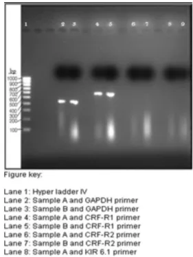
performed using savant PS250 power pack for 60 minutes at 105 V. The PCR products from control and treated cell samples were also run on an agarose gel and visualized in UVipro light box visualizer; using UVtec gel analysis software (Figures III and IV).
Densitometric analysis
for densitometric analysis. The band sizes provided an
approximate idea of the amount of PCR product formed.
The amount of product formed gave an estimate of the
extent of receptor expression (Tables I and II).
CRF receptor expression
For the determination of relative expression of
CRF receptors to GAPDH gene, the ratio of band areas (determined by densitometry) of GAPDH gene (PCR product) to CRF receptor gene (PCR product) were calculated for respective cell samples (untreated and treated with Ucn).
RESULTS AND DISCUSSION
ND7/23 cells exhibited slow growth during the irst passage, whereas growth was much better in later
passages. Over all ND7/23 cells displayed very good growth in RPMI 1640 media within 3-4 days.
MTT assay performed on cells treated with Ucn was used to determine cell viability. Mean values of percentage cell survival for 4 wells of each concentration of Ucn and
hydrogen peroxide have been plotted in Figure 1 and 2,
respectively. The percentage cell survival of each sample was calculated relative to the absorbance of control well.
The igure shows that maximum cell viability is observed
when cells were treated with Ucn at 1×10⁻⁸ µM dose, which is in accordance with the concentration by Brat et al. (2000), in the case of cardiac myocytes. Hence, this
dose was used for all proceeding experiments.
Second MTT assay was performed to determine
sub-lethal dose of hydrogen peroxide that could be used to induce oxidative stress to cells without causing complete
cell death. 0.00375% hydrogen peroxide was found to cause signiicant stress to the cells and was therefore was chosen for all proceeding experiments.
In Figure 3 clear bands are visible in lane 2 and 3 that contain PCR products of control and GAPDH primer, but the band in lane 3 appears to be brighter than the band in
lane 2. Lane 3 contained the PCR product of RNA sample
from cells treated with Ucn. Clear bands are visible in lane 4 and 5, which contained PCR product of CRF-R1 primer and controls; here as well the PCR product of RNA sample
extracted from cells treated with Ucn is brighter than the
FIGURE 1 – Effect of different concentrations of Ucn on ND7/23 cells survival.
FIGURE 2 – Effect of different concentrations of hydrogen
peroxide on ND7/23 cells survival.
with the band area produced by the housekeeping gene,
which was GAPDH. Relative expression of CRF receptor
to GAPDH was calculated using the values obtained by
densitometry. Calculation of relative expression (Table III)
of the CRF receptors to GAPDH products clearly indicates
a higher level of expressions of CRF-R1 and CRF-R2 in
samples treated with Ucn as compared to those, which were not treated with Ucn.
The current study has established the presence and
expression of both CRF receptors in ND7/23 cell line, but the results also clearly suggest that CRF-R1 is expressed in
much greater quantity as compared to CRF-R2. It has been
conirmed by previous studies that Ucn binds to CRF-R2
(Bale, Wale, 2004), but unlike CRF-R1, its effect on cell survival when working via CRF-R2 has not yet been
TABLE III - Relative expression of CRF receptors to GAPDH
receptor in all cell samples
Sample Relative expression
A + CRFR1 primer (untreated) 1.07
B + CRFR1 primer (treated with Ucn) 1.10
C + CRFR1 primer (untreated) 0.00
D + CRFR1 primer (treated with Ucn) 0.0877
C + CRFR2 primer (untreated) 0.00
D + CRFR2 primer (treated with Ucn) 0.7
TABLE II - Showing band areas by densitometric analysis of Figure 4
BAND BAND AREA
Lane 2 649
lane 3 989
Lane 4 Too small to calculate
Lane 5 868
Lane 7 693
Note: band area is mentioned in arbitrary units
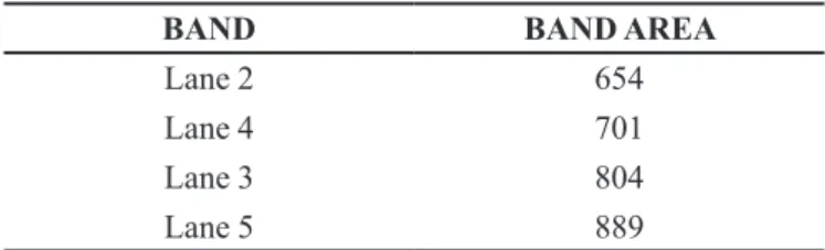
TABLE I – Showing band areas by densitometry analysis of Figure 3
BAND BAND AREA
Lane 2 654
Lane 4 701
Lane 3 804
Lane 5 889
Note: band area is mentioned in arbitrary units
one not treated with Ucn. No bands are visible in lanes 6 to 9; therefore, it can believed that no PCR product was formed with CRF-R2 and KIR6.1 primers, which could have been due to non-homologous primers, improper
reaction conditions or that the cells do not express the
genes at all. Visually it is evident that samples treated with Ucn resulted in brighter bands.
In Figure 4 clear bands are visible in lane 2 and 3 that contained PCR products of GAPDH primer and controls. The band in lane 3 is considerably brighter than the band
in lane 2. Lane 3 contained PCR product of RNA sample extracted from the cell sample treated with Ucn. Clear
band is visible in lane 5 that contains PCR product of RNA sample from cells treated with Ucn and CRF-R1 primer. A faint band is visible in lane 4 which contained RNA
extracted from the cell sample that was not treated with Ucn. Lane 6 and 7 contained PCR products with CRF-R2
primers and a band is only visible in lane 7 that contained
RNA sample extracted from cell sample treated with Ucn.
No bands are visible in lane 8 and 9 that contained PCR products with KIR6.1 primer, which could have been due to reasons as discussed earlier.
Densitometry analysis (Table I and II) was performed to determine the band area of PCR product obtained on the gel. The band area of CRFR primers was compared
FIGURE 4 - Agarose gel photograph showing PCR products of
samples treated with 0.00375% H2O2 with GAPDH, CRF-R1
established (Sawada, Fukui, Hawkes, 2008). Therefore,
greater expression of CRF-R1 certainly increase the
chance of attaining more cell viability after treatment, but
further experiments need to be conducted to conirm this phenomenon. The irst indication of higher expression of CRF receptors in both igures was the brightness of
the bands obtained from PCR products of all samples
pre-treated with Ucn and the later calculations conirmed
the hypothesis. Since it is known that Ucn binds to CRF receptors, and all other components in each sample treated with Ucn were same as in samples not treated with Ucn; thus the presence of higher amount of CRF receptor protein in the cell samples indicates that Ucn has up-regulated the CRF gene. The increased amount of CRF receptor protein in samples could be either because of an
increased expression or the presence of higher number
of viable cells. The PCR product of GAPDH gene from cell sample treated with Ucn is brighter as compared to untreated sample, which could have been due to increased survival of cells in samples treated with Ucn.
Gene expression experiment was performed as a unique experiment due to lack of funds and for future studies it would be more useful if the experiment could be replicated to further conirm the results.
CONCLUSION
The results obtained from PCR confirmed that
ND7/23 cells express CRF-R1 and CRF-R2. CRF-R1 was found to be expressed more strongly as compared to CRF-R2. The results obtained from experiments showing
increased concentration of RNA in samples treated with Ucn are suggestive that treatment of cells with Ucn has
positive effect on CRF receptor expression. Amongst the PCR products, higher expression of CRF receptors was
observed in samples that were treated with Ucn. From the
set of experiments performed in this study there is evidence
to suggest that Ucn has up regulated CRF receptors. Since CRF receptors are involved in the stress management of
cells in the body, therefore increased expression of CRFR
should increase the survival of cells by protecting against
oxidative stress. The results obtained would be helpful
in determining the use of Ucn for the treatment of neuro-degenerative disorders like PD.
ACKNOWLEDGMENTS
The authors would like to thank the School of Biosciences at University of Westminster for providing technical support throughout the study.
REFERENCES
ABUIRMEILEH, A.; HARKAVYI, A.; LEVER, R.; BIGGS
C. S.; WHITTON, P. S. Urocortin, a CRF-like peptide, restores key indicators of damage in the substantia nigra
in a neuroinlammatory model of Parkinson’s disease. J.
Neuroinlamm., v.4, n.19, p.1-5, 2007.
BALE, T. L.; WALE, W. W. CRF and CRF receptors: role
in stress responsivity and other behaviors. Annu. Rev.
Pharmacol. Toxicol., v.44, p.525-527, 2004.
BLOCK, M. L.; ZECCA, L.; HONG, J. S. Microglia-mediated
neurotoxicity: uncovering the molecular mechanism. Nat.
Rev. Neurosci., v.8, n.1, p.57-69, 2007.
BRAR, B. K.; JONASSEN, A. K.; STEPHANOU, A.; SANTILLI, G.; RAILSON, J.; KNIGHT, R. A.; YELLON, D. M.; LATCHMAN, D. S. Urocortin protects against
ischemic and reperfusion injury via a MAPK-dependant
pathway. J. Biol. Chem., v.275, n.12, p.8508-8514, 2000.
BRAR, B.K.; RAILSON, J.; STEPHANOU, A.; KNIGHT, R. A.; LATCHMAN, D. S. Urocortin increases the expression
of heat shock protein 90 in rat cardiac myocytes in a
MEK1/2-dependent manner. J. Endocrinol., v.172, n.2,
p.283-293, 2002.
CHINTA, S. J.; KUMAR, M. J.; HSU, M.; RAJAGOPALAN,
S.; KAUR, D.; RANE, A.; NICHOLLS, D. G.; CHOI, J.;
ANDERSEN, J. K.Inducible alterations of glutathione
levels in adult dopaminergic midbrain neurons result in
nigrostriatal degeneration. J. Neurosci., v.27, n.51,
p.13997-14006, 2007.
CHOMCZYNSKI, P.; MACKEY, K. Modification of
the TRI reagent™ procedure for isolation of RNA from polysaccharide - and proteoglycan - rich sources. Biotechniques, v.19, n.6, p.942-945, 1995.
GANDHI, S.; WOOD, N. W. Molecular pathogenesis of
Parkinson’s disease. Hum. Mol. Genet., v.14, n.2,
p.2749-2755, 2005.
GRIGORIADIS, D. E.; HADDACH, M.; LING, N.; SAUNDERS, J. The CRF receptor structure, function and
potential for therapeutic intervention. Curr. Med. Chem.:
HEROLD, K. F.; NAU, C.; OUYANG, W.; HEMMINGS Jr., H.C. Isolurane inhibits the tetrodotoxin-resistant
voltage-gated sodium channel Nav1.8. Anesthesiology, v.111, n.3,
p.591-599, 2009.
KWITEK, A. E.; TONELLATO, P. J.; CHEN, D.; HANDLEY,
J. G.; CHENG, Y. S.; TWIGGER, S.; SCHEETZ ,T. E.;
CASAVANT, T. L.; STOLL, M.; NOBREGA, M. A.; SHIOZAWA, M.; SOARES, M. B.; SHEFFIELD, V. C.; JACOB, H. J. Automated construction of high-density
comparative maps between rat, human, and mouse. Genome
Res., v.11, n.11, p.1935-1943, 2001.
MARTIN, B.; LOPEZ DE MATURANA, R.; BRENNEMAN, R.; WALENT, T.; MATTSON, M. P.; MAUDSLEY, S.
Class II G protein-coupled receptors and their ligands in
neuronal function and protection. Neuromol. Med., v.7,
n.1/2, p.3-36, 2005.
MEANEY, M. J.; DIORIO, J.; FRANCIS, D.; WIDDOWSON, J.; LaPLANTE, P.; CALDJI, C.; SHARMA, S.; SECKL, J. R.; PLOTSKY, P. M. Early environmental regulation of forebrain glucocorticoid receptor gene expression:
implications for adrenocortical responses to stress. Dev.
Neurosci., v.18, n.1/2, p.49-72, 1996.
OLIVEIRA, H.; PIRES, L. R.; FERNANDEZ, R.; MARTINS,
M. C.; SIMÕES, S.; PÊGO, A. P. Chitosan-based gene delivery vectors targeted to the peripheral nervous system. J. Biomed. Mater. Res. A, v.95, n.3, p.801-810, 2010.
PACHNER, A. R.; RICALTON, N. S. Western blotting in
evaluating Lyme seropositivity and the utility of a gel
densitometric approach. Neurology, v.42, n.11,
p.2185-2192, 1992.
QIU, D. L.; CHU, C. P.; SHIRASAKA, T.; TSUKINO, H.;
NAKAO, H.; KATO, K.; KUNITAKE, T.; KATOH, T.; KANNAN, H.Corticotrophin-releasing factor augments the
IH in rat hypothalamic paraventricular nucleus parvocellular
neurons in vitro. J. Neurophysiol., v.94, n.1, p.226-234,
2005.
ROLOFF, B.; FECHNER, K.; SLOMINSKI, A.; FURKERT, J.; BOTCHKAREV, V. A.; BULFONE-PAUS, S., ZIPPER, J.; KRAUSE, E.; PAUS, R. Hair cycle-dependent expression of
corticotrophin-releasing factor (CRF) and CRF receptors in
murine skin. FASEB J., v.12, n.3, p.287-297, 1998.
SAWADA, K.; FUKUI, Y.; HAWKES, R. Spatial distribution
of corticotrophin-releasing factor immunopositive climbing
ibers in the mouse cerebellum: analysis by whole mount
immunohistochemistry. Brain Res., v.1222, p.106-117,
2008.
SREDNI, B.; GEFFEN-ARICHA, R.; DUAN, W.; ALBECK,
M.; SHALIT, F.; LANDER, H. M.; KINOR, N.; SAGI,
O.; ALBECK, A.; YOSEF, S.; BRODSKY, M.; SREDNI-KENIGSBUCH, D.; SONINO, T.; LONGO, D. L.; MATTSON, M. P.; YADID, G. Multifunctional tellurium
molecule protects and restores dopaminergic neurons in
Parkinson’s disease models. FASEB J., v.21, n.8,
p.1870-1883, 2007.
TEZVAL, H.; JURK, S.; ATSCHEKZEI, F.; BECKER, J. U.;
JAHN, O.; SERTH, J.; KUCZYK, M.A. Urocortin and
corticotropin-releasing factor receptor 2 in human renal cell carcinoma: disruption of an endogenous inhibitor of
angiogenesis and proliferation. World J. Urol., v.27, n.6,
p.825-830, 2009.
WANG, J.; XU, Y.; XU, Y.; ZHU, H.; ZHANG, R.; ZHANG, G.; LI, S. Urocortin’s inhibition of tumor growth and
angiogenesis in hepatocellular carcinoma via
corticotrophin-releasing factor receptor 2. Cancer Invest., v.26, n.4,
p.359-368, 2008.
Received for publication on 13th December 2013