Serum Levels of Monocyte Chemoattractant
Protein-1 and All-Cause and Cardiovascular
Mortality among Patients with Coronary
Artery Disease
Ding Ding1,2, Dongfang Su1, Xinrui Li1, Zhongxia Li1, Yujie Wang2, Jian Qiu3, Puqing Lin3, Yuan Zhang3, Pi Guo4, Min Xia1, Dan Li1*, Yan Yang1, Gang Hu2*, Wenhua Ling1
1Guangdong Provincial Key Laboratory of Food, Nutrition and Health, Department of Nutrition, School of Public Health, Sun Yat-sen University, Guangzhou, Guangdong, China,2Chronic Disease Epidemiology Laboratory, Pennington Biomedical Research Center, Baton Rouge, Louisiana, United States of America,3
Department of Cardiology, General Hospital of Guangzhou Military Command of People’s Liberation Army, Guangdong, China,4Department of Medical Statistics and Epidemiology, School of Public Health, Sun Yat-sen University, Guangzhou, Guangdong, China
*dolphin_danli@yahoo.com.cn(DL); gang.hu@pbrc.edu(GH)
Abstract
Background
Monocyte chemoattractant protein-1 (MCP-1) is an important chemokine at multiple phases of atherosclerosis in animals, but human studies are few and inconsistent. The aim of this study is to investigate the association of serum MCP-1with all-cause and cardiovascular disease (CVD) mortality among coronary artery disease (CAD) patients and determine whether this biomarker can add secondary prognostic value to standard risk predictors.
Methods
MCP-1 was measured at baseline in 1411 CAD patients who were 40–85 years of age. Cox proportional hazards regression models were used to estimate the association of MCP-1 levels with death risk.
Results
During a median follow-up of 3.3 years, 117 deaths were recorded, 88 of which were due to CVD. The multivariable-adjusted hazard ratios across tertiles of MCP-1 were 1.51 (95% confidence intervals [CI] 0.89–2.58), 1.00, and 2.11 (95% CI 1.31–3.40) for all-cause mor-tality, and 1.50 (95% CI 0.80–2.81), 1.00, and 2.21 (95% CI 1.27–3.87) for CVD mortality. The addition of serum MCP-1 to the fully adjusted model increased the C-index by 0.009 (p<0.0001) for all-cause mortality and 0.008 (p<0.0001) for CVD mortality and significantly
improved the predictive ability by 12.1% (P = 0.006) on all-cause mortality and 12.6% (P = 0.003) on CVD mortality using the net reclassification improvement method.
OPEN ACCESS
Citation:Ding D, Su D, Li X, Li Z, Wang Y, Qiu J, et al. (2015) Serum Levels of Monocyte
Chemoattractant Protein-1 and All-Cause and Cardiovascular Mortality among Patients with Coronary Artery Disease. PLoS ONE 10(3): e0120633. doi:10.1371/journal.pone.0120633
Academic Editor:Christian Herder, German Diabetes Center, Leibniz Center for Diabetes Research at Heinrich Heine University Duesseldorf, GERMANY
Received:July 22, 2014
Accepted:January 27, 2015
Published:March 18, 2015
Copyright:© 2015 Ding et al. This is an open access article distributed under the terms of the
Creative Commons Attribution License, which permits unrestricted use, distribution, and reproduction in any medium, provided the original author and source are credited.
Data Availability Statement:All relevant data are within the paper and its Supporting Information files.
Funding:This work was supported by the Key Project of National Natural Science Fund [grant number 81130052], China. The funders had no role in study design, data collection and analysis, decision to publish, or preparation of the manuscript.
Conclusions
Both lower and higher MCP-1 levels are associated with an increased risk of all-cause and CVD mortality among CAD patients. More research is needed to confirm its
clinical relevance.
Introduction
Cardiovascular disease (CVD) is the leading cause of death and disability in China and world-wide, and it is projected by the World Health Organization to be the greatest cause of death in the world within the next 15 years [1]. Since coronary artery disease (CAD) patients are at high risk of recurrent cardiovascular events and premature deaths, more attention is needed on the risk prediction and secondary prevention of CAD patients.
It is widely accepted that inflammation plays a crucial role in both initiation and progres-sion of CAD, and a lot of immune cells and chemokines are involved in the inflammatory path-way. C-reactive protein (CRP) and IL-6 have been widely studied as effective biomarkers and independent risk factors for CAD [2]. Although CRP is a reliable marker of inflammation, sev-eral other inflammatory mediators are critically involved in the pathogenesis of CAD and may serve as potential biomarkers providing additional prognostic information, i.e. monocyte che-moattractant protein-1 (MCP-1) [3,4]. MCP-1 is a chemokine responsible for the recruitment of monocytes to sites of inflammation, while monocytes play a key role in various phrases of CAD, including initiation of the fatty streak, promotion of plaque instability, as well as remod-eling and restenosis after myocardial infarction [5,6]. Animal experiments have found that the expression level of MCP-1 is directly associated with the extent of atherosclerosis and the infil-tration of monocytes into the atherosclerotic lesion [7,8]. However, population studies on the association of MCP-1 with CVD or death risk among CAD patients are still limited. Several small case-control studies find MCP-1 level is higher among CAD patients than in healthy sub-jects [9,10]. Only 4 cohort studies have analyzed the association between MCP-1 levels and death risk, and 3 of them recruited acute coronary syndrome (ACS) patients, and the other one collected patients with ischemic chest pain [3,4,11,12]. None of them recruited stable CAD patients at baseline. Thus, the aim of the present study is to evaluate whether MCP-1 can add prognostic value to traditional risk factors for Chinese patients with ACS and stable CAD.
Methods
Participants
artery bypass grafting. Generally, when patients entered the Department of Cardiology, eligibil-ity was reviewed by one pathologist and two cardiologists in the specific hospital. After exclud-ing 569 participants because of missexclud-ing MCP-1 measurements, the final sample comprised 1411 CAD patients aged 40 to 85 years (S1 Fig.).S1 Tableshowed the comparison between re-tained and excluded participants. Compared with the rere-tained participants, those excluded from the present analysis were generally younger (62.4 vs. 64.1 years). There were no signifi-cant differences of other relative baseline characteristics between included and excluded partic-ipants. The study was approved by the Sun Yat-sen University ethics committee, and all participants signed the informed consent.
Clinical measurements
A standardized questionnaire on general information of examination date, birth date, gender, education level, leisure-time physical activity, smoking habits, alcohol consumption, family history of CAD, medication history, and a validated food frequency questionnaire [17] were conducted through a face-to-face interview. Smoking was defined as at least one cigarette a day and lasting more than six months. Alcohol drinking was defined as drinking any type of alco-holic beverage at least once a week and lasting more than six months. Smoking and drinking status was classified as never, past, or current.
Clinical characteristics, clinical tests’results and treatment of participants were collected from an electronic case record system. At admission, trained nurses measured height, weight and blood pressure using a standard protocol. BMI was calculated by dividing weight in kilo-grams by the square of height in meters. Glomerular filtration rate (GFR) was used to assess renal function according to the most recent Modification of Diet in Renal Disease Study equa-tion [18], which is estimated at GFR = 175 × (standardized serum creatinine in mg/dL)−1.154×
Age−0.203× 0.742 (if female). Treatment information of CAD included percutaneous coronary
intervention and coronary artery bypass graft. Venous blood samples were drawn in the next morning after hospital admission with at least 12 hours fasting. Lipids and fasting plasma glu-cose (FPG) were determined by standard methods immediately after collection. Blood samples were stored at−80° C until thawed and then analyzed. Serum levels of MCP-1 and CRP were
measured with a FlowCytomix technique using FlowCytomix Human Basic Kit (BMS8420FF, eBioscience, USA) together with Human MCP-1 FlowCytomix Simplex Kit (BMS8281FF, eBioscience, USA) and Human CRP FlowCytomix Simplex Kit (BMS82288FF, eBioscience, USA) on a BD FACSCalibur instrument (BD Biosciences, USA). Data were obtained from Cell-Quest software (BD Biosciences, USA) and calculated by the FlowCytomix Program
(eBioscience, USA). The limit of detection was 18.2pg/mL for MCP-1 and 0.1mg/L for CRP. The mean intra-assay and inter-assay coefficients of variation were 10%, 13% for MCP-1and 9%, 14% for CRP.
Prospective follow-up
Statistical analysis
Serum MCP-1 levels were log-transformed and then classified into tertiles(<33.3%, 33.3 to <66.7% [reference group], and66.7%). Differences in normally distributed continuous
vari-ables among three groups were analyzed by the general linear model after adjustment for age. A chi-square test was used for categorical variables and a Kruskal-Wallis one-way ANOVA was used for continuous variables that were not normally distributed. The associations between baseline serum MCP-1 levels and the risks of all-cause and CVD mortality were analyzed by using Cox proportional hazards models. All analyses were adjusted for age and gender, and fur-ther for education, leisure-time physical activity, smoking, alcohol drinking, BMI, systolic blood pressure, FPG, low-density lipoprotein cholesterol, GFR, history of heart failure, and use of antihypertensive, anti-diabetic, cholesterol-lowering, and anti-platelet drugs. CRP (log transformed) was included in the final models. We used restricted cubic splines in Cox models to test whether there was a dose-response or non-linear association of MCP-1 as a continuous variable with all-cause and CVD mortality risk. The C-index associated with the risk-estima-tion model was calculated based on all the classic risk factors listed above, and the likelihood ratio test was used to compare the discrimination of the models including and excluding MCP-1 [19,20]. Net reclassification improvement (NRI) was further used to assess the contribution of MCP-1 [21]. We stratified patients into four risk categories (<5%, 5 to<10%, 10 to<15%, and15%) based on the clinical variables. Statistical significance was considered to be P<0.05. All statistical analyses were performed with IBM SPSS Statistics 20.0 (IBM SPSS Inc, Chicago, III), SAS for Windows, version 9.3 (SAS Institute, Cary, NC), and R for Windows, version 3.0.1.
Results
At baseline, only age, FPG, CRP, CAD types, GFR, history of heart failure, and treatment with percutaneous coronary intervention were different among MCP-1 tertiles (Table 1).
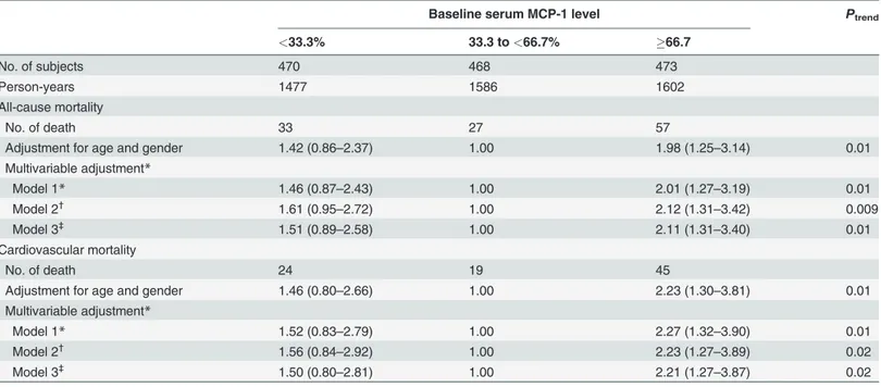
During a median follow-up of 3.3 years, 117 deaths were recorded, 88 of which were due to CVD. The 29 non-CVD deaths comprised 14 deaths of cancer, 10 deaths of asphyxia due to lung disease, and 5 deaths of other reasons. Since the interactions between gender and MCP-1 levels on the risk of all-cause and CVD mortality were not statistically significant, data for males and females were combined in the analyses to maximize the statistical power. After ad-justment for major conventional CVD risk factors, serum MCP-1 showed non-linear associa-tions with all-cause and CVD mortality (Table 2). The multivariable-adjusted hazard ratios (HRs) across three MCP-1 categories were 1.61 (95% confidence intervals [CI] 0.95–2.72), 1.00, and 2.12 (95% CI 1.31–3.42) for all-cause mortality (Ptrend= 0.009), and 1.56 (95% CI 0.84–2.92), 1.00, and 2.23 (95% CI 1.27–3.89) for CVD mortality (Ptrend= 0.02). After further adjustment for serum CRP levels, MCP-1 remained related to risks of all-cause and CVD mor-tality with this non-linear association. The addition of serum MCP-1 to the fully adjusted model increased the C-index from 0.811 to 0.820 (P<0.0001) for all-cause mortality, and 0.827 to 0.835 (P<0.0001) for CVD mortality. When we added MCP-1 to the clinical variables to predict all-cause and CVD mortality risk, 17.9% and 14.8% of dead patients were correctly re-classified to higher risk category and 6.0% and 2.3% incorrectly rere-classified to lower risk cate-gory. Similarly, 2.3% and 2.6% of survive patients were correctly reclassified to lower risk category and 2.1% and 2.5% incorrectly reclassified to higher risk category. So the estimated NRI was 12.1% (95% CI 3.5–20.9%, P = 0.006) for all-cause mortality and 12.6% (95% CI 4.3– 20.9%, P = 0.003) for CVD mortality by including MCP-1 (Table 3).
Table 1. Baseline characteristics according to serum MCP-1 level among coronary artery disease patients.
Baseline serum MCP-1 level P for
difference
<33.3% 33.3 to<66.7% 66.7
MCP-1 (pg/mL) <200.4 (male)<231.1
(female)
200.4–354.5 (male)231.1– 360.6 (female)
>354.5 (male)>360.6
(female)
No. of participants (%) 470 468 473
Male (%) 65.3 65.4 65.1 1.00
Age at baseline (yrs) 62.7 (0.5) 64.6 (0.5) 65.1 (0.5) 0.002
Body mass index (kg/m2) 23.9 (0.2) 23.9 (0.2) 23.9 (0.2) 0.97
Fasting plasma glucose (mmol/L) 6.70 (0.12) 6.36 (0.12) 6.32 (0.12) 0.04
Systolic blood pressure (mm Hg) 135 (1.0) 134 (1.0) 134 (1.0) 0.84
Diastolic blood pressure (mm Hg) 77 (0.6) 77 (0.6) 76 (0.6) 0.27
Low-density lipoprotein cholesterol (mmol/L) 3.00 (0.05) 2.93 (0.05) 3.01 (0.05) 0.32 High-density lipoprotein cholesterol (mmol/L) 1.10 (0.01) 1.07 (0.01) 1.06 (0.01) 0.06 C-reactive protein (mg/L) 5.72 (1.24–17.3) 2.55 (0.75–10.2) 3.33 (1.15–10.6) <0.001
Duration of CAD (yrs)
First diagnosed CAD (n = 767) - -
-History of CAD (n = 644) 2.91 (0.87–8.03) 2.53 (0.70–7.50) 2.96 (0.75–8.00) 0.83
Married (%) 92.0 90.7 91.3 0.85
Years of education (%) 0.67
9 62.3 62.3 61.7
10–12 22.1 18.7 19.6
13 15.6 19.0 18.6
Smoking (%) 0.63
Never 60.2 61.5 62.7
Past 8.8 10.9 9.0
Current 31.0 27.6 28.4
Alcohol drinking (%) 0.80
Never 77.3 77.1 80.5
Past 7.6 7.8 6.2
Current 15.0 15.1 13.3
Leisure-time physical activity (%) 0.70
None 35.6 32.0 32.7
<30 minutes/day 22.7 21.6 20.8
30 minutes/day 41.7 46.3 46.5
Type of CAD (%) <0.001
Acute coronary syndrome 68.5 53.2 55.2
Chronic CAD 31.5 46.8 44.8
No. of diseased vessels in coronary angiography (n = 914)
0.32
0 6.8 6.0 8.3
One-vessel disease 24.4 22.4 17.4
Two-vessel disease 23.4 23.7 20.2
Three-vessel disease 45.5 47.8 54.1
Glomerularfiltration rate (mL/min/1.73m2), (%) <0.001
90 36.5 27.1 24.2
60–89 45.1 49.1 48.9
30–59 17.4 22.1 22.4
with BMI below 24 kg/m2or CRP above 3.0 mg/L (data not shown). We further checked the in-teractions between CAD subtypes and MCP-1 levels on the risks of all-cause and CVD mortali-ty. Although the interactions were statistically significant, we didn’t stratify the data by CAD subtypes due to the small number of events in the subgroups.
Discussion
In this large and hospital-based population of Chinese CAD patients, we found a non-linear as-sociation between MCP-1 levels and the risks of all-cause and CVD mortality, independent of conventional CVD risk factors and CRP. CAD patients with higher MCP-1 levels were signifi-cantly associated with increased risks for all-cause and CVD mortality.
Previous research found that MCP-1 levels were associated with several traditional risk fac-tors of CAD and subclinical atherosclerosis [4,22]. Several case-control studies with small sam-ple sizes (from 50 to 76 subjects) also found the MCP-1 level was higher in CAD patients than in healthy subjects, especially among patients with ACS [10,23,24]. An Italian study of 50 CAD patients who underwent percutaneous transluminal coronary angioplasty (PTCA) found that MCP-1 levels significantly increased among restenotic patients compared with nonreste-notic patients, and the increase was more significant in the samples collected after 15 and 180 days after PTCA compared with that in the samples collected as early as 24 hours after the pro-cedure [24]. This suggested that the induction and high expression of MCP-1 might be Table 1. (Continued)
Baseline serum MCP-1 level P for
difference
<33.3% 33.3 to<66.7% 66.7
15–29 0.9 1.1 3.1
<15 0.2 0.7 1.3
History of diseases (%)
Hypertension 61.1 61.5 59.0 0.70
Diabetes 23.0 20.1 27.1 0.04
Dyslipidemia 29.1 31.4 31.1 0.72
Heart failure 48.7 37.8 41.9 0.003
Use of medication before admission (%)
Anti-diabetic drugs 15.0 15.0 19.6 0.10
Antihypertensive drugs 48.1 52.8 52.3 0.28
ACE inhibitors 20.7 14.2 15.5 0.02
Angiotensin II antagonists 18.6 22.7 21.0 0.29
Calcium antagonists 25.2 24.7 24.2 0.93
β-blockers 29.1 30.0 29.9 0.94
Diuretics 7.5 7.9 8.9 0.72
Lipid-lowering drugs 11.8 14.6 14.6 0.34
Anti-platelet drugs 18.4 24.9 25.0 0.02
Treatment of CAD (%)
Coronary artery bypass graft 3.0 2.1 2.5 0.72
Percutaneous coronary intervention 60.6 51.9 53.1 0.01
Continuous data are reported as mean (standard error) if normally distributed and median (25th, 75th percentile) if non-normally distributed, categorical data are reported as percentages. All normally distributed continuous variables are adjusted for age.
MCP-1, monocyte chemoattractant protein-1; CAD, coronary artery disease.
sustained to the late phase of acute coronary event compared with CRP which increased markedly in the acute phase only.
Four cohort studies have evaluated the association between baseline MCP-1 levels and death risk among CAD patients. Two posthoc analyses of clinical trials gave inconsistent find-ings [3,4]. MCP-1 level was positively associated with the risk of all-cause mortality among ACS patients in the A to Z trial, but there was no significant association between MCP-1 levels and death risk in the Oral Glycoprotein IIb/IIIa Inhibition with Orbofiban in Patients with Un-stable Coronary Syndromes (OPUS-TIMI 16) trial. Other two cohort studies with small sample sizes also found a positive association between baseline MCP-1 level and death risk [11,12]. However, all these four studies only recruited ACS patients and excluded CAD patients in the stable phase. In the A to Z trial, they further measured MCP-1 levels at 4 months after ACS onset to reflex chronic and stable phase of CAD, and still found a positive association between MCP-1 levels and death risk [3]. The result from the A to Z trial suggested that the positive as-sociation of MCP-1 levels with death risk was the same among CAD patients at both acute and stable phases. However, we found a non-linear association between MCP-1 levels and death risk among CAD patients, which was different from previous findings. Several differences be-tween our study and above studies limit a direct comparison of all the results. First, study sub-jects in two trials were treated with particular drugs which had an influence on both MCP-1 levels and the subsequent events; second, the two previous trials had short follow-up time (10– 18 months) compared with our study (mean 3.3 years); third, two cohorts had smaller study sample sizes (183 and 216 subjects) and lower death rates, which limit their applicability to find the association.
Table 2. Hazard ratios for all-cause and cardiovascular mortality according to serum MCP-1levels.
Baseline serum MCP-1 level Ptrend
<33.3% 33.3 to<66.7% 66.7
No. of subjects 470 468 473
Person-years 1477 1586 1602
All-cause mortality
No. of death 33 27 57
Adjustment for age and gender 1.42 (0.86–2.37) 1.00 1.98 (1.25–3.14) 0.01
Multivariable adjustment*
Model 1* 1.46 (0.87–2.43) 1.00 2.01 (1.27–3.19) 0.01
Model 2†
1.61 (0.95–2.72) 1.00 2.12 (1.31–3.42) 0.009
Model 3‡
1.51 (0.89–2.58) 1.00 2.11 (1.31–3.40) 0.01
Cardiovascular mortality
No. of death 24 19 45
Adjustment for age and gender 1.46 (0.80–2.66) 1.00 2.23 (1.30–3.81) 0.01
Multivariable adjustment*
Model 1* 1.52 (0.83–2.79) 1.00 2.27 (1.32–3.90) 0.01
Model 2† 1.56 (0.84–2.92) 1.00 2.23 (1.27–3.89) 0.02
Model 3‡
1.50 (0.80–2.81) 1.00 2.21 (1.27–3.87) 0.02
*Model 1 was adjusted for age, gender, education, leisure-time physical activity, smoking, and alcohol drinking.
†
Model 2 was adjusted for model 1 covariates plus history of heart failure, body mass index, systolic blood pressure, fasting plasma glucose, low-density lipoprotein cholesterol, glomerularfiltration rate, use of antihypertensive, anti-diabetic, cholesterol-lowering, and anti-platelet drugs.
‡
Model 3 was adjusted for model 2 covariates plus C-reactive protein. MCP-1, monocyte chemoattractant protein-1.
Previous basic and animal studies have confirmed that MCP-1 plays an important role in the initiation, development, and progression of CAD. Produced mainly by monocytes, smooth muscle cells, and endothelial cells within atherosclerotic plaques, MCP-1recruits monocytes or macrophages and induces them to migrate to the sites of inflammation [8,25]. Besides the in-flammation reaction, MCP-1 also regulates tissue factor expression and interferes the nitric oxide synthesis, proliferates and migrates the smooth muscle cells, neovascularizes the athero-matous plaque and makes it instable, and participates in the oxidative stress reaction [5–7,26,
27]. Through all these possible mechanism pathways, MCP-1 is involved in the pathogenesis from the early stage of atherosclerosis development to the reperfusion injury and ventricular remodeling after myocardial infarction. However, the non-linear association from the present study reflected that inflammation was not entirely deleterious or entirely beneficial. This can be explained by the finding of Nahrendorf M et al [28]. The monocyte response after myocar-dial infarction is temporally biphasic, and a well-coordinated biphasic monocyte response is necessary for proper healing. MCP-1 is released during phase 1(days 1 to 4) after myocardial infarction and recruits proinflammatory monocytes to promote digestion of infracted tissue and removal of necrotic debris, which is followed by active resolution of inflammation and Table 3. Reclassification of predicted risk with the addition of MCP-1 in coronary artery disease patients.
Predicted risk (without MCP-1) Reclassified predicted risk (with MCP-1) % (N) of subjects reclassified
<5% 5 to<10% 10 to<15% 15% Increased risk Decreased risk Net correctly reclassified (%)
All-cause mortality Dead patients (117)
<5% 40 8 0 0 17.9 6.0 11.9
5 to<10% 4 21 9 2 (21) (7)
10 to<15% 0 0 9 2
15% 0 0 3 19
Survival patients(1294)
<5% 1134 13 1 0 2.1 2.3 0.2
5 to<10% 13 70 8 0 (27) (30)
10 to<15% 0 11 16 5
15% 0 0 6 17
NRI (95% CI) 12.1 (3.5–20.9)
P = 0.006 Cardiovascular mortality
Dead patients (88)
<5% 36 5 0 0 14.8 2.3 12.5
5 to<10% 0 19 4 1 (13) (2)
10 to<15% 0 1 5 3
15% 0 0 1 13
Survival patients(1323)
<5% 1159 19 0 0 2.5 2.6 0.1
5 to<10% 19 68 10 0 (33) (34)
10 to<15% 0 10 11 4
15% 0 0 5 18
NRI (95% CI) 12.6 (4.3–20.9)
P = 0.003
MCP-1, monocyte chemoattractant protein-1; NRI, net reclassification improvement.
tissue repair in phase 2. Thus, insufficient numbers of MCP-1 and proinflammatory monocytes may delay wound healing. However, if inflammatory monocytes persist too long, the reparative functions of monocytes may be impaired. Therefore, there exists an optimum amount of in-flammatory monocytes recruitment due to MCP-1to sites of injury for better prognosis.
Although the present study indicates that MCP-1 levels may be valuable for prediction of secondary prognosis on CAD patients, these findings require further confirmation from other populations. Since the association was non-linear, future studies are needed to find out the proper cut point for risk prediction in the clinical practice. Recently, a number of animal inter-vention studies including lipid-lowering, anti-diabetic drugs, hormone replacement, and red wine have been shown to effectively reduce the MCP-1 levels [29–32]. Thus, more studies are needed to confirm this effectiveness in human clinical trials.
There are several limitations in our study. First, our subjects were enrolled from hospitals which may bring selection bias. In general, in-patients are considered having severer disease status than non-hospitalized people. However, we included both acute CAD patients and those with stable manifestation, and some of them were electively admitted patients with mild status. Thus we can reduce the bias. Besides, since our participants were all CAD patients, this limited the generalizability of our findings. Second, we cannot completely exclude the effects of residu-al confounding resulting from measurement error in the assessment of confounding factors or some unmeasured factors. Third, the present study was a cohort study based on patients al-ready with CAD. We could not get causal inference from the present data. Fourth, age was sig-nificantly different between excluded and retained participants (62.4 vs. 64.1 years), which may bring selection bias.
In conclusion, there was a non-linear association between MCP-1 and death risk among CAD patients. The addition of MCP-1 to the fully multivariable-adjusted models modestly but significantly improved the discrimination for all-cause and CVD mortality. Thus, MCP-1 may be considered as a biomarker for risk prediction for CAD patients and more investigations of MCP-1 as a therapeutic target are needed.
Supporting Information
S1 Fig. Flow chart illustrating the recruitment of the patients for the study. (DOCX)
S2 Fig. Spline plots displaying the risk cardiovascular mortality over the range of MCP-1 (log transformed).
(TIF)
S1 Table. Characteristics of included and excluded participants. (DOCX)
Author Contributions
Conceived and designed the experiments: DD JQ GH WL. Performed the experiments: DD DS XL ZL YZ WL. Analyzed the data: DD YW PG GH. Contributed reagents/materials/analysis tools: DS PL MX DL YY. Wrote the paper: DD GH WL.
References
1. World Health Organization. Global Health Observatory Data Repository: Mortality and global health es-timates. Available at:http://apps.who.int/gho/data/node.main.686?lang = en.
3. de Lemos JA, Morrow DA, Blazing MA, Jarolim P, Wiviott SD, Sabatine MS, et al. Serial measurement of monocyte chemoattractant protein-1 after acute coronary syndromes: results from the A to Z trial. J Am Coll Cardiol. 2007; 50(22):2117–24. Epub 2007/11/27. doi:10.1016/j.jacc.2007.06.057PMID:
18036447.
4. de Lemos JA, Morrow DA, Sabatine MS, Murphy SA, Gibson CM, Antman EM, et al. Association be-tween plasma levels of monocyte chemoattractant protein-1 and long-term clinical outcomes in patients with acute coronary syndromes. Circulation. 2003; 107(5):690–5. Epub 2003/02/13. PMID:12578870.
5. Birdsall HH, Green DM, Trial J, Youker KA, Burns AR, MacKay CR, et al. Complement C5a, TGF-beta 1, and MCP-1, in sequence, induce migration of monocytes into ischemic canine myocardium within the first one to five hours after reperfusion. Circulation. 1997; 95(3):684–92. Epub 1997/02/04. PMID:
9024158.
6. Libby P. Molecular bases of the acute coronary syndromes. Circulation. 1995; 91(11):2844–50. Epub 1995/06/01. PMID:7758192.
7. Gu L, Okada Y, Clinton SK, Gerard C, Sukhova GK, Libby P, et al. Absence of monocyte chemoattrac-tant protein-1 reduces atherosclerosis in low density lipoprotein receptor-deficient mice. Mol Cell. 1998; 2(2):275–81. Epub 1998/09/12. PMID:9734366.
8. Namiki M, Kawashima S, Yamashita T, Ozaki M, Hirase T, Ishida T, et al. Local overexpression of monocyte chemoattractant protein-1 at vessel wall induces infiltration of macrophages and formation of atherosclerotic lesion: synergism with hypercholesterolemia. Arterioscler Thromb Vasc Biol. 2002; 22 (1):115–20. Epub 2002/01/15. PMID:11788470.
9. Economou E, Tousoulis D, Katinioti A, Stefanadis C, Trikas A, Pitsavos C, et al. Chemokines in patients with ischaemic heart disease and the effect of coronary angioplasty. Int J Cardiol. 2001; 80(1):55–60. Epub 2001/09/05. PMID:11532547.
10. Nishiyama K, Ogawa H, Yasue H, Soejima H, Misumi K, Takazoe K, et al. Simultaneous elevation of the levels of circulating monocyte chemoattractant protein-1 and tissue factor in acute coronary syn-dromes. Jpn Circ J. 1998; 62(9):710–2. Epub 1998/10/10. PMID:9766714.
11. Kavsak PA, Ko DT, Newman AM, Palomaki GE, Lustig V, MacRae AR, et al. Risk stratification for heart failure and death in an acute coronary syndrome population using inflammatory cytokines and N-termi-nal pro-brain natriuretic peptide. Clin Chem. 2007; 53(12):2112–8. Epub 2007/10/13. doi:10.1373/ clinchem.2007.090613PMID:17932131.
12. Kervinen H, Manttari M, Kaartinen M, Makynen H, Palosuo T, Pulkki K, et al. Prognostic usefulness of plasma monocyte/macrophage and T-lymphocyte activation markers in patients with acute coronary syndromes. Am J Cardiol. 2004; 94(8):993–6. Epub 2004/10/13. doi:10.1016/j.amjcard.2004.06.052
PMID:15476610.
13. Ding D, Qiu J, Li X, Li D, Xia M, Li Z, et al. Hyperglycemia and mortality among patients with coronary artery disease. Diabetes Care. 2014; 37(2):546–54. Epub 2013/10/04. doi:10.2337/dc13-1387PMID:
24089546.
14. Li X, Zhang Y, Wang M, Lv X, Su D, Li Z, et al. The prevalence and awareness of cardiometabolic risk factors in Southern Chinese population with coronary artery disease. ScientificWorldJournal. 2013; 2013:416192. Epub 2013/11/14. doi:10.1155/2013/416192PMID:24222736; PubMed Central PMCID: PMC3810187.
15. Gibbons RJ, Chatterjee K, Daley J, Douglas JS, Fihn SD, Gardin JM, et al. ACC/AHA/ACP-ASIM guide-lines for the management of patients with chronic stable angina: executive summary and recommenda-tions. A Report of the American College of Cardiology/American Heart Association Task Force on Practice Guidelines (Committee on Management of Patients with Chronic Stable Angina). Circulation. 1999; 99(21):2829–48. Epub 1999/06/03. PMID:10351980.
16. Braunwald E, Antman EM, Beasley JW, Califf RM, Cheitlin MD, Hochman JS, et al. ACC/AHA guide-lines for the management of patients with unstable angina and non-ST-segment elevation myocardial infarction: executive summary and recommendations. A report of the American College of Cardiology/ American Heart Association task force on practice guidelines (committee on the management of pa-tients with unstable angina). Circulation. 2000; 102(10):1193–209. Epub 2000/09/07. PMID:10973852.
17. Zhang B, Wang P, Chen CG, He QQ, Zhuo SY, Chen YM, et al. Validation of an FFQ to estimate the in-take of fatty acids using erythrocyte membrane fatty acids and multiple 3d dietary records. Public Health Nutr. 2010; 13(10):1546–52. Epub 2009/12/19. S1368980009992849 [pii] doi:10.1017/
S1368980009992849PMID:20018122.
19. Harrell FE Jr., Lee KL, Mark DB. Multivariable prognostic models: issues in developing models, evaluat-ing assumptions and adequacy, and measurevaluat-ing and reducevaluat-ing errors. Stat Med. 1996; 15(4):361–87. doi:10.1002/(SICI)1097-0258(19960229)15:4<361::AID-SIM168>3.0.CO;2–4PMID:8668867.
20. Pencina MJ, D'Agostino RB. Overall C as a measure of discrimination in survival analysis: model spe-cific population value and confidence interval estimation. Stat Med. 2004; 23(13):2109–23. doi:10. 1002/sim.1802PMID:15211606.
21. Pencina MJ, D'Agostino RB Sr., D'Agostino RB Jr., Vasan RS. Evaluating the added predictive ability of a new marker: from area under the ROC curve to reclassification and beyond. Stat Med. 2008; 27 (2):157–72; discussion 207–12. doi:10.1002/sim.2929PMID:17569110.
22. Deo R, Khera A, McGuire DK, Murphy SA, Meo Neto Jde P, Morrow DA, et al. Association among plas-ma levels of monocyte chemoattractant protein-1, traditional cardiovascular risk factors, and subclinical atherosclerosis. J Am Coll Cardiol. 2004; 44(9):1812–8. Epub 2004/11/03. doi:10.1016/j.jacc.2004.07. 047PMID:15519012.
23. Hokimoto S, Ogawa H, Saito T, Oshima S, Noda K, Soejima H, et al. Increased plasma antigen levels of monocyte chemoattractant protein-1 in patients with restenosis after percutaneous transluminal coro-nary angioplasty. Jpn Circ J. 2000; 64(11):831–4. Epub 2000/12/08. PMID:11110426.
24. Cipollone F, Marini M, Fazia M, Pini B, Iezzi A, Reale M, et al. Elevated circulating levels of monocyte chemoattractant protein-1 in patients with restenosis after coronary angioplasty. Arterioscler Thromb Vasc Biol. 2001; 21(3):327–34. Epub 2001/03/07. PMID:11231910.
25. Nelken NA, Coughlin SR, Gordon D, Wilcox JN. Monocyte chemoattractant protein-1 in human athero-matous plaques. J Clin Invest. 1991; 88(4):1121–7. Epub 1991/10/01. doi:10.1172/JCI115411PMID:
1843454; PubMed Central PMCID: PMC295565.
26. Sakamoto T, Ishibashi T, Sakamoto N, Sugimoto K, Egashira K, Ohkawara H, et al. Endogenous NO blockade enhances tissue factor expression via increased Ca2+ influx through MCP-1 in endothelial cells by monocyte adhesion. Arterioscler Thromb Vasc Biol. 2005; 25(9):2005–11. Epub 2005/07/16. doi:10.1161/01.ATV.0000178171.61754.cdPMID:16020745.
27. Lee PC, Ho IC, Lee TC. Oxidative stress mediates sodium arsenite-induced expression of heme oxyge-nase-1, monocyte chemoattractant protein-1, and interleukin-6 in vascular smooth muscle cells. Toxicol Sci. 2005; 85(1):541–50. Epub 2005/02/04. doi:10.1093/toxsci/kfi101PMID:15689417.
28. Nahrendorf M, Pittet MJ, Swirski FK. Monocytes: protagonists of infarct inflammation and repair after myocardial infarction. Circulation. 2010; 121(22):2437–45. doi:10.1161/CIRCULATIONAHA.109. 916346PMID:20530020; PubMed Central PMCID: PMC2892474.
29. Martinez-Gonzalez J, Alfon J, Berrozpe M, Badimon L. HMG-CoA reductase inhibitors reduce vascular monocyte chemotactic protein-1 expression in early lesions from hypercholesterolemic swine indepen-dently of their effect on plasma cholesterol levels. Atherosclerosis. 2001; 159(1):27–33. Epub 2001/11/ 02. PMID:11689203.
30. Ghanim H, Garg R, Aljada A, Mohanty P, Kumbkarni Y, Assian E, et al. Suppression of nuclear factor-kappaB and stimulation of inhibitor factor-kappaB by troglitazone: evidence for an anti-inflammatory effect and a potential antiatherosclerotic effect in the obese. J Clin Endocrinol Metab. 2001; 86(3):1306–12. Epub 2001/03/10. doi:10.1210/jcem.86.3.7309PMID:11238525.
31. Stork S, Baumann K, von Schacky C, Angerer P. The effect of 17 beta-estradiol on MCP-1 serum levels in postmenopausal women. Cardiovasc Res. 2002; 53(3):642–9. Epub 2002/02/28. PMID:11861035.