Evaluation of TGF-
b
1 and MCP-1 expression and
tubulo-interstitial fibrosis in children with Henoch-Scho¨nlein
purpura nephritis and IgA nephropathy: A clinical
correlation
Zhao Shuiai, Shen Huijun, Gu Weizhong, Liu Aimin, Mao Jianhua*
The Children’s Hospital of Zhejiang University School of Medicine, Department of Nephrology, Hangzhou 310003, Zhejiang Province, China.
OBJECTIVES:Henoch-Scho¨nlein purpura nephritis and immunoglobulin A nephropathy are two diseases with similar clinical presentations but very different prognoses. Transforming growth factorb1 and monocyte chemo-attractant protein-1 have been associated with the development of tissue fibrosis. We examined the development of tubulointerstitial fibrosis and its relationship with Transforming growth factor b1 and monocyte chemo-attractant protein-1 expression in these patients.
METHODS: Renal tissue samples were collected by renal biopsy from 50 children with Henoch-Scho¨nlein purpura nephritis and 50 children with immunoglobulin A nephropathy. Hematoxylin and eosin and Masson’s trichrome-stained tissues were examined using light microscopy. Tubulointerstitial fibrosis was graded using the method described by Bohle et al. (1). The immunohistochemical detection of Transforming growth factorb1 and monocyte chemoattractant protein-1 expression was correlated with the tubulointerstitial fibrosis grade. Clinical Trial registration number: ZJCH-2012-0105.
RESULTS: Transforming growth factor b1 and monocyte chemoattractant protein-1 expression in the renal tissues was significantly greater in the patients with immunoglobulin A nephropathy than in the patients with Henoch-Scho¨nlein purpura nephritis (both po0.001). The immunoglobulin A nephropathy patients had a higher tubulointerstitial fibrosis grade than the Henoch-Scho¨nlein purpura nephritis patients (po0.001). The tubulointerstitial fibrosis grade was in accordance with the Transforming growth factor b1 and monocyte chemoattractant protein-1 expression levels in both diseases (bothpo0.001).
CONCLUSION: Transforming growth factor b1 and monocyte chemoattractant protein-1 expression was associated with the development of immunoglobulin A nephropathy and Henoch-Scho¨nlein purpura nephritis. Further studies are needed to better evaluate this association.
KEYWORDS: Henoch-Scho¨nlein Purpura Nephritis; IgA Nephropathy; Immunohistochemistry; Monocyte Chemoattractant Protein-1; Transforming Growth Factorb1; Fibrosis.
Shuiai Z, Huijun S, Weizhong G, Aimin L, Jianhua M. Evaluation of TGF-b1 and MCP-1 expression and tubulointerstitial fibrosis in children with Henoch-Scho¨nlein purpura nephritis and IgA nephropathy: A clinical correlation. Clinics. 2017;72(2):95-102
Received for publication onOctober 5, 2016;First review completed onNovember 24, 2016;Accepted for publication onNovember 24, 2016
*Corresponding author. E-mail: [email protected]
’ INTRODUCTION
Henoch-Schönlein purpura (HSP) is one of the most com-mon childhood vasculitides (2). Approximately 30 to 50% of children with HSP have nephritis (HSPN), which most commonly presents with hematuria and/or proteinuria. The microscopic findings of this disease include mesangial cell proliferation, crescent formation in epithelial cells, and mesangial
IgA deposition (3). Most HSPN patients have a good prog-nosis (4). Although HSPN has been related to infection and allergies (5), the exact etiology and pathogenesis of this disease are unknown.
Immunoglobulin A nephropathy (IgAN) is the most com-mon primary glomerular disease worldwide. IgAN is char-acterized by IgA or IgA immune complex deposition in the glomerular mesangial region with mesangial cell prolifera-tion (6). The main clinical findings are isolated hematuria or proteinuria. A total of 15 to 20% of IgAN patients develop chronic renal failure within 10 years of their diagnosis. IgAN is one of the leading causes of end-stage renal disease in China (7). Although HSPN and IgAN share some manifesta-tions (8), their prognoses are very different.
Tubulointerstitial damage is a key factor in determining the prognosis of IgAN patients (9,10). Tubulointerstitial damage DOI:10.6061/clinics/2017(02)05
Copyright&2017CLINICS–This is an Open Access article distributed under the terms of the Creative Commons License (http://creativecommons.org/licenses/by/ 4.0/) which permits unrestricted use, distribution, and reproduction in any medium or format, provided the original work is properly cited.
in the setting of IgAN includes tubular epithelial cell degen-eration, necrosis, or atrophy, interstitial inflammatory cell infiltration and fibrosis (11), tubular atrophy, interstitial lym-phatic and mononuclear cell infiltration and fibrosis. The degree of tubulointerstitial fibrosis has been associated with the severity of renal impairment and the patient prognosis in both disease states (12-14). However, whether tubulointersti-tial fibrosis is more severe in IgAN patients than in HSPN patients is unknown.
The causes of tubulointerstitial fibrosis are not entirely clear; however the association between transforming growth factorb1 (TGF-b1) and monocyte chemoattractant protein-1 (MCP-1) expression and the development of tissue fibrosis is well documented (15). TGF-b1 is a cytokine with powerful stimulatory effects on collagen productionin vivo. Hypoxia, high serum glucose and high plasma protein levels stimulate TGF-b1 expression in human proximal tubular epithelial cells (16) and have been linked to the development of tubuloin-terstitial fibrosis (8,17). MCP-1 is a chemotactic factor that activates monocytes and macrophages and mediates the interstitial inflammation that precedes fibrosis. However, no reports have compared TGF-b1 or MCP-1 expression in the renal tissues from children with HSPN and IgAN.
We examined renal tissues from children with HSPN and IgAN and determined the association between TGF-b1 and MCP-1 expression and tubulointerstitial fibrosis.
’ METHODS
Study Subjects
Fifty children with HSPN and 50 children with IgAN were admitted to the Department of Nephrology, The Children’s Hospital, Zhejiang University School of Medicine Hospital, between May 2012 and April 2014. The patients had similar clinical presentations with proteinuria and hematuria. The patients underwent rheumatic and anti-neutrophil cytoplas-mic antibody testing to exclude vasculitis and autoimmune diseases. The HSPN diagnosis was based on the criteria recom-mended by the Nephrology Group of the Chinese Medical Society (18) in November 2000. A total of 32 males and 18 females with HSPN were enrolled with a mean age of 8.3±2.5 years (range: 4–13 years). The IgAN diagnosis was based on the primary glomerular nephropathy classification criteria recommended by the World Health Organization (WHO) in 1995 (18). A total of 34 males and 16 females with IgAN were enrolled with a mean age of 8.8±2.1 years (range: 4–15 years). The two diagnostic groups had similar age and gender distributions. Light microscopic, immunofluorescence and electron microscopic examinations of the renal tissues were performed to confirm all diagnoses (data not shown). Patients with kidney dysfunction secondary to other diseases were excluded from this study. Informed consent was obtained from the parents of all patients. The study protocol was approved by the local institutional ethnics committee and was in accordance with the tenets of the Declaration of Helsinki. Written informed consent was obtained from all study parti-cipants after the nature of the study was explained. Clinical Trial registration number: ZJCH-2012-0105.
Renal Tissue Collection
All patients underwent ultrasound guided renal biopsy. Two renal cores were collected from each patient. Each speci-men was 1 cm to 2 cm in length. The tissues were fixed with neutral formalin solution for 24 hr and embedded in paraffin.
One specimen underwent hematoxylin and eosin (H&E) and Masson’s trichrome staining for the pathological diagnosis, and the second specimen was used for TGF-b1 and MCP-1 immunohistochemical testing.
Glomerular Pathological Classification
The HSPN glomerular findings were graded according to the International Study of Kidney Disease in Children (ISKDC) classifications as follows: grade I, minimal glomerular abnor-malities; grade II, mesangial proliferation without crescents; grade III, focal segmental (IIIa) or diffuse (IIIb) mesangial pro-liferation with less than 50% crescents; grade IV, mesangial proliferation with 50 to 75% crescents; grade V, mesangial proliferation with greater than 75% crescents; and grade VI, membranoproliferative-like lesions.
IgAN was classified into five grades as follows: grade I, mostly normal glomeruli under the light microscope and mild mesangial proliferation without cellular proliferation; grade II, mild mesangial widening and mesangial cell proliferation with no more than three mesangial cells in each mesangial area; grade III, focal segmental glomerulonephritis with less than 50% focal or segmental glomerular, mesangial cell pro-liferation, and minor lesions with occasional adhesions and small crescents in the remaining glomeruli; grade IV, diffuse mesangial proliferative glomerulonephritis with significant glomerular diffuse mesangial proliferation and sclerosis and varying degrees of cell proliferation, common abandoned glo-meruli, and adhesions and crescents in less than 50% of the glomeruli; and grade V, diffuse sclerosing glomerulonephritis in more than 80% of the glomeruli and crescents in more than 50% of the glomeruli (19).
Grading of Tubulointerstitial Fibrosis
An experienced pathologist reviewed the H&E and Masson’s trichrome-stained tissues (Supplementary Figure 1A–D) to grade the level of tubulointerstitial fibrosis. Tubulointerstitial fibrosis was graded using the method described by Bohle et al. (1). Grade+was defined as normal tubulointerstitial tissue with mild tubular degeneration, grade++as tubulointerstitial fibrosis with less than 20% tubular atrophy and scattered inflamma-tory cell infiltrates, grade+++as tubulointerstitial fibrosis with 30 to 40% tubular atrophy and scattered and/or diffuse inflammatory cell infiltrates, and grade++++as tubuloin-terstitial fibrosis with more than 50% tubular atrophy and scattered and/or diffuse inflammatory cell infiltrates (1).
Renal TGF-b1 and MCP-1 Expression
Immunohistochemical (IHC) staining of paraffin sections was performed using the EnVisionTM+Kit (Dako, Denmark) according to the manufacturer’s instructions. Briefly, 1) the paraffin sections were dewaxed in xylene and then rinsed in graduated absolute ethanol solutions (95%, 80%, and 70% ethanol) and water. 2) The sections were washed with distilled water. 3) Antigen retrieval was performed using high temper-ature and high pressure for 100 sec in a 0.01 M (pH 6.0) citrate buffer solution. 4) The sections were washed with distilled water and then with PBS 3 times for 5 min per wash. 5) Endogenous peroxidase was blocked using 3% H2O2 for 10 min. 6) The tissue sections were washed with PBS 3 times for 5 min per wash. 7) An appropriately diluted primary antibody was added to the sections and incubated at 37o
and then at 4o
C overnight. The substitution of PBS for the primary antibody was used as a negative control. 8) The slides were washed in PBS 3 times for 5 min per wash. 9) A sec-ondary antibody (i.e., goat anti-rabbit IgG antibody-HRP multimers) was added to the sections and incubated at 37o
C for 40 min. 10) The slides were washed in PBS 3 times for 5 min per wash. 11) Color development was performed using a DAB chromogenic agent for 1–3 min; the staining intensity was controlled under microscopic observation. The reaction was terminated by washing the slide with tap water. 12) The slides were stained using the Harris hematoxylin nuclear stain for 1 min and mounted with neutral resin.
TGF-b1 expression was observed in the cytoplasm of the renal tubular epithelial cells, some interstitial inflammatory cells, glomerular endothelial cells, mesangial cells and epithelial cells. MCP-1 was expressed in cytoplasm of the glomerular endothelial cells, mesangial cells, epithelial cells, tubular epithelial cells, and endothelial cells of small interstitial blood vessels. The appearance of yellow or brown particles in the cytoplasm after IHC staining was considered positive expres-sion. The expression levels were compared to the negative controls. Expression was graded according to the scope and intensity of the staining as follows (20): negative ( ), no positive staining (Supplementary Figure 1E, 1I); weak positive (+), occasional positive staining with pale yellow in less than 25% of the cells (Supplementary Figure 1F, 1J); positive (++), dark yellow staining in 26% to 50% of the cells (Supplementary Figure 1G, 1L); and strong positive (+++), brown staining in more than 50% of the cells (Supplementary Figure 1H, 1K).
Statistical Analysis
Normally distributed data were expressed as the mean± standard deviation. Non-normally distributed data were expressed as the median. Fisher’s exact test, the Chi-square test or a T test were used for comparisons between groups. The Mann-Whitney rank sum test was used to compare the TGF-b1 and MCP-1 expression levels between the two diagnostic groups. The Spearman rank correlation analysis was used to evaluate correlations between the TGF-band MCP-1 expression levels and the grade of tubulointerstitial fibrosis. The SPSS 11.5 software was used for the statistical analysis. A p value less than 0.05 was considered statistically significant.
’ RESULTS
Evaluation of Demographic Findings in Patients with HSPN and IgAN
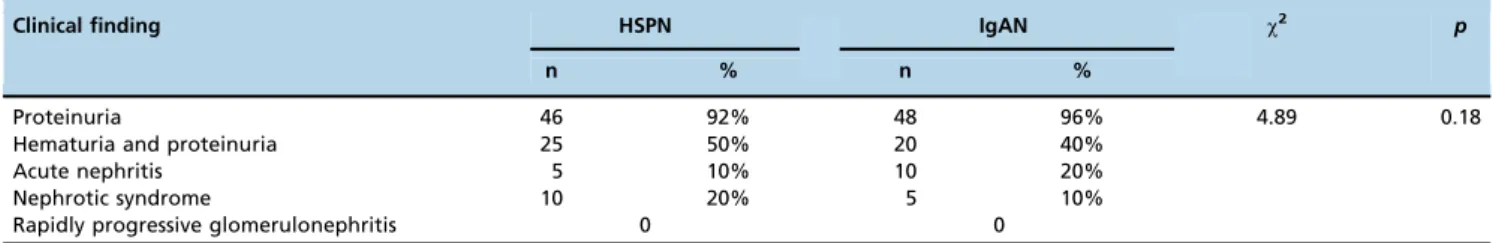
The clinical features and intensity of proteinuria in 50 children with HSPN and 50 children with IgAN are sum-marized in Tables 1 and 2. No differences were observed in the clinical findings of the two groups of patients (p=0.18; Table 1), but a significant difference was detected in the proteinuria intensity (p=0.026; Table 2).
Evaluation of Tubulointerstitial Fibrosis
The IgAN patients had significantly more severe tubu-lointerstitial fibrosis than the HSPN patients (Ppo0.001; Table 3) (Supplementary Figure 1A–D).
Correlation between the Tubulointerstitial Fibrosis Grade and the Glomerular Findings in Children with HSPN and IgAN
The tubulointerstitial fibrosis grade was moderately cor-related with the glomerular findings in both disease groups (HSPN, Spearman correlation coefficient=0.55, po0.001; IgAN, Spearman correlation coefficient=0.63, po0.001) (Table 4).
Table 2-Intensity of proteinuria in patients with HSPN and IgAN.
24 h proteinuria HSPN IgAN Z p
n % n %
o25 mg/kg 19 41.3% 8 16.7% 2.23 0.026
25-50 mg/kg 17 36.9% 25 52.1% 50 mg/kg 10 21.7% 15 31.2% Table 1-Clinical findings in patients with HSPN and IgAN.
Clinical finding HSPN IgAN w2 p
n % n %
Proteinuria 46 92% 48 96% 4.89 0.18
Hematuria and proteinuria 25 50% 20 40%
Acute nephritis 5 10% 10 20%
Nephrotic syndrome 10 20% 5 10%
Rapidly progressive glomerulonephritis 0 0
Table 3-Grading of tubulointerstitial fibrosis in patients with HSPN and IgAN.
Fibrosis grade HSPN IgAN
I 38 5
II 9 17
III 2 20
IV 1 8
Total 50 50
Z=6.74,po0.001, by Mann–Whitney–Wilcoxon test.
Table 4-Correlation between the tubulointerstitial fibrosis grade and the glomerular pathological findings in patients with HSPN and IgAN.
Disease grade Grade of fibrosis in HSPNa
Grade of fibrosis in IgANb
I–II III IV–V I–II III IV–V
I 26 12 0 4 1 0
II 2 6 1 13 3 1
III 0 0 2 3 13 4
IV 0 0 1 1 8 5
V 0 0 0 0 0 0
a
w2=21.44,Ppo0.001 for children with HSPN, andbw2=17.83,po0.001 for
TGF-b1 and MCP-1 Expression in HSPN and IgAN Patients
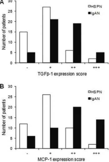
TGF-b1 and MCP-1 expression was significantly greater in the IgAN patients than in the HSPN patients (po0.001 for both) (Figure 1).
Correlation between the Tubulointerstitial Fibrosis Grade and TGF-b1 and MCP-1 Expression
The tubulointerstitial fibrosis grade was moderately cor-related with TGF-b1 and MCP-1 expression in both disease groups (HSPN: Spearman correlation coefficient for TGF-b1= 0.56,po0.001; Spearman correlation coefficient for MCP-1= 0.63, po0.001; IgAN: Spearman correlation coefficient for TGF-b1=0.72,po0.001; Spearman correlation coefficient for MCP-1=0.62,po0.001) (Tables 5–8).
’ DISCUSSION
Increased TGF-b1 and MCP-1 expression has been reported in many fibrotic diseases (21). TGF-b1 is a cytokine that is expressed in the kidney in association with the development of tubulointerstitial fibrosis (22). MCP-1 plays a role in the chemotaxis and activation of monocytes and macrophages.
Tubulointerstitial injury is related to monocyte and macro-phage infiltration, which stimulates the ingrowth of activated neutrophils, mononuclear cells, and subsequent T cell infiltra-tion and in turn stimulates the growth of fibroblasts and keratinocytes (23). MCP-1 also stimulates the expression of IL-6, cell adhesion molecules, and other inflammatory factors that contribute to renal tubular fibrosis (24).
TGF-b1 has been hypothesized to participate in the initia-tion and progression of early immune renal tubular injury and the formation of tubulointerstitial fibrosis (25). Yamumoto et al. (17). examined renal tissues from patients with different types of renal disease. Patients with thin basement membrane nephropathy or minimal change nephropathy had TGF-b1 Table 5-Correlation between the tubulointerstitial fibrosis grade and MCP-1 expression in children with HSPN.
Fibrosis grade
n MCP-1 expression
- + ++ +++
I 38 12 23 3 0
II 9 0 3 6 0
III 2 0 0 1 1
IV 1 0 0 0 1
Total 50 12 26 10 2
Spearman correlation coefficient for MCP-1=0.63,po0.001.
Table 6-Correlation between the tubulointerstitial fibrosis grade and TGF-b1 expression in HSPN.
Fibrosis grade
n TGF-b1 expression
- + ++ +++
I 38 15 21 2 0
II 9 0 6 3 0
III 2 0 0 1 1
IV 1 0 0 0 1
Total 50 15 27 6 2
Spearman correlation coefficient for TGF-b1=0.56,po0.001.
Table 7-Correlation between the tubulointerstitial fibrosis grade and MCP-1 expression in IgAN.
Fibrosis grade
n MCP-1 expression
- + ++ +++
I 5 4 1 0 0
II 17 2 7 4 4
III 20 0 2 14 4
IV 8 0 0 2 6
Total 50 6 10 20 14
Spearman correlation coefficient for MCP-1=0.62,po0.001.
Table 8-Correlation between the tubulointerstitial fibrosis grade and TGF-b1 expression in IgAN.
Fibrosis grade
n TGF-b1 expression
- + ++ +++
I 5 3 2 0 0
II 17 2 11 4 0
III 20 0 8 12 0
IV 8 0 0 3 5
Total 50 5 21 19 5
Spearman correlation coefficient for TGF-b1=0.72,po0.001. Figure 1 - Immunohistochemical staining scores for TGF-b1 (A)
mRNA expression levels that were similar to the normal con-trol patients. Conversely, patients with IgAN, HSPN, lupus nephritis or crescent nephritis and glomerular or tubulointer-stitial disease had significantly greater renal TGF-b1 mRNA expression levels than the normal control patients, which was consistent with our findings. We found that the IHC expres-sion of TGF-b1 in the renal tubular epithelial cells was posi-tively correlated with the tubulointerstitial fibrosis grade in children with IgAN. Together, these findings suggest that renal TGF-b1 expression may be related to tubulointerstitial disease and impaired renal tubular functions in IgAN patients. A study of 25 adult IgAN cases by Celie et al. (26) demonstrated an association between MCP-1 expression and the degree of glomerular and tubulointerstitial injury. MCP-1 expression in the tubulointerstitial tissues was positively correlated with the amount of proteinuria. MCP-1 expression in the glomerular and tubulointerstitial tissues was sug-gested to be a possible indicator of renal disease severity in IgAN patients. We found a positive correlation between MCP-1 expression and the grade of tubulointerstitial fibrosis. Together, these findings suggest that MCP-1 expression is related to the tubulointerstitial fibrosis and impaired tubular function found in IgAN patients.
Patients with HSPN typically present with acute episodes of glomerular inflammation consisting of mesangial pro-liferation, fibrin deposits and epithelial crescents that heal spontaneously or lead to chronic lesions. Sick children with HSPN suffer from humoral immune abnormalities, T cell subset dysfunction, and abnormal cytokine production. These findings may lead to TGF-b1 expression, which can mediate fibrosis formation. In contrast, patients with IgAN typically have an IgA-based granular sediment deposited in the glomerulus mesangial region. These patients develop slowly progressive mesangial lesions from the continuous low-grade deposition of macromolecular IgA1. Abnormal IgA1 glycosylation in these patients is associated with renal deposition and complement system activation. The children with HSPN examined in this study had low renal TGF-b1 and MCP-1 expression, whereas the IgAN patients com-monly had high expression levels of these cytokines. The mechanism of inflammation initiation appears to be more active in patients with IgAN, which may explain why these patients have more fibrosis and higher TGF-b1 and MCP-1 expression levels than the HSPN patients.
Increased renal TGF-b1 and MCP-1 expression was asso-ciated with increased tubulointerstitial fibrosis in both disease states. Tubulointerstitial fibrosis was observed in both diag-nostic groups but was more common in the IgAN group. We found a similar positive moderate correlation between the tubulointerstitial fibrosis grade and the intensity of protei-nuria and TGF-b1 and MCP-1 expression in both diseases. The relative frequency of cytokine expression suggests that TGF-b1 and MCP-1 are likely linked to tubulointerstitial fibrosis formation in both disease groups. Several studies have shown that higher proteinuria levels are related to the progression of renal diseases in patients with different types of glomerulopathy (27), which is consistent with the results of our study (Table 2). One possible mechanism is that the increase in protein reabsorption by the tubule cells leads to changes in these cells, which start to produce cytokines and chemoattractant factors for lymphocytes and macrophages; then, these cells migrate to the interstitial area to induce inflammation and fibrosis (28-30). Further work is needed to better understand the development of inflammation and
TGF-b1 and MCP-1 expression in these patients and the progression to tubulointerstitial fibrosis.
In summary, we observed tubulointerstitial fibrosis in the renal tissues of children with HSPN and IgAN. Greater tubulointerstitial fibrosis was observed in the IgAN patients compared to the HSPN patients. Increased amounts of tubu-lointerstitial fibrosis were associated with increased TGF-b1 and MCP-1 expression. These findings suggest that TGF-b1 and MCP-1 may be involved in the development of tubu-lointerstitial fibrosis in both these disease states, possibly through different inflammatory mechanisms.
’ ACKNOWLEDGMENTS
This study was supported by the National Natural Foundation of China (81470939 and 81270792), the Specialized Research Fund for the Doctoral Program of Higher Education (20120101110018), the Natural Science Foundation of Zhejiang Province (LH14H050002 and LY15H050001) and the Medicine & Health Technology Innovation Project of Zhejiang Pro-vince (2014KYA123). We thank all patients and their family members for participating in this study.
’ AUTHOR CONTRIBUTIONS
All authors were involved in drafting the manuscript or critically revising it for important intellectual content, and all authors approved thefinal version for publication. Jianhua M has full access to all data in the study and takes responsibility for the integrity of the data and the accuracy of the data analysis. Shuiai Z and Jianhua M conceived and designed the study and were responsible for the data analysis and interpretation. Shuiai Z, Huijun S and Weizhong G were responsible for the data acquisition. Aimin L and Jianhua M were responsible for the clinical data collection and patients diagnosis.
’ REFERENCES
1. Bohle A, Muller GA, Wehrmann M, Mackensen-Haen S, Xiao JC. Pathogenesis of chronic renal failure in the primary glomerulopathies, renal vasculopathies, and chronic interstitial nephritides. Kidney Int Suppl. 1996;54:S2-9.
2. Muller D, Greve D, Eggert P. Early tubular proteinuria and the develop-ment of nephritis in Henoch-Schonlein purpura. Pediatr Nephrol. 2000; 15(1-2):85-9.
3. Davin JC, Coppo R. Henoch-Schonlein purpura nephritis in children. Nat Rev Nephrol. 2014;10(10):563-73, http://dx.doi.org/10.1038/nrneph. 2014.126.
4. Dedeoglu F, Sundel RP. Vasculitis in children. Pediatr Clin North Am. 2005;52(2):547-75, http://dx.doi.org/10.1016/j.pcl.2005.01.006. 5. Pohl M. Henoch-Schonlein purpura nephritis. Pediatr Nephrol. 2015;
30(2):245-52, http://dx.doi.org/10.1007/s00467-014-2815-6.
6. Barratt J, Feehally J. IgA nephropathy. J Am Soc Nephrol. 2005;16(7): 2088-97, http://dx.doi.org/10.1681/ASN.2005020134.
7. Zhang C, Zeng X, Li Z, Wang Z, Li S. Immunoglobulin A nephropathy: current progress and future directions. Transl Res. 2015;166(2):134-44, http://dx.doi.org/10.1016/j.trsl.2015.02.007.
8. Wada J, Sugiyama H, Makino H. Pathogenesis of IgA nephropathy. Semin Nephrol. 2003;23(6):556-63, http://dx.doi.org/10.1053/S0270-9295(03) 00134-7.
9. Davin JC, Ten Berge IJ, Weening JJ. What is the difference between IgA nephropathy and Henoch-Schonlein purpura nephritis? Kidney Int. 2001;59(3):823-34, http://dx.doi.org/10.1046/j.1523-1755.2001.0590 03823.x.
10. Roberts IS. Pathology of IgA nephropathy. Nat Rev Nephrol. 2014; 10(8):445-54, http://dx.doi.org/10.1038/nrneph.2014.92.
11. Floege J. The pathogenesis of IgA nephropathy: what is new and how does it change therapeutic approaches? Am J Kidney Dis. 2011;58(6):992-1004, http://dx.doi.org/10.1053/j.ajkd.2011.05.033.
12. Kim CH, Lim BJ, Bae YS, Kwon YE, Kim YL, Nam KH, et al. Using the Oxford classification of IgA nephropathy to predict long-term outcomes of Henoch-Schonlein purpura nephritis in adults. Mod Pathol. 2014; 27(7):972-82, http://dx.doi.org/10.1038/modpathol.2013.222.
14. Tanaka S, Ninomiya T, Katafuchi R, Masutani K, Tsuchimoto A, Noguchi H, et al. Development and validation of a prediction rule using the Oxford classification in IgA nephropathy. Clin J Am Soc Nephrol. 2013;8(12): 2082-90, http://dx.doi.org/10.2215/CJN.03480413.
15. Wynn TA. Cellular and molecular mechanisms of fibrosis. J Pathol. 2008;214(2):199-210, http://dx.doi.org/10.1002/path.2277.
16. Fukasawa H, Yamamoto T, Suzuki H, Togawa A, Ohashi N, Fujigaki Y, et al. Treatment with anti-TGF-beta antibody ameliorates chronic pro-gressive nephritis by inhibiting Smad/TGF-beta signaling. Kidney Int. 2004;65(1):63-74, http://dx.doi.org/10.1111/j.1523-1755.2004. 00393.x.
17. Yamamoto T, Noble NA, Cohen AH, Nast CC, Hishida A, Gold LI, et al. Expression of transforming growth factor-beta isoforms in human glomerular diseases. Kidney Int. 1996;49(2):461-9, http://dx.doi.org/ 10.1038/ki.1996.65.
18. Chinese Medical Association Branch of Pediatrics Nephrology Group. Draft of the diagnosis and treatment of Henoch-Schönlein purpura nephritis. Chin J Pediatr. 2001;39:746-8.
19. Chinese Medical Association Branch of Pediatrics Nephrology Group. Glomerular disease classification, diagnosis and treatment. Chin J Pediatr. 2001;39:746-9.
20. Kliem V, Johnson RJ, Alpers CE, Yoshimura A, Couser WG, Koch KM, et al. Mechanisms involved in the pathogenesis of tubulointerstitial fibrosis in 5/6-nephrectomized rats. Kidney Int. 1996;49(3):666-78, http:// dx.doi.org/10.1038/ki.1996.95.
21. Li X, Kimura H, Hirota K, Sugimoto H, Yoshida H. Hypoxia reduces constitutive and TNF-alpha-induced expression of monocyte chemoat-tractant protein-1 in human proximal renal tubular cells. Biochem Biophys Res Commun. 2005;335(4):1026-34, http://dx.doi.org/10.1016/j.bbrc. 2005.07.175.
22. Strehlau J, Schachter AD, Pavlakis M, Singh A, Tejani A, Strom TB. Activated intrarenal transcription of CTL-effectors and TGF-beta1 in
children with focal segmental glomerulosclerosis. Kidney Int. 2002; 61(1):90-5, http://dx.doi.org/10.1046/j.1523-1755.2002.00090.x. 23. Segerer S, Cui Y, Hudkins KL, Goodpaster T, Eitner F, Mack M, et al.
Expression of the chemokine monocyte chemoattractant protein-1 and its receptor chemokine receptor 2 in human crescentic glomerulonephritis. J Am Soc Nephrol. 2000;11(12):2231-42.
24. Schwarz M, Wahl M, Resch K, Radeke HH. IFNgamma induces functional chemokine receptor expression in human mesangial cells. Clin Exp Immunol. 2002;128(2):285-94, http://dx.doi.org/10.1046/j.1365-2249.2002. 01829.x.
25. Kipari T, Cailhier JF, Ferenbach D, Watson S, Houlberg K, Walbaum D, et al. Nitric oxide is an important mediator of renal tubular epithelial cell death in vitro and in murine experimental hydronephrosis. Am J Pathol. 2006;169(2):388-99, http://dx.doi.org/10.2353/ajpath.2006.050964. 26. Celie JW, Reijmers RM, Slot EM, Beelen RH, Spaargaren M, Ter Wee PM, et al.
Tubulointerstitial heparan sulfate proteoglycan changes in human renal diseases correlate with leukocyte influx and proteinuria. Am J Physiol Renal Physiol. 2008;294(1):F253-63, http://dx.doi.org/10.1152/ajprenal.00429.2007.
27. Remuzzi G, Benigni A. Progression of proteinuric diabetic and non-diabetic renal diseases: a possible role for renal endothelin. Kidney Int Suppl. 1997;58:S66-8.
28. Zoja C, Donadelli R, Colleoni S, Figliuzzi M, Bonazzola S, Morigi M, et al. Protein overload stimulates RANTES production by proximal tubular cells depending on NF-kappa B activation. Kidney Int. 1998;53(6):1608-15, http://dx.doi.org/10.1046/j.1523-1755.1998.00905.x.
29. Abbate M, Remuzzi G. Proteinuria as a mediator of tubulointerstitial injury. Kidney Blood Press Res. 1999;22(1-2):37-46, http://dx.doi.org/ 10.1159/000025907.