Small RNA sX13: A Multifaceted Regulator of Virulence in
the Plant Pathogen
Xanthomonas
Cornelius Schmidtke1*, Ulrike Abendroth1, Juliane Brock1, Javier Serrania2, Anke Becker2, Ulla Bonas1*
1Institute for Biology, Department of Genetics, Martin-Luther-Universita¨t Halle-Wittenberg, Halle, Germany,2Loewe Center for Synthetic Microbiology and Department of Biology, Philipps-Universita¨t Marburg, Marburg, Germany
Abstract
Small noncoding RNAs (sRNAs) are ubiquitous posttranscriptional regulators of gene expression. Using the model plant-pathogenic bacteriumXanthomonas campestrispv.vesicatoria(Xcv), we investigated the highly expressed and conserved sRNA sX13 in detail. Deletion ofsX13impinged onXcvvirulence and the expression of genes encoding components and substrates of the Hrp type III secretion (T3S) system. qRT-PCR analyses revealed that sX13 promotes mRNA accumulation of HrpX, a key regulator of the T3S system, whereas the mRNA level of the master regulator HrpG was unaffected. Complementation studies suggest that sX13 acts upstream of HrpG. Microarray analyses identified 63 sX13-regulated genes, which are involved in signal transduction, motility, transcriptional and posttranscriptional regulation and virulence. Structure analyses ofin vitrotranscribed sX13 revealed a structure with three stable stems and three apical C-rich loops. A computational search for putative regulatory motifs revealed that sX13-repressed mRNAs predominantly harbor G-rich motifs in proximity of translation start sites. Mutation of sX13 loops differentially affected Xcvvirulence and the mRNA abundance of putative targets. Using a GFP-based reporter system, we demonstrated that sX13-mediated repression of protein synthesis requires both the C-rich motifs in sX13 and G-rich motifs in potential target mRNAs. Although the RNA-binding protein Hfq was dispensable for sX13 activity, thehfqmRNA and Hfq::GFP abundance were negatively regulated by sX13. In addition, we found that G-rich motifs in sX13-repressed mRNAs can serve as translational enhancers and are located at the ribosome-binding site in 5% of all protein-codingXcvgenes. Our study revealed that sX13 represents a novel class of virulence regulators and provides insights into sRNA-mediated modulation of adaptive processes in the plant pathogen Xanthomonas.
Citation:Schmidtke C, Abendroth U, Brock J, Serrania J, Becker A, et al. (2013) Small RNA sX13: A Multifaceted Regulator of Virulence in the Plant Pathogen Xanthomonas. PLoS Pathog 9(9): e1003626. doi:10.1371/journal.ppat.1003626
Editor:Matthew K. Waldor, Harvard University, United States of America
ReceivedFebruary 5, 2013;AcceptedAugust 1, 2013;PublishedSeptember 12, 2013
Copyright:ß2013 Schmidtke et al. This is an open-access article distributed under the terms of the Creative Commons Attribution License, which permits unrestricted use, distribution, and reproduction in any medium, provided the original author and source are credited.
Funding:This work was supported by grants from the Deutsche Forschungsgemeinschaft to UB and AB. (SPP 1258; ‘‘Sensory and Regulatory RNAs in Prokaryotes’’) and the ‘‘Graduiertenkolleg’’ (GRK 1591) to UB, and by the Bundesministerium fu¨r Bildung und Forschung (grant 0313105) to AB. The funders had no role in study design, data collection and analysis, decision to publish, or preparation of the manuscript.
Competing Interests:The authors have declared that no competing interests exist.
* E-mail: [email protected] (CS); [email protected] (UB)
Introduction
The survival and prosperity of bacteria depends on their ability to adapt to a variety of environmental cues such as nutrient availability, osmolarity and temperature. Besides the adaptation to the environment by transcriptional regulation of gene expression bacteria express regulatory RNAs that modulate expression on the posttranscriptional level [1,2]. Small regulatory RNAs (sRNAs; ,50–300 nt) have been intensively studied in the enterobacteria
Escherichia coli and Salmonella spp. and, in most cases, regulate translation and/or stability of target mRNAs through short and imperfect base-pairing (10 to 25 nucleotides) [1,3,4,5]. The majority of characterized sRNAs inhibits translation of target mRNAs by pairing near or at the ribosome-binding site (RBS) [1,6]. In addition, sRNAs can promote target mRNA translation, e. g., the sRNAs ArcZ, DsrA and RprA activate translation of sigma factor RpoS [7,8,9]. Regulation of multiple rather than single genes has emerged as a major feature of sRNAs affecting processes like iron homeostasis, carbon metabolism, stress responses and quorum sensing (QS) [1,2,6]. In numerous cases, sRNAs are under transcriptional control of two-component systems (TCS), which themselves are often controlled by sRNAs
[10]. The activity and stability of most enterobacterial sRNAs requires the hexameric RNA-binding protein Hfq, which facili-tates the formation of sRNA-mRNA duplexes and their subse-quent degradation by the RNA degradosome [1,11]. Hfq is present in approximately 50% of all bacterial species and acts in concert with sRNAs to regulate stress responses and virulence of a number of animal- and human-pathogenic bacteria [5,12].
To date, little is known about sRNAs in plant-pathogenic bacteria. Only recently, high throughput RNA-sequencing approaches uncovered potential sRNAs in the plant-pathogenic a-proteobacterium Agrobacterium tumefaciens [13], the c -proteo-bacteria Pseudomonas syringae pv. tomato [14] and Xanthomonas campestrispv.vesicatoria(Xcv) [15,16]. Additional studies identified four and eight sRNAs in X. campestrispv.campestris (Xcc) andX. oryzae pv. oryzae (Xoo), respectively [17,18]. So far, only few sRNAs of plant-pathogenic bacteria were characterized with respect to potential targets. Examples include theA. tumefaciens
the activity of the translational repressor protein RsmA, which impacts on QS, the production of extracellular enzymes and virulence [21,22,23]. In Xcv, sX12 was reported to be required for full virulence [16].
Xanthomonads are ubiquitous plant-pathogenic bacteria that infect approximately 120 monocotyledonous and 270 dicotyle-donous plant species, many of which are economically important crops [24,25]. These pathogens, usually only found in association with plants and plant parts, differ from most other Gram-negative bacteria in their high G+C content (,65%), and high numbers of TonB-dependent transporters and signaling proteins [26]. Pathogenicity of most Xanthomonas
spp. and other Gram-negative plant- and animal-pathogenic bacteria relies on a type III secretion (T3S) system which translocates bacterial effector proteins into the eukaryotic host cell [27,28]. In addition, other protein secretion systems, degradative enzymes and QS regulation contribute to virulence of Xanthomonas spp. [29,30].
One of the models to study plant-pathogen interactions isXcv, the causal agent of bacterial spot disease on pepper and tomato [31,32]. The T3S system of Xcv is encoded by the hrp2
[hypersensitive response (HR) and pathogenicity] gene cluster and translocates effector proteins into the plant cell where they interfere with host cellular processes to the benefit of the pathogen [29,33,34]. The mutation of hrp-genes abolishes bacterial growth in the plant tissue and the induction of the HR in resistant plants. The HR is a local and rapid programmed plant cell death at the infection site and coincides with the arrest of bacterial multipli-cation [33,35,36]. The expression of the T3S system is transcrip-tionally induced in the plant and in the synthetic medium XVM2, and is controlled by the key regulators HrpG and HrpX [37,38,39,40]. The OmpR-type regulator HrpG induces tran-scription of hrpXwhich encodes an AraC-type activator [39,41]. HrpG and HrpX control the expression ofhrp, type III effector and other virulence genes [16,29,40,42,43]. Recently, dRNA-seq identified 24 sRNAs in Xcvstrain 85-10, expression of eight of
which is controlled by HrpG and HrpX, including the aforemen-tioned sX12 sRNA [15,16].
In this study, we aimed at a detailed characterization of sX13 fromXcvstrain 85-10, which is an abundant and HrpG-/HrpX-independently expressed sRNA [16]. Using mutant and comple-mentation studies, we discovered that sX13 promotes the expression of the T3S system and contributes to virulence of
Xcv. Microarray and quantitative reverse transcription PCR (qRT-PCR) analyses identified a large sX13 regulon and G-rich motifs in presumed sX13-target mRNAs. Selected putative targets were analyzed by site-directed mutagenesis of sX13 and mRNA::gfp
fusions. Furthermore, we provide evidence that sX13 acts Hfq-independently. Our study provides the first comprehensive characterization of a trans-encoded sRNA that contributes to virulence of a plant-pathogenic bacterium.
Results
sX13contributes to bacterial virulence
The sRNA sX13 (115 nt; [16]) is encoded in a 437-bp intergenic region of the Xcv 85-10 chromosome, i. e., 175 bp downstream of the stop codon of the DNA polymerase I-encoding genepolAand 148 bp upstream of the translation start site (TLS) of
XCV4199, which encodes a hypothetical protein. Sequence comparisons revealed that thesX13 gene is exclusively found in members of theXanthomonadaceaefamily, i. e., in the genomes of plant-pathogenic Xanthomonas and Xylella species, the human pathogenStenotrophomonas maltophiliaand non-pathogenic bacteria of the genus Pseudoxanthomonas. Interestingly, sX13 homologs are highly conserved [16] and always located downstream ofpolA. By contrast,sX13-flanking sequences are highly variable.
To characterize sX13 in Xcv strain 85-10, we introduced an unmarkedsX13deletion into the chromosome (see ‘Materials and Methods’). Analysis of bacterial growth of thesX13mutant strain (DsX13) revealed a significantly reduced stationary-phase density compared to theXcvwild-type strain 85-10 in complex medium (NYG; Figure 1A) and in minimal medium A (MMA; Figure 1B). The mutant phenotype of XcvDsX13 was complemented by chromosomal re-integration ofsX13into the sX13locus, termed DsX13+sX13ch(Figure 1A, B; see ‘Materials and Methods’).
To address a potential role ofsX13in virulence, we performed plant infection assays. As shown in Figure 1C, theXcvstrains 85-10 and DsX13 grew similarly in leaves of susceptible pepper plants (ECW). Strikingly, infection with the sX13 mutant resulted in strongly delayed disease symptoms in susceptible and a delayed HR in resistant pepper plants (ECW-10R) (Figure 1D). Ectopic expression of sX13 under control of the lac promoter (psX13), which is constitutive inXcv[38], and re-integration ofsX13into the DsX13 locus fully complemented the mutant phenotype of
XcvDsX13(Figure 1D; data not shown). StrainXcv85-10 carrying psX13induced an accelerated HR in comparison to the wild type (data not shown).
Deletion ofsX13derogateshrp-gene expression
As the HR induction in ECW-10R plants depends on the recognition of the bacterial type III effector protein AvrBs1 by the plant disease resistance geneBs1[44,45], thein plantaphenotype of
XcvDsX13 suggested a reduced activity of the T3S system. To address this question, we investigated protein accumulation of selected components of the T3S system. Given that T3S apparatus proteins are not detectable in NYG-grown bacteria, we incubated the bacteria for 3.5 hours in the hrp-gene inducing XVM2 medium [38,40]. Western blot analysis revealed reduced amounts of the translocon protein HrpF, the T3S-ATPase HrcN and the Author Summary
T3S-apparatus component HrcJ in XcvDsX13 compared to the wild type, DsX13(psX13) (Figure 2A) and strain DsX13+sX13ch
(selectively tested for HrcJ; Figure 2B). Thus, sX13 positively affects the synthesis of T3S components.
As HrpG controls the expression of thehrp-regulon [39], we analyzed whether the reduced virulence of strainDsX13is due to a reduced activity of HrpG. Therefore, we ectopically expressed a constitutively active version of HrpG (HrpG*; phrpG*; [41]) in
XcvDsX13 and performed plant-infection assays. The disease symptoms induced by XcvDsX13 and the wild type were comparable in presence of phrpG*, whereas with low inoculum ofXcv85-10DsX13the HR was slightly delayed (Figure 1D). This suggests that HrpG* suppresses the 85-10DsX13phenotype. HrpF, HrcN and HrcJ protein accumulation in strainDsX13(phrpG*) was identical to the wild type suggesting full complementation (Figure 2A, B).
To investigate whether the reduced protein amounts of T3S-system components inXcvDsX13are due to altered mRNA levels, we performed qRT-PCR analyses. mRNA accumulation ofhrpF,
hrcJand the type III effector genesavrBs1andxopJwas two-fold lower in XcvDsX13 than in the wild type and the complemented strainDsX13+sX13ch(Figure 2C). In addition, the mRNA amount
of hrpX, but not of hrpG, was reduced in the sX13 mutant (Figure 2C). In presence of phrpG*, comparable mRNA amounts of hrpG, hrpX, hrpF, hrcJ and xopJ were detected in Xcv 85-10, DsX13 and DsX13+sX13ch, whereas theavrBs1 mRNA
accumu-lation was significantly reduced in strain 85-10DsX13(Figure 2C). Taken together, our data suggest that the reduced virulence of the 85-10DsX13 mutant is caused by a lower expression of compo-nents and substrates of the T3S system (Figure 1D; Figure 2A–C).
The deletion and chromosomal re-insertion of sX13 in
XcvDsX13 and DsX13+sX13ch, respectively, were verified by
Northern blot using an sX13-specific probe (Figure S1). The sX13 abundance was not affected by expression of HrpG*, which confirms our previous findings [16] and suggests that expression of sX13 is independent of HrpG and HrpX (Figure S1).
sX13 accumulates under stress conditions
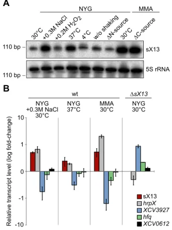
The expression of known bacterial sRNAs depends on a variety of environmental stimuli, which often reflect the physiological functions of sRNAs [2,46], e. g., the E. coli sRNA Spot42 is repressed in the absence of glucose and regulates carbon metabolism [47,48]. Northern blots revealed similar sX13 amounts in bacteria incubated in NYG medium at 30uC (standard condition), in presence of H2O2, at 4uC and in NYG medium
lacking nitrogen (Figure 3A). By contrast, sX13 accumulation was increased in presence of high salt (NaCl), 37uC and in MMA (Figure 3A). Hence, sX13 is differentially expressed in different growth conditions and might contribute to environmental adaptation ofXcv.
Microarray analyses suggest a large sX13 regulon To gain an insight into the sX13 regulon we performed microarray analyses. For this, cDNA derived fromXcvstrains 85-10 andDsX13grown in NYG and MMA, respectively, was used as a probe. The hybridization data were evaluated using EMMA 2.8.2 [49] (see ‘Materials and Methods’). InXcvDsX13grown in NYG, 23 mRNAs were upregulated and 21 mRNAs were downregulated by a factor of at least 1.5 compared to the wild type (Table S2). In the MMA-grownsX13mutant, 23 upregulated Figure 1.sX13contributes to bacterial growth in culture and virulence.Growth ofXcvwild type 85-10 (wt), thesX13deletion mutant (DsX13) andDsX13containing chromosomally re-integratedsX13(DsX13+sX13ch) in (A) complex medium NYG and (B) minimal medium MMA, respectively. Error bars represent standard deviations. Asterisks indicate statistically significant differences compared to wt (t-test;P,0.05). (C) Growth ofXcv85-10 (wt) andDsX13in leaves of susceptible ECW pepper plants. Data points represent the mean of three different samples from three different plants of one experiment. Standard deviations are indicated by error bars. (D) Plant infection assay.Xcvstrains 85-10 (wt) andDsX13carrying the empty vector (pB) or the sX13 expression construct (psX13) and strains additionally expressing HrpG* (phrpG*) were inoculated at a density of 46108(left panel)
and 108cfu/ml (right panel), respectively, into leaves of susceptible ECW and resistant ECW-10R pepper plants. Disease symptoms in ECW were photographed 9 days post inoculation (dpi). The HR was visualized by ethanol bleaching of the leaves 3 dpi (left panel) and 18 hours post inoculation (right panel), respectively. Dashed lines indicate the inoculated areas. All experiments were performed at least three times with similar results. doi:10.1371/journal.ppat.1003626.g001
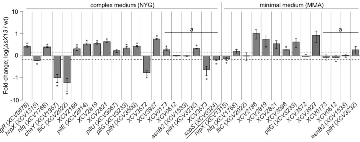
mRNAs were detected, four of which were also upregulated in NYG-grown bacteria, whereas no downregulated genes were identified (Table S2). With respect to both growth conditions, 42 and 21 genes were upregulated and downregulated, respectively, in thesX13mutant. qRT-PCR analyses of 11 selected upregulated
and four downregulated genes confirmed the microarray data (Table 1; Figure 4).
sX13 negatively affectshfqand type IV pilus-biosynthesis mRNAs
Based on the annotated genome sequence of Xcv 85-10 [32], genes upregulated in XcvDsX13 can be grouped (Table S2): 18 genes encode proteins with unknown function, e. g., the putative LysM-domain protein XCV3927. 14 genes encode proteins involved in type IV pilus (Tfp) biogenesis, e. g., the putative Tfp assembly protein XCV2821, the pilus component PilE and the TCS response regulator PilG. Tfp enable twitching motility, i. e., adhesion to and movement on solid surfaces [50,51]. Three genes encode proteins assigned to signal transduction, i. e., the TCS regulator AlgR, the GGDEF-domain protein XCV2041 and the chemotaxis regulator XCV2186. Moreover,hfqmRNA accumu-lation was two-fold increased inXcvDsX13.
The microarray data suggested that most upregulated genes in
XcvDsX13were only expressed in NYG- or MMA-grown bacteria (Table S2), which might be explained by theP-value and signal-strength thresholds applied for data evaluation. qRT-PCR analyses showed that the mRNA accumulation ofhfq,XCV2186,
pilGand XCV3927was increased in both the NYG- and MMA-Figure 2. Deletion ofsX13 derogates virulence gene
expres-sion.(A)Xcvstrains 85-10 (wt) and thesX13deletion mutant (DsX13) carrying the empty vector (pB) or the sX13 expression construct (psX13) and strains additionally expressing HrpG* (phrpG*) were incubated for 3.5 hours inhrp-gene inducing medium XVM2. Total protein extracts were analyzed by immunoblotting using antibodies directed against HrpF, HrcN, HrcJ and GroEL. The experiment was repeated twice with similar results. (B)Xcv85-10 (wt),DsX13andDsX13+sX13chand strains additionally expressing HrpG* were incubated for 3.5 hours inhrp-gene inducing medium XVM2. Total protein extracts were analyzed by immunoblotting using antibodies directed against HrcJ and GroEL. The experiment was repeated twice with similar results. (C) Indicated genes were analyzed by qRT-PCR using RNA from cultures described in (B). The amount of each RNA inXcv85-10 was set to 1. Data points and error bars represent mean values and standard deviations obtained with three independent biological samples. Asterisks indicate statisti-cally significant differences compared to wt (t-test;P,0.03).
doi:10.1371/journal.ppat.1003626.g002
Figure 3. sX13 accumulation is altered under stress conditions inXcv85-10.(A) Northern blot analysis of sX13. Exponential phase cultures of NYG-grownXcv85-10 were transferred to NYG medium or MMA containing the indicated additives or lacking a nitrogen or carbon source (DN;DC). Cultures were shaken for three hours at 30uC unless otherwise indicated. 5S rRNA was probed as loading control. (B) sX13 and selected sX13-regulated genes (see Table 1) were analyzed by qRT-PCR using RNA fromXcv85-10 (wt) cultures shown in (A) and NYG-grownDsX13. Bars represent fold-changes (log10) of mRNA amounts compared to Xcv 85-10 grown in NYG at 30uC. Experiments were performed twice with similar results.
grownsX13mutant compared to the wild type (Figure 4; Table 1). qRT-PCR analyses also revealed an upregulation ofpilH in the NYG- and MMA-grown XcvDsX13 compared to the wild type (Figure 4; Table 1). Because pilHis the second gene in the pilG
operon and was not detected as expressed in the microarray data, the number of mRNAs affected bysX13deletion might be higher than suggested by the microarray data.
sX13 positively affectshrpXand chemotaxis-regulating mRNAs
Five of 21 genes downregulated in XcvDsX13 presumably encode proteins involved in flagellum-mediated chemotaxis, e. g., the sensor kinase CheA1, the corresponding response regulator CheY and the flagellum components FliD and FliC (Table S2). qRT-PCR analyses revealed 17-fold lowerfliCmRNA abundance inXcvDsX13grown in NYG compared to the wild type, whereas the accumulation in MMA-grown cells was identical (Figure 4; Table 1). Similarly,XCV3572, which encodes a TonB-dependent receptor, was downregulated in NYG- but not in MMA-grown
XcvDsX13(Figure 4; Table 1). GeneXCV3573, which is encoded adjacent to XCV3572and encodes an AraC-type regulator, was also downregulated (Figure 4; Table 1). As mentioned above, sX13 positively affected the mRNA accumulation of hrpX in XVM2 medium (see Figure 2C), which was also true for bacteria grown in NYG and MMA (Figure 4; Table 1). Since HrpX controls the expression of many type III effector genes, we analyzedxopS[52] by qRT-PCR and detected similarly decreased levels in NYG-grownXcvDsX13as forhrpX(Figure 4; Table 1). Taken together, our data suggest that the sX13 regulon comprises genes involved in signal transduction, motility, transcriptional and posttranscrip-tional regulation and virulence.
Accumulation of potential target mRNAs correlates with sX13 abundance
To address whether differential expression of sX13 under different conditions (see Figure 3A) affects the mRNA abundance of sX13-regulated genes, we performed qRT-PCR. We detected
elevated sX13 levels in Xcv strain 85-10 cultivated in high salt conditions, at 37uC and in MMA compared to standard conditions and an increased hrpX and decreased XCV3927
mRNA accumulation (Figure 3B). In addition, low amounts of the hfq mRNA were detected in presence of high sX13 levels, whereas the abundance of the sX13-independent XCV0612
mRNA (see Table 1) was not altered (Figure 3B).
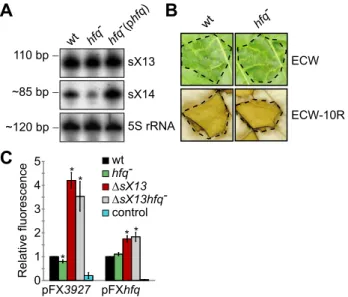
sX13 activity does not require Hfq
ThehfqmRNA accumulation inXcvDsX13(Figure 3B; Figure 4; Table 1) prompted us to test whether sX13 activity depends on the RNA-binding protein Hfq. For this, we introduced a frameshift mutation into thehfqgene of Xcvstrains 85-10 and 85-10DsX13. Northern blot analyses revealed comparable sX13 accumulation in both strains and the complemented hfq mutant, which ectopically expressed Hfq (phfq) (Figure 5A). By contrast, the accumulation of the sRNA sX14 [16] was strongly reduced in the
hfqmutant; this was restored by phfq(Figure 5A). Unexpectedly, thehfqmutant strain was not altered in the induction ofin planta
phenotypes, i. e., in virulence (Figure 5B).
To investigate whether sX13 affects translation of putative target mRNAs, we established a GFP-basedin vivoreporter system for Xcv similar to the one described for E. coli [53]. The promoterless broad-host range plasmid pFX-P permits generation of translationalgfpfusions in a one-step restriction-ligation reaction (Golden Gate cloning [54]; see ‘Materials and Methods’). We cloned the promoter, 59-UTRs, and 10 and 25 codons of
XCV3927andhfq, respectively, into pFX-P resulting in pFX3927
and pFXhfq.XCV3927was selected because of a strongly increased mRNA accumulation inXcvDsX13compared to the wild type (see Table 1). In presence of pFX3927 or pFXhfq, fluorescence of XCV3927::GFP or Hfq::GFP fusion proteins was comparable in theXcvwild type andhfqmutant (Figure 5C). The XCV3927::GFP and Hfq::GFP fluorescence was about 4-fold and 2-fold increased, respectively, inXcvDsX13 compared to strain 85-10 (Figure 5C), suggesting that the synthesis of the fusion proteins is repressed by sX13. Interestingly, the XCV3927::GFP and Hfq::GFP fluores-cence was similarly increased inXcvDsX13and thesX13hfqdouble Figure 4. qRT-PCR analysis of sX13-regulated genes.Selected sX13-regulated genes (see Table 1) were analyzed by qRT-PCR using RNA from NYG- and MMA-grownXcvstrains 85-10 (wt) andDsX13. The amount of each mRNA in the wt was set to 1. Bars represent fold-changes of mRNA amounts in strainDsX13compared to 85-10 on a logarithmic scale (log10). Data points and error bars represent mean values and standard deviations obtained with at least three independent biological samples. Asterisks denote statistically significant differences (t-test;P,0.05). Dashed lines indicate a 1.5-fold change. Transcripts not detected in the microarray analyses are marked with ‘a’.
doi:10.1371/journal.ppat.1003626.g004
mutant (Figure 5C). As abundance and activity of sX13 were not affected by the hfq mutation, we assume that sX13 acts Hfq-independently.
sX13 activityin plantadepends on C-rich loop motifs The predicted secondary structure of sX13 obtained by mfold [55] displays an unstructured 59-region and three stable stem-loops, termed stem 1 to 3, and loop 1 to 3 (Figure 6A). Interestingly, loop 1 and loop 2 contain a ‘CCCC’ (4C) motif, whereas loop 3 harbors a ‘CCCCC’ (5C) motif (Figure 6A). To experimentally verify the predicted structure, we performed structure analyses ofin vitrotranscribed and radioactively-labeled sX13 treated with RNase V1 or RNase T1. While RNase T1 cleaves single-stranded RNA with a preference for G residues, RNase V1 randomly cleaves double-stranded RNA. We detected RNase T1-cleavage products for the 59-region and RNase V1-cleavage products for stem 1 and 2, which is in good agreement with the predicted structure (Figure 6A; Figure S2). Moreover, RNase V1-cleavage products were less abundant for the 4C-motif of loop 1 and loop 2, suggesting single-stranded sequences
(Figure 6A; Figure S2). The results did not allow conclusions about stem 3 and loop 3 structures.
To assess the contribution of the 4C-/5C-motifs to sX13 function, we mutated psX13 in loop 1 and 2, respectively, to ‘GCGC’, and the 5C-motif in loop 3 to ‘GCGCC’ resulting in pL1, pL2and pL3(Figure 6A). In addition, loop mutations were combined (pL1/2, pL1/3, pL2/3) and analyzed for their ability to complement thein planta phenotype of strain DsX13. As shown above,XcvDsX13induced a delayed HR, which was complement-ed by psX13(Figure 1D). Similar phenotypes were observed with
sX13 mutants carrying pL1 or psX13D59, which encodes a 59 -truncated sX13 derivative lacking the terminal 14 nucleotides (Figure 6B). The HR induced by thesX13mutant containing pL2
or pL1/2was intermediate, whereas pL3, pL1/3and pL2/3failed to complement XcvDsX13 (Figure 6B). Northern blot analyses revealed expression of all sX13-loop mutant derivatives (Figure S3). The different RNA species derived from ectopically expressed sX13 and derivatives compared to chromosomally encoded sX13 might be due to alternative transcription termination of plasmid-derived sX13 and derivatives.
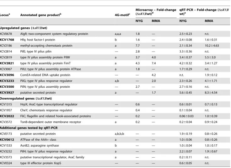
Table 1.Selected sX13-regulated genes validated by qRT-PCR analysis.
Locusa Annotated gene productb 4G-motifc
Microarray – Fold-change (DsX13/wt)d
qRT-PCR – Fold-change (DsX13/ wt)e
NYG MMA NYG MMA
Upregulated genes (DsX13/wt)
XCV0678 AlgR; two-component system regulatory protein a,a,a 1.8 — 2.560.23 n.t.
XCV1768 Hfq; host factor-I protein b 1.6 — 2.460.08 1.660.31
XCV2186 methyl-accepting chemotaxis protein a 7.7 — 2.160.34 10.264.63
XCV2814 PilE; type IV pilus pilin — 2.8 — 3.360.36 n.t.
XCV2819 type IV pilus assembly protein PilW a 3.7 4.0 3.460.37 5.563.0
XCV2821 type IV pilus assembly protein FimT a 4.3 7.4 4.260.32 3.461.27
XCV3067 PilU; type IV pilus assembly protein ATPase a 1.8 — 1.760.29 n.t.
XCV3096 ComEA-related DNA uptake protein — — 4.2 n.t. 1.960.12
XCV3233 PilG; type IV pilus response regulator a,b — 2.0 2.360.26 4.161.71
XCV3500 PilN; type IV pilus assembly protein — 2.7 — 2.760.16 n.t.
XCV3927 putative secreted protein a — 1.7 5.660.45 8.364.54
Downregulated genes (DsX13/wt)
XCV1315 HrpX; AraC-type transcriptional regulator — 0.6 — 0.660.01 0.760.13
XCV1957 CheY; chemotaxis response regulator — 0.4 — 0.160.04 n.t.
XCV2022 FliC; flagellin and related hook-associated proteins — 0.2 — 0.0660.03 1.060.39
XCV3572 TonB-dependent outer membrane receptor a 0.2 — 0.260.04 0.960.24
Additional genes tested by qRT-PCR
XCV0173 putative secreted protein a,b,b,b — — 1.960.19 0.860.26
XCV0612 ATPase of the AAA+class a — — 1.060.06 0.860.26
XCV1533 AsnB2; asparagine synthase b — — 1.060.04 1.060.17
XCV3232 PilH; type IV pilus response regulator a — — 2.260.07 1.960.67
XCV3573 putative transcriptional regulator, AraC family a — — 0.260.11 n.t.
XCV0324 type III effector protein XopS — — — 0.660.05 n.t.
a, bold letters indicate genes with known TSS [16].
b, refers to Thiemeet al.(2005) [32]. c, presence of a 4G-motif within the 5
9-UTR or 100 bp upstream of translation start codon if TSS is unknown (a) and within 100 bp downstream of start codon (b) (see Figure S4).
d, genes not detected as expressed are marked with —.
e, values represent mean fold-change and standard deviation (see Figure 4);
n.t. - not tested.
sX13 loops differentially contribute to mRNA accumulation
As mutation of sX13 loops impinged on Xcv virulence (Figure 6B), we addressed by qRT-PCR whether loop mutations affect the mRNA abundance ofXCV2821,XCV3927,hfqandpilH, which were upregulated inXcvDsX13 (see Figure 4; Table 1). In addition, we analyzed a downregulated gene, XCV3572, and
XCV0612, which was not affected bysX13deletion. As shown in Figure 7A–E, sX13 negatively affected the mRNA abundance of
XCV2821, XCV3927, hfq and pilH, whereas sX13 promoted mRNA accumulation of XCV3572. Mutation of sX13 loops differentially affected the mRNA abundance of the tested genes: pL2and pL1/2failed to complementXcvDsX13with respect to the mRNA abundance ofXCV2821,XCV3927andhfq(Figure 7A–C). Intermediate mRNA amounts ofXCV3927andhfqwere detected inXcvDsX13carrying pL1/3or pL2/3compared to pB and psX13
(Figure 7B, C). Taken together, the mRNA abundance of
XCV2821,XCV3927and hfqappears to be mainly controlled by sX13-loop 2. In contrast, pilH mRNA accumulation appears to depend on multiple sX13 loops as only psX13 and pL1
complemented XcvDsX13 (Figure 7D). The reduced mRNA amount of XCV3572 in XcvDsX13 was complemented by pL1
and pL3but not by pL1/3 (Figure 7E), which suggests redundant
roles of sX13-loops. In presence of pL2, pL1/2 or pL2/3 in
XcvDsX13, theXCV3572 mRNA levels were intermediate com-pared toXcvDsX13carrying pB or psX13(Figure 7E). As expected, the mRNA abundance ofXCV0612was identical in the different strains (Figure 7F).
Figure 5. sX13 activity does not require Hfq.(A) Northern blot analysis. Total RNA from NYG-grownXcv strains 85-10 (wt), the hfq
frameshift mutant (hfq2) and thehfqmutant ectopically expressing Hfq (phfq) was analyzed using sX13- or sX14-specific probes. 5S rRNA was probed as loading control. The experiment was performed twice with two independent mutants and with similar results. (B) Plant infection assay. TheXcvwild-type 85-10 (wt) andhfqmutant strain (hfq2) were inoculated at 26108cfu/ml into leaves of susceptible ECW and resistant
ECW-10R plants. Disease symptoms were photographed 6 dpi. The HR was visualized 2 dpi by ethanol bleaching of the leaves. Dashed lines indicate the inoculated areas. The experiment was repeated three times with similar results. (C) GFP fluorescence of NYG-grownXcv85-10 (wt), thehfqmutant (hfq2), thesX13deletion mutant (DsX13) and the double mutant (DsX13hfq2) carrying pFX3927or pFXhfq.Xcvautofluorescence was determined byXcv85-10 carrying pFX0 (control). Data points and error bars represent mean values and standard deviations obtained with at least four independent experiments. GFP fluorescence of the wt was set to 1. Asterisks denote statistically significant differences (t-test;
P,0.01).
doi:10.1371/journal.ppat.1003626.g005
Figure 6. sX13 loops impact on Xcvvirulence. (A) Secondary structure of sX13 based on prediction and probing (see Figure S2). sX13 consists of an unstructured 59-, three double-stranded regions (S1; S2; S3) and three loops (loop 1–3). 4C-/5C-motifs are highlighted in red. Bold letters indicate unpaired bases and bars mark double-stranded regions deduced from structure probing. Mutations in loops are boxed, exchanged nucleotides are underlined. (B) Derivatives mutated in loops 2 and 3 fail to complement the plant phenotype ofDsX13. Leaves of resistant ECW-10R plants were inoculated at 108cfu/ml withXcv85-10 (wt) and DsX13 carrying pBRS (pB), psX13 or one of the following derivatives: sX13 lacking 14 terminal nucleotides (psX13D59), sX13 mutated in single loops (pL1, pL2, pL3) or in two loops (pL1/2, pL1/3, pL2/3). The HR was visualized by ethanol bleaching of the leaves 2 dpi. Dashed lines indicate the inoculated areas. The experiment was performed four times with similar results.
doi:10.1371/journal.ppat.1003626.g006
Identification of putative sX13-binding sites
To identify potential regulatory motifs in sX13-regulated mRNAs, a discriminative motif search was performed using DREME [56]. For this, sequences surrounding the TLSs of the 42 up- and 21 downregulated genes identified by microarray analyses (Table S2) were compared. More precisely, sequences spanning from known transcription start sites (TSSs) to 100 bp downstream of TLSs or, in case of unknown TSSs, 100 bp up- and 100 bp downstream of the TLS were inspected.
We found that up- and downregulated genes differ in the presence of ‘GGGG’ (4G) motifs. In the NYG-grown sX13
mutant, 15 out of 23 (65%) upregulated genes contain up to three 4G-motifs which are predominantly located upstream of the TLS (Figure S4A; Table S2). 70% of the genes upregulated in MMA (16 out of 23), but only 14% of the genes downregulated in NYG medium (3 out of 21) contain 4G-motifs (Figure S4A; Table S2). Thus, 4G-motifs appear to be enriched in sX13-repressed mRNAs. However, the position of the motifs and flanking nucleotides are not conserved among sX13-regulated genes. Note that the term ‘4G-motif’ also refers to motifs containing more than four G-residues in a row. The complementarity of C-rich sX13-loop sequences and G-rich mRNA motifs suggests sX13-mRNA interactions via antisense-base pairing (Figure 6A; Table 1; Table S2).
Compared to the occurrence of 4G-motifs in approximately 70% of sX13-repressed genes, only 30.71% of all chromosomally encoded Xcv genes (1,378 out of 4,487) carry 4G-motifs in
proximity of their TLS (Figure S4A). Interestingly, 4G-motifs in 241 of the chromosomally encoded genes (5.37%) are located between nucleotide position 8 and 15 upstream of the TLS (Figure S4B). This position corresponds to the presumed location of the RBS and suggests a role of 4G-motifs in translation control.
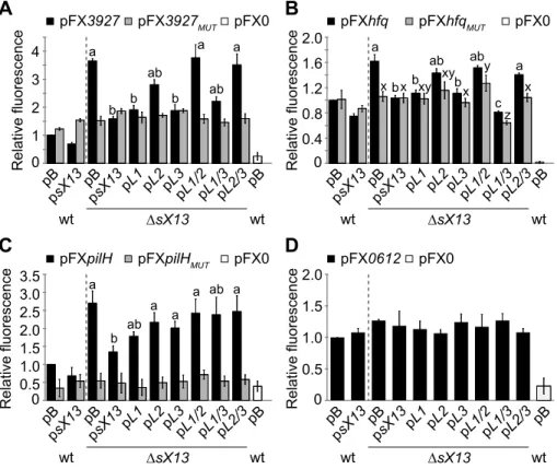
sX13 dependency of target::GFP synthesis requires both 4C- and 4G-motifs
To study the effect of sX13 on translation of selected putative targets, i. e., XCV3927 and hfq, we used the above-mentioned GFP-reporter plasmids pFX3927 and pFXhfq. In addition, we generatedpilH::gfp(pFXpilH) andXCV0612::gfp(pFX0612) fusions (see ‘Materials and Methods’). All mRNA::gfpfusions contain a G-rich motif in the proximity of their TLS which is complementary to C-rich sX13-loop regions (see ‘Materials and Methods’). The fluorescence of the sX13 deletion mutant carrying pFX3927, pFXhfq and pFXpilH was about 3.5-, 1.6- and 2.5-fold higher, respectively, compared to the Xcv wild type (Figure 8A–C). In presence of psX13, pL1, pL3 or pL1/3 in XcvDsX13, the XCV3927::GFP and Hfq::GFP fluorescence levels were compa-rable to the Xcv wild type (Figure 8A, B). By contrast, the XCV3927::GFP and Hfq::GFP fluorescence of strain DsX13
carrying pL2, pL1/2 or pL2/3 was similarly increased as compared toXcvDsX13carrying pB (Figure 8A, B). This suggests that the 4C-motif in sX13-loop 2 is required to repress XCV3927::GFP and Hfq::GFP synthesis. The increased PilH::GFP fluorescence of XcvDsX13 was complemented by Figure 7. sX13 loops differentially contribute to abundance of putative mRNA targets.Relative transcript levels of (A)XCV2821, (B)
XCV3927, (C)hfq, (D)pilH, (E)XCV3572and (F)XCV0612were analyzed by qRT-PCR in total RNA of NYG-grownXcvstrains 85-10 (wt) andDsX13
carrying pBRS (pB), psX13or mutated sX13-derivatives (see Figure 6). The mRNA abundance in the wt was set to 1. Data points and error bars represent mean values and standard deviations obtained with at least three independent biological samples. Statistically significant differences are indicated (t-test;P,0.015).
psX13and pL1, in contrast to othersX13-loop mutant derivatives (Figure 8C). Fluorescence values of all analyzed Xcv strains carrying pFX0612 were comparable confirming sX13 -indepen-dency (Figure 8D).
To address whether the G-rich motif in presumed target mRNAs is required for sX13 dependency of mRNA::gfp transla-tion, we introduced compensatory mutations. i. e., mutated the motif to ‘GCGC’. Xcv strains carrying the resulting plasmids, pFX3927MUT, pFXhfqMUT or pFXpilHMUT, exhibited a similar fluorescence in absence and presence of sX13 and mutated sX13 derivatives (Figure 8A–C). This suggests that the G-rich motif is required for sX13-dependency of target::GFP synthesis. However, mutation of the C-rich motifs in sX13 and the G-rich motifs in mRNA::gfpfusions did not restore sX13 dependency (Figure 8A– C). Unexpectedly, the fluorescence detected for Xcv strains containing pFX3927MUT or pFXhfqMUTwas comparable to the fluorescence of Xcv 85-10 carrying the non-mutated plasmids pFX3927and pFXhfq, respectively (Figure 8A, B). The mutation of the 5G-motif in pilH abolished the fluorescence of strains containing pFXpilHMUTindicating an essential role of the motif in
pilHtranslation (Figure 8C).
Because sX13 was more abundant in MMA- than NYG-grown bacteria (Figure 3), we also analyzed the fluorescence of MMA-grown Xcv strains containing pFX-derivatives. XCV3927::GFP and PilH::GFP synthesis in MMA was sX13-dependently repressed to a greater extent than in NYG (Figure S5; see Figure 8).
Because sX13 negatively affected both the mRNA accumulation of chromosomally encoded XCV3927, hfq and pilH genes and accumulation of the corresponding GFP-fusion proteins, we exemplarily analyzed whether this is due to an altered mRNA abundance. However, qRT-PCR analyses revealed that the
XCV3927::gfp mRNA accumulation was sX13-independent sug-gesting that sX13 posttranscriptionally affects the synthesis of XCV3927::GFP (Figure S6).
To discriminate between transcriptional and posttranscriptional effects of sX13 on target::GFP synthesis we generated reporter fusions controlled by plac(see ‘Materials and Methods’). Note that the activity of thelacpromoter is not affected by deletion ofsX13
(data not shown). As shown in Figure S7, the fluorescence of
XcvDsX13carrying pFXpl-3927(XCV3927) and pFXpl-pilH(pilH) was 2.5- and 4-fold higher, respectively, compared to theXcvwild type and the complemented sX13 mutant strain. Interestingly, mutation of the 4G-motif in theXCV392759-UTR did not only abolish sX13-dependency but also led to a significantly reduced fluorescence compared to theXcvwild type which carried the non-mutated reporter plasmid (Figure S7). This suggests that the 4G-motif in theXCV392759-UTR promotes translation, i. e., acts as translational enhancer element. In presence of pFXpl-pilH, the fluorescence of the fusion protein was only detectable in thesX13
mutant but not in the wild type or complemented strain, confirming that PilH::GFP synthesis is repressed by sX13 (Figure S7). Overall, the data confirm that sX13 represses the synthesis of XCV3927 and PilH on the posttranscriptional level.
Figure 8. sX13-dependency of mRNA target::GFP synthesis requires a G-rich motif.GFP fluorescence of NYG-grownXcvstrains 85-10 (wt) andDsX13carrying pB, psX13or mutated sX13-derivatives (see Figure 6) and carrying GFP-reporter plasmids (A) pFX3927, (B) pFXhfq, (C) pFXpilHor (D) pFX0612. pFXMUTderivatives contain a mutated 4G-motif.Xcvautofluorescence was determined using pFX0. GFP fluorescence of the wt was set to
1. Data points and error bars represent mean values and standard deviations obtained from at least four independent experiments. Statistically significant differences are indicated (t-test;P,0.015).
doi:10.1371/journal.ppat.1003626.g008
Discussion
sX13 controls Xcvvirulence
This study provides a first insight into the posttranscriptional modulation of clade-specific adaptive processes in a plant-pathogenic c-proteobacterium. We identified sX13 as a major regulator ofXcvvirulence in that it promotes expression of genes in thehrp-regulon, i. e., components and substrates of the T3S system (Figure 2A–C). This finding is remarkable because thehrp-regulon is only expressed under certain conditions, whereas sX13 is constitutively expressed (Figure S1) [16]. The sX13 gene is exclusively found and highly conserved in members of the
Xanthomonadaceae family of Gram-negative bacteria [16]. Intrigu-ingly, several species with ansX13homolog lack a T3S system, e. g., the plant pathogen X. fastidiosa and the opportunistic human pathogen S. maltophilia. This suggests a role of sX13 apart from regulation of thehrp-regulon in these organisms.
The expression of thehrp-regulon depends on HrpG and HrpX [39,40]. HrpG is presumably posttranslationally activated in the plant and in XVM2 medium and induces the expression ofhrpX
[38,39,40,41]. As the XVM2-grown sX13 mutant displayed decreased mRNA amounts ofhrpX but not ofhrpG(Figure 2C), we suppose that sX13 acts upstream of HrpG. This idea is supported by the finding that constitutively active HrpG (HrpG* [41]) suppressed thesX13mutation with respect to virulence and the expression of hrpX and downstream genes (Figure 1D; Figure 2A–C). In addition, sX13 affected the basal expression level and, hence, the activity of HrpX under non-inducing conditions, which might impact on the efficiency of hrp-gene induction during infection. Based on the fact that HrpG::GFP and HrpX::GFP synthesis was sX13-independent (Figure S8) we assume that sX13 indirectly controls the expression of the hrp -regulon.
Physiological roles of sX13
Deletion ofsX13affected the mRNA abundance of more than 60 genes involved in signaling, motility, transcriptional and posttranscriptional regulation (Table S2). sX13 negatively regu-lated mRNAs involved in Tfp biogenesis but promoted the accumulation of mRNAs involved in flagellum-mediated chemo-taxis in a growth-phase dependent manner (Table 1; Table S2). This, together with the fact that sX13 is differentially expressed under certain stress conditions (Figure 3), implies a central role of sX13 in the transduction of environmental signals into compre-hensive cellular responses affecting virulence gene expression, motility and QS regulation. The latter is corroborated by the reduced stationary-phase cell density of the sX13 mutant compared to the Xcv wild type (Figure 1A, B) and the sX13-dependency of theXCV2041mRNA (Table S2), which encodes a GGDEF-/EAL-domain protein. Such domains play a role in the control of cyclic-di-GMP levels and QS regulation [57]. Interest-ingly, XCV2041 shares 94% identity with theXccprotein XC2226 which is a repressor of Tfp-mediated motility [58].
Another remarkable finding of our study was the sX13-dependent accumulation of the hfqmRNA. To the best of our knowledge, sX13 is the first sRNA which affects expression of this conserved RNA-binding protein (Table 1). TheXcv hfqmutant was unaltered in virulence on its host plant (Figure 5B), which is in good agreement with recent findings forXoo[18]. By contrast, Hfq contributes to virulence in a number of other bacteria, including the plant pathogenA. tumefaciens, and is also involved in symbiotic plant interactions of Sinorhizobium meliloti [5,12,59,60]. In Vibrio cholerae, four redundantly acting and Hfq-dependent sRNAs (Qrr) destabilize hapR mRNA, which encodes the master regulator of
QS, the T3S system and other virulence genes [61,62]. In the Gram-positive human pathogen Staphylococcus aureus, the Hfq-independent RNAIII is induced by the agr QS system and mediates the switch between the expression of surface proteins and excreted toxins through translational repression of Rot (repressor of toxins) [63,64,65].
sX13 activity depends on C-rich loop regions
Xcv sRNAs are strongly structured and lack extended single-stranded regions [15,16,32]. In contrast, enterobacterial sRNAs commonly exhibit a modular structure consisting of a single-stranded mRNA-targeting domain, often located at the 59-end, an A/U-rich Hfq-binding site and a Rho-independent terminator [1]. The sX13 structure suggests that direct sRNA-mRNA interactions are energetically confined to the unstructured 59-region and its three C-rich loops (Figure 6A). However, the 59-region of sX13 was dispensable for full virulence ofXcvand sX13 activity appears to be exerted via loops 2 and 3 (Figure 6B). Although loops 1 and 2 just differ in the 39-adjacent nucleotide (U/A) (Figure 6A), only loop 2 was required to repress the synthesis of XCV3927::GFP and Hfq::GFP, which might depend on the position of stem-loops in the sRNA and, thus, accessibility. By contrast, repression of PilH::GFP appears to depend on multiple sX13 regions (Figure 8C).
An important question is whether sX13 controls target gene expression on the level of mRNA stability or translation. On one hand, sX13-loop mutant derivatives affected the mRNA levels of presumed targets (Figure 7). On the other hand, protein levels, but not the mRNA level of anXCV3927::gfpfusion, harboring only the 59-UTR and 10 codons of XCV3927, were sX13-dependent (Figure 8A; Figure S6). This suggests that the impact of sX13 on
XCV3927mRNA abundance and translation are separate events and hints at the presence of additional regulatory sites in the
XCV3927mRNA. It should be noted that the assessment of RNA stability by rifampicin treatment is hampered by the fact that our
Xcvstrains are rifampicin resistant.
The sX13 loops are reminiscent of regulatory RNAs inS. aureus, many of which contain ‘UCCC’-motifs in loops [66]. For example, RNAIII contains C-rich stem-loops, which interact with the RBS of target mRNAs [63,65,67]. RNAIII represses Rot synthesis through formation of kissing complexes between two stem-loops of each RNAIII and rot mRNA [64,65]. Such multiple loop interactions are also employed by the E. coli sRNA OxyS to target fhlA [68]. In Helicobacter pylori, the sRNA HPnc5490 represses the synthesis of the chemotaxis regulator TlpB [69]. Interestingly, the central part of the HPnc5490-loop sequence is identical to the ‘UCCCCCU’-motif of loop 3 in sX13 [69].
G-rich enhancer motifs confer sX13-dependency of target mRNAs
Similarly to RNAIII targets inS. aureusand thetlpBmRNA in
two 4G-motifs close to the TLSs (Figure S4), we assume that sX13 loops can interact with multiple 4G-motifs in certain mRNAs. As positively regulated mRNAs lack G-rich motifs, sX13 presumably acts indirectly on the corresponding genes (Figure S4; Table S2). Direct sRNA-mRNA interactions are commonly validated by compensatory mutant studies [1]. However, in case of theE. coli
sRNA RyhB, mutations were suggested to interfere with Hfq-binding and rendered compensatory mutants non-functional [70]. Here, mutation and deletion of sX13 increased the synthesis of XCV3927::GFP and Hfq::GFP fusions, whereas mutation of corresponding 4G-motifs resulted in similar fluorescence values as non-mutated mRNA::gfpfusions inXcvwild type. In addition, the reduced fluorescence of mutated target::GFP fusions was unaf-fected by compensatory sX13-mutant derivatives (Figure 8). This suggests that G-rich motifs in sX13-repressed mRNAs play a role besides mediation of sRNA interactions. While Xanthomonasspp., like other G+C-rich bacteria, lack a consensus RBS [16,71], 5% of the chromosomalXcvcoding sequences (241 of 4,487) contain a G-rich motif 8–15 nucleotides upstream of their TLS (Figure S4). As anticipated, mutation of the 5G-motif at the RBS position ofpilH
abolished translation (Figure 8; Figure S5; Figure S7). By contrast, the 4G-motifs inXCV3927andhfqmRNAs, located 21 nucleotides upstream and nine nucleotides downstream of the AUG, respectively, confer sX13-dependency but were not essential for translation (Figure 8). Thus, G-rich motifs confer sX13-depen-dency and mRNA translation in a position-dependent manner. As mutation of the 4G-motif inXCV3927reduced protein synthesis, the motif appears to function as translational enhancer (Figure S7). We suggest that sequestration of a G-rich motif by sX13 as well as mutation of the motif precludes the binding of an unknown factor, which promotes mRNA translation. Such a factor could be RNA, protein or the ribosome.
The presumed sX13 mode of action is reminiscent of the
Salmonella sRNA GcvB, which inhibits translation of mRNAs by targeting C/A-rich enhancer elements [72,73]. By increasing the ribosome-binding affinity, C/A-rich motifs enhance mRNA translation, irrespective of their localization upstream or down-stream of the TLS [72,74].
sRNAs encoded at thepolAlocus in other bacteria Homologs ofXcvsRNAs are predominantly found in members of theXanthomonadaceae family but not in other bacteria [15,16]. The sX13 gene is located adjacent to the DNA polymerase I-encodingpolAgene, which resembles a locus encoding the Spot42 sRNA in E. coli and members of the ar7 sRNA family in a -proteobacteria [75,76,77,78]. In contrast to sX13, Spot42 requires Hfq and regulates targets involved in carbon metabolism [48,79], e. g., the discoordinated expression of genes within the gal
galactose utilization operon [47], which is absent inXcv. Although sX13 lacks sequence similarity to Spot42 and ar7 sRNAs, the latter also contain three stem-loops and apical C-rich motifs [80] suggesting that sRNAs in distantly related bacteria evolved divergently but retained structural conservation. Thus, it will be interesting to see whether the polA locus in other bacteria also encodes sRNAs, and whether sX13 and structurally related sRNAs act in a similar manner.
Materials and Methods
Bacterial strains and growth conditions
For bacterial strains, plasmids and oligonucleotides used in this study see Table S1.E. colistrains were grown at 37uC in lysogeny broth (LB),Xcvstrains at 30uC in nutrient-yeast-glycerol (NYG) [81], XVM2 [40] or minimal medium A (MMA) [82], which was
supplemented with casamino acids (0.3%) and sucrose (10 mM). Plasmids were introduced intoE. coliby chemical transformation and into Xcv by tri-parental conjugation, using pRK2013 as helper plasmid [83]. Antibiotics were added to the final concentrations: ampicillin, 100mg/ml; gentamycin, 15mg/ml; kanamycin, 25mg/ml; rifampicin, 100mg/ml; spectinomycin, 100mg/ml.
Generation of constructs
To generate the sRNA-expression vector pBRS, a 28-bp fragment between thelacpromoter and the EcoRI cloning site in pBBR1mod1 [84] was replaced by a truncated fragment, amplified by PCR from pBBR1mod1 using primers pBRS-EcoRI-fw and pBRS-NcoI-rev. For generation of constructs expressing sX13 (psX13) and sX13D59 (psX13D59; lacking 14 nt at the 59-end), respective fragments were PCR-amplified from Xcv 85-10 using primers sX13-fw/-rev or sX13d5-fw/-rev. PCR products were digested with EcoRI/HindIII and cloned into pBRS. The sX13 -mutant plasmids pL1, pL2, pL3and pL2/3were generated by PCR amplification of plasmid psX13using primers L1-fw/-rev, L2-fw/-rev, L3-fw/-rev and L3-fw/L2/3-L2-fw/-rev, respectively; plasmid pL1/3
was generated with primers L3-fw/-rev and pL1as template; pL1/2
was generated with primers L2-fw/L1/2-rev and pL2as template. For ectopic expression ofhfqunder control of its own promoter, a PCR product obtained fromXcv85-10 using primers pMphfq-fw/-rev was cloned into the promoterless vector pBRM-P [84].
The GFP-based promoter-less reporter plasmid pFX-P permits
BsaI-mediated cloning of PCR amplicons (Golden Gate cloning) in a one-step restriction-ligation reaction [54] and was generated as follows: pDSK602 [85] was digested withPstI/BamHI to remove thelac promoter and multiple-cloning site. To allow blue-white selection, a dummy module containing 59- and 39-BsaI recognition sites, placandlacZwas PCR-amplified from pBRM-P [84] using primers pFX-lz-fw/-rev. A fragment containing both the gfp
coding sequence without translation start codon and the rrnB
terminator was PCR-amplified from pXG-1 [53] using primers pFXgfp-fw/-rev. After blunt-end ligation of dummy- and gfp -module, the fragment was digested with Mph1103I/BglII and ligated into the PstI/BamHI sites of the pDSK602 backbone, resulting in pFX-P. For generation of the GFP-control plasmid pFX0, a promoterless fragment (138 bp) of the sRNA genesX6
[16] was PCR-amplified fromXcv85-10 using primers pFX0-fw/-rev and cloned into pFX-P.
To generate mRNA::gfp expression constructs, fragments containing the promoter, 59-UTR and 10 to 25 codons of the respective genes were PCR-amplified from Xcv 85-10 using corresponding pFX-fw/-rev primers (Table S1) and cloned into pFX-P. Plasmids pFX3927, pFXhfqand pFX0612were generated by cloning of nucleotide sequences298 to+30,2160 to+75 and
2116 to+33 relative to the translation start codon ofXCV3927,
hfq and XCV0612, respectively. pFXpilH was constructed by cloning a fragment spanning nucleotides299 upstream of thepilG
translation start codon to nucleotide +60 within the coding sequence ofpilH.
The 4G-motif inXCV3927::gfpandhfq::gfpis located 21–24 bp upstream and 9–12 bp downstream of the ATG, respectively.
pilH::gfp and XCV0612::gfp contain a 5G-motif at nucleotide positions 10–14 and 8–12 upstream of the ATG, respectively. Plasmids pFXMUT were constructed as follows: to mutate the ‘GGGG’ motif to ‘GCGC’, sequences upstream and downstream of the motif were PCR-amplified fromXcv85-10 using primers pFX-fw/pFX-mut-L-rev and pFX-mut-R-fw/pFX-rev, respective-ly. Primers pFX-mut-L-rev and pFX-mut-R-fw contain the mutation flanked by aBsaI-recognition site. pFX and
ing pFXMUTderivatives only differ in the sequence of the G-rich motif at nucleotide positions relative to the translation start codons:224/222 in pFX3927MUT,+10/+12 in pFXhfqMUTand
212/210 in pFXpilHMUT.
Plasmids pFXpl, which express plac-driven mRNA::gfpfusions, were constructed by cloning respective fragments into pFX-P: plac
was PCR-amplified from pBRM-P [84] using primers plac-fw/rev; sequences259 to+54 and2147 to+54 relative to the ATG ofhrpX
andhrpG, respectively, were PCR-amplified fromXcv85-10 using primers pFXpl-hrpX-fw/-rev and pFXpl-hrpG-fw/-rev; fragments ofXCV3927andpilHwere PCR-amplified from respective pFX and pFXMUTplasmids using primers pFXpl3927-fw/pFXpl3927mut-fw/pFX3927-rev and pFXplpilH-fw/pFXpilH-rev.
Generation ofXcvmutant strains
To generate a chromosomal sX13 deletion mutant, flanking sequences of ,650 bp up- and downstream of sX13 [16] were amplified by PCR fromXcv85-10 using primers d13L-fw/-rev and d13R-fw/-rev. PCR products were digested withBamHI/HindIII andHindIII/XbaI, respectively, and ligated into the suicide vector pOK1 [86].XcvDsX13+sX13ch, which carries ansX13copy at the
DsX13locus, was created as follows: two PCR fragments amplified from Xcv85-10 using primers int13L-fw/rev and int13R-fw/rev were digested withPsp1406I, ligated and cloned into theBamHI/
XbaI site of pOK1. Conjugation of pOKDsX13intoXcv85-10 and pOKint13 into XcvDsX13 and selection of the correct double crossing-over events were performed as described [86].
XcvDsX13+sX13ch was identified by PCR amplification of the
sX13locus andPsp1406I restriction.
To introduce a frameshift mutation into chromosomalhfq, PCR products obtained fromXcv85-10 using primers hfqL-fw/-rev and hfqR-fw/-rev were digested with BamHI/BsaI and BsaI/XbaI, respectively, and cloned into pOK1. Conjugation of pOK-fshfq
into Xcv and selection of double crossing-over events were performed as described [86]. The resulting hfq mutant strain carries a 4 bp deletion in anMnlI site (nucleotides 33–36 in hfq) and was identified by PCR using primers seqhfq-fw/-rev followed by digestion withMnlI.
Plant material and plant inoculations
Pepper (Capsicum annuum) plants of the near-isogenic cultivars ECW and ECW-10R [87] were grown at 25uC with 60–70% relative humidity and 16 hours light. For infection assays, Xcv
bacteria were resuspended in 10 mM MgCl2and inoculated with
a needleless syringe into the intercellular spaces of leaves using concentrations of 1–46108colony-forming units (CFU) per ml for scoring plant reactions and 104CFU/ml for in planta growth curves. For better visualization of the HR, leaves were bleached in 70% ethanol.In plantagrowth curves were performed as described [33]. All experiments were repeated at least two times.
Protein detection and measurement of GFP fluorescence inXcv
Xcv cells grown overnight in NYG medium were washed, incoculated at OD600= 0.2 into XVM2 medium and incubated for
3.5 hours at 30uC. Total cell extracts were analyzed by sodium dodecyl sulfate-polyacrylamide gel electrophoresis and immuno-blotting using specific polyclonal antibodies directed against HrpF [88], HrcN [89], HrcJ [89] and GroEL (Stressgen). A horseradish peroxidase-labeled anti-rabbit antibody (Amersham Pharmacia Biotech) was used as secondary antibody. Antibody reactions were visualized by enhanced chemiluminescence (Amersham Pharma-cia Biotech).
To determine GFP fluorescence, bacteria were adjusted to OD600= 1.0 in 10 mM MgCl2. Fluorescence was measured at
485-nm excitation and 535-nm emission using a microplate reader (SpectraFluor Plus; Tecan Trading AG).
In vitrotranscription and structure probing
sX13 [16] was PCR-amplified from Xcv 85-10 using primers sX13T7-fw, containing the T7-promoter, and sX13T7-rev and cloned into pUC57 (Thermo Fisher Scientific), resulting in pUC-13T7. Template DNA forin vitrotranscription was amplified from pUC-13T7 using primers sX13-ITC-fw/-rev.sX13 transcription and DNase treatment were performed according to manufactur-er’s instructions (MEGAscriptHKit; Invitrogen). RNA labeling using [c-32P]-ATP, treatment with RNase T1 (1 Pharmacia unit; Ambion) or RNase V1 (0.01 to 0.0002 Pharmacia units; Ambion) and generation of nucleotide ladders were performed as described [90]. Samples were analyzed on 12% polyacrylamide gels containing 7 M urea. Signals were visualized with a phosphoima-ger (FLA-3000 Series; Fuji).
RNA preparation, Northern blot and qRT-PCR analysis Bacteria were grown overnight in NYG and inoculated at OD600= 0.2 into NYG or XVM2 medium. XVM2 cultures were
incubated for 3.5 hours at 30uC. NYG-grown cells were harvested at exponential growth phase (OD600= 0.5–0.7) or used to
inoculate the following media at OD600= 0.5: NYG containing
0.3 M NaCl, 0.2 M H2O2 or NYG lacking a nitrogen source,
MMA or MMA lacking a carbon source followed by incubation for 3 hours.
RNA isolation and Northern blot hybridization was performed as described [16,91]. Oligonucleotide probes for detection of sX13 and 5S rRNA are described in [16].
For qRT-PCR analyses, cDNA was synthesized using Rever-tAid H Minus First Strand cDNA-Synthesis Kit according to manufacturer’s instructions (Fermentas). qRT-PCR was per-formed using 2 ng cDNA and ABsolute BlueSYBR Green Fluorescein (Thermo Scientific) and analyzed on MyiQ2 and CFX Connect systems (Bio-Rad). The efficiency and specificity of PCR amplifications was determined by standard curves derived from a dilution series of template cDNA and melting curve analysis, respectively. Mean transcript levels were calculated based on values obtained from technical duplicates of at least three independent biological replicates and the levels of 16S rRNA (reference gene) as described (ABI user bulletin 2; Applied Biosystems).
Microarray analysis
For isolation of total RNA, NYG-grown cells were harvested at exponential growth phase (OD600= 0.5–0.7) or used to inoculate
MMA at OD600= 0.5 followed by incubation for 3 hours.
Fluorescently labeled cDNA was prepared as described [92]. Starting from 10–15mg total RNA, aminoallyl-modified first
strand cDNA was synthesized by reverse transcription using random hexamer primers, Bioscript reverse transcriptase (Bioline) and 0.5 mM dNTP, dTTP:aminoallyl-dUTP (1:4). After hydro-lysis and clean up using Nucleotide removal kit (Qiagen), Cy3- and Cy5-N-Hydroxysuccinimidyl ester dyes (GE Healthcare) were coupled to the aminoallyl-labeled first strand cDNA. Uncoupled dye was removed using the Nucleotide removal kit (Qiagen). For RNA from NYG- and MMA-grown bacteria, four and three microarray hybridizations were performed, respectively, using labeled cDNA obtained from independent bacterial cultures.
each oligonucleotide spotted in three technical replicates per microarray [93]. Preprocessing of microarrays was performed as described [94]. Hybridization was performed in EasyHyb hybrid-ization solution (Roche) supplemented with sonicated salmon sperm DNA at 50mg/ml in a final volume of 130ml for 90 min at 45uC using the HS400 Pro hybridization station (Tecan Trading AG). Labeled cDNA samples were denatured for 5 min at 65uC prior hybridization. After hybridization microarrays were washed as described [94].
Mean signal and mean local background intensities were obtained for each spot on the microarray images using ImaGene 8.0 software for spot detection, image segmentation and signal quantification (Biodiscovery Inc.). Spots were flagged as empty if
R#0.5 in both channels, where R= (signal mean2background mean)/background standard deviation. Remaining spots were analyzed further. The log2 value of the ratio of intensities was
calculated for each spot using the formula Mi= log2(Ri/Gi).
Ri=Ich1(i)-Bgch1(i)andGi=Ich2(i)-Bgch2(i), whereIch1(i)orIch2(i)is the
intensity of a spot in channel 1 or channel 2, andBgch1(i)orBgch2(i)is
the background intensity of a spot in channel 1 or channel 2. The mean intensity was calculated for each spot,Ai= log2(RiGi)0.5[95].
For data normalization (Median), significance test (Holm) andt -statistics analysis, the EMMA 2.8.2 open source platform was used [49]. Genes were accounted as differentially expressed ifP adjusted
#0.05,A$8, and if the ratio showed at least a 1.5-fold difference between the two experimental conditions.
Biocomputational analyses
Homology searches were performed using BLASTn and the NCBI genome database (http://blast.ncbi.nlm.nih.gov; http:// www.ncbi.nlm.nih.gov/genome; date: 11/22/2012).
The secondary structure of sX13 [16] was predicted using Mfold version 3.5 (http://mfold.rna.albany.edu/?q = mfold/ RNA-Folding-Form) and default folding parameters [55]. To identify putative regulatory motifs in the 59-regions of sX13-regulated mRNAs, a discriminative motif search was performed using DREME version 4.9.0 (http://meme.nbcr.net/meme/cgi-bin/dreme.cgi) [56]. Sequences of regulated genes comprising nucleotide positions 2100 to +100 relative to translation start codons or in case of known TSSs [16] (see Table S2), sequences comprising the 59-UTR to position +100 downstream of translation start codons, were extracted from the genome ofXcv
strain 85-10 (NC_007508 and NC_007507) [32]. DREME motif search was performed with negatively regulated genes as input and positively regulated genes as comparative sequences and an E -value of#5.
Accession numbers
YP_363045.1; YP_363046.1; YP_362142.1; YP_362163.1; YP_362160.1; YP_361663.1; YP_363499.1; YP_365931.1; YP_363887.1; YP_365930.1; YP_365658.1; YP_364552.1; YP_363772.1; YP_363917.1; YP_364964.1; YP_364963.1; YP_364545.1; YP_365303.1; YP_365304.1; YP_362343.1; YP_362302.1; YP_362409.1; YP_364550.1; YP_364798.1; YP_364827.1; YP_365231.1; YP_363688.1; YP_363753.1; YP_361904.1; YP_363264.1; YP_362055.1; YP_363957.1
Supporting Information
Figure S1 sX13 abundance is not affected by expression of HrpG*.Xcv85-10 (wt),DsX13andDsX13+sX13chand strains
additionally expressing HrpG* were incubated for 3.5 hours in
hrp-gene inducing medium XVM2 (see Figure 2B). Total RNA was analyzed by Northern blot using an sX13-specific probe. 5S rRNA
was probed as loading control. The experiment was performed twice with similar results.
(EPS)
Figure S2 Structure probing of sX13.In vitrotranscribed sX13 was 59-labeled and treated with RNase T1 (T1) or alkaline hydroxyl (OH2) buffer for generation of nucleotide ladders and RNase V1 (V1) for mapping of base-paired regions. Lane ‘C’ contains untreated sX13; triangle indicates decreasing concentrations of RNase V1; ‘#G’ indicates positions of G residues; the deduced secondary structure is indicated on the right hand side (see Figure 6A). (EPS)
Figure S3 Expression of sX13 derivatives.Total RNA of NYG-grownXcvstrains 85-10 (wt) andDsX13carrying pBRS (pB), psX13or expressing mutated sX13-derivatives (see Figure 6) was analyzed by Northern blot using an sX13-specific probe. 5S rRNA was probed as loading control. The experiment was performed twice with similar results.
(EPS)
Figure S4 Distribution of 4G-motifs among sX13-regu-lated genes and chromosomally encodedXcvgenes. (A) Percentage of sX13-regulated genes identified by microarray analyses (see Table S2) and chromosomal CDSs inXcvcontaining one or more 4G-motifs in region2100 to+100 relative to the TLS or in case of known TSSs, in the sequence comprising the 59-UTR to position+100. The number of genes analyzed (n) is given below. (B) Distribution of 4G-motifs found in region2100 to +100 bp relative to the TLSs of 1,378 chromosomal CDSs [see (A)]. (EPS)
Figure S5 sX13-dependency of mRNA target::GFP syn-thesis in MMA-grown Xcv strains. GFP fluorescence of MMA-grown Xcv strains 85-10 (wt) and DsX13 carrying pB or psX13 and carrying GFP-reporter plasmids pFX3927, pFX3927MUT, pFXpilH or pFXpilHMUT. pFX3927MUT and pFXpilHMUTcontain a mutated 4G- and 5G-motif, respectively.
Xcvautofluorescence was determined using pFX0. GFP fluores-cence of the wt was set to 1. Data points and error bars represent mean values and standard deviations obtained from three independent experiments. Statistically significant differences compared to the wt are indicated by an asterisk (t-test;P,0.03). For comparison, see Figure 8A and C.
(EPS)
Figure S6 mRNA amount of XCV3927::gfp is sX13-independent.TheXCV3927::gfpmRNA amount in NYG-grown
Xcvstrains 85-10 (wt) andDsX13carrying pB, psX13or mutated sX13-derivatives and containing pFX3927or pFX3927MUTwas analyzed by qRT-PCR using gfp-specific oligonucleotides. The RNA level in the wt was set to 1. Data points and error bars represent mean values and standard deviations obtained with three independent biological samples. For comparison, see Figure 7B and Figure 8A.
(EPS)
Figure S7 sX13 posttranscriptionally affects XCV3927::GFP and PilH::GFP synthesis.GFP fluorescence of NYG-grown Xcv strains 85-10 (wt), DsX13 and DsX13
containing chromosomally re-integrated sX13 (DsX13+sX13ch);
strains expressXCV3927::gfp(pFXpl-3927) orpilH::gfp(pFXpl-pilH) under control of plac. pFXMUTderivatives contain a mutated 4G-motif. Xcv autofluorescence was determined using pFX0 and is indicated by dashed line. GFP fluorescence of the wt carrying pFXpl-3927or pFXpl-pilHwas set to 1. Data points and error bars represent mean values and standard deviations obtained from