Transcriptional Regulation of the
β-Type
Carbonic Anhydrase Gene
bca
by RamA in
Corynebacterium glutamicum
Adnan Shah, Bernhard J. Eikmanns*
Institute of Microbiology and Biotechnology, University of Ulm, D-89069 Ulm, Germany
*bernhard.eikmanns@uni-ulm.de
Abstract
Carbonic anhydrase catalyzes the reversible hydration of carbon dioxide to bicarbonate and maintains the balance of CO2/HCO3-in the intracellular environment, specifically for carboxylation/decarboxylation reactions. InCorynebacterium glutamicum, two putative genes, namely thebca(cg2954) andgca(cg0155) genes, coding forβ-type andγ-type car-bonic anhydrase, respectively, have been identified. We here analyze the transcriptional organization of these genes. The transcriptional start site (TSS) of thebcagene was shown to be the first nucleotide“A”of its putative translational start codon (ATG) and thus,bca codes for a leaderless transcript. The TSS of thegcagene was identified as an“A”residue located at position -20 relative to the first nucleotide of the annotated translational start codon of the cg0154 gene, which is located immediately upstream ofgca. Comparative expression analysis revealed carbon source-dependent regulation of thebcagene, with 1.5- to 2-fold lower promoter activity in cells grown on acetate as compared to glucose as sole carbon source. Based on higher expression ofbcain a mutant deficient of the regulator of acetate metabolism RamA as compared to the wild-type ofC.glutamicumand based on the binding of His-tagged RamA protein to thebcapromoter region, we here present evi-dence that RamA negatively regulates expression ofbcainC.glutamicum. Functional char-acterization of agcadeletion mutant ofC.glutamicumrevealed the same growth
characteristics ofC.glutamicumΔgcaas that of wild-typeC.glutamicumand no effect on expression of thebcagene.
Introduction
Carbonic anhydrase (CA) (EC 4.2.1.1) catalyzes the reversible hydration of carbon dioxide (CO2) to bicarbonate (HCO3-) and plays an important role in various biochemical and
physio-logical processes in prokaryotic and eukaryotic organisms [1,2]. CAs are ubiquitously found in eukarya, bacteria and archaea domains of life [2,3,4] and five genetically distinct CA families are known to date, namely theα-,β-,γ-,δ-, andz-CAs [5]. Theα-class is predominant in mammals whereas theδ- andz-classes have been found in marine diatoms [6]. Theβandγare the ancient classes of CAs, predominantly found in prokaryotes and their presence in species of archaea and bacteria indicate their fundamental role in prokaryotic biology [6,7,8]. The
a11111
OPEN ACCESS
Citation:Shah A, Eikmanns BJ (2016) Transcriptional Regulation of theβ-Type Carbonic Anhydrase Genebcaby RamA inCorynebacterium glutamicum. PLoS ONE 11(4): e0154382. doi:10.1371/journal.pone.0154382
Editor:Patrick C. Cirino, University of Houston, UNITED STATES
Received:January 31, 2016
Accepted:April 12, 2016
Published:April 27, 2016
Copyright:© 2016 Shah, Eikmanns. This is an open access article distributed under the terms of the Creative Commons Attribution License, which permits unrestricted use, distribution, and reproduction in any medium, provided the original author and source are credited.
Data Availability Statement:All relevant data are within the paper and its Supporting Information files.
Funding:AS obtained a scholarship grant by German Academic Exchange Service (DAAD) and Higher Education Commission (HEC) Pakistan.
bacterialβ-CAs are zinc metalloenzymes that maintain CO2/HCO3-balance in the intracellular
environment [9,10]. By keeping a given balance, the CAs also represent important“accessory enzymes”for other enzymes that use CO2or HCO3-[10], such as ribulose-1,5-bisphosphate
carboxylase/oxygenase (RuBisCO) in chloroplasts, carbamate hydro-lyase (cyanase) in Escheri-chia coli[11], urease inHelicobacter pylori[12] and HCO3--dependent carboxylases in a variety
of eukaryotes and prokaryotes [10,13–17]. In several bacteria, CA has been shown to be essen-tial during aerobic growth under normal atmospheric conditions [14–17] and Ueda et al. [18] suggested that microorganisms that are lacking CA can persist in nature only by choosing niches with higher CO2concentrations.
Corynebacterium glutamicumis a Gram-positive, facultative anaerobic organism, able to use a variety of sugars, alcohols, and organic acids as carbon and energy source [19–21]. The organism has a long tradition in biotechnology and is used as an“industrial workhorse”for the production of amino acids, mainly L-glutamate and L-lysine [22,23]. In addition, the use ofC.
glutamicumin the production of other amino acids [24–30], different organic acids [31–34], vitamins [35], diamines [36–40], ethanol and higher alcohols [41–45], 2-ketoacids [46–49], lycopene [50], and polymers [51,52] has further widened the industrial importance ofC. gluta-micum. Besides,C.glutamicumis also regarded as a model organism for the Corynebacteri-neae, such as the genusMycobacterium[53].
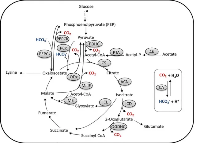
The PEP (phosphoenolpyruvate)-pyruvate-oxaloacetate node inC.glutamicum(Fig 1) has attracted specific attention due to its importance in carbon flux distribution within the central metabolism and in particular for supply of precursors required for the production of various amino acids (reviewed in [54,4]), especially those of the aspartate and glutamate amino acid families.C.glutamicumpossesses two C3-carboxylating anaplerotic enzymes, namely the PEP carboxylase and pyruvate carboxylase, converting phosphoenolpyruvate (PEP) and pyruvate to oxaloacetate, respectively [54]. Apart from these C3-carboxylating enzymes,C.glutamicum
possesses three C4-decarboxylating enzymes, i.e., PEP carboxykinase, converting oxaloacetate to PEP, and oxaloacetate decarboxylase and malic enzyme, converting oxaloacetate and malate, respectively, to pyruvate (reviewed in [54]) (Fig 1). Whereas these decarboxylating enzymes [and also those of the tricarboxylic acid (TCA) cycle] liberate CO2, the carboxylating PEP and
pyruvate carboxylases require HCO3-as substrate [4,17] which highlights the importance of
intracellular CO2/HCO3-balance for the central metabolism, especially the reactions at
meta-bolic switch-points of carbon flux distribution. As HCO3-is needed as substrate of metabolism,
its significant source is the hydration of CO2. Due to the low tension of CO2in the
environ-ment and its diffusion out of the cell, the spontaneously formed HCO3-obviously is not
suffi-cient to meet metabolic demands of the cell and thus, enzymatic hydration of CO2might be
necessary, especially under conditions when the intracellular CO2generation is low [15,17].
InC.glutamicum, two genes putatively coding for beta-type CA (β-CA) and gamma-type CA (γ-CA) have been identified and designated asbca(locus-tag cg2954) andgca(cg0155), respectively [17]. Thebcagene is located betweenmutY, encoding an adenine glycosylase, and cg2953, encoding putatively a benzaldehyde dehydrogenase. Thegcagene is directly preceeded by cg0154, encoding also a so far unknown protein and followed bycysR, encoding the dual transcriptional regulator CysR, which is involved in control of sulfur metabolism inC. glutami-cum[55]. Though, agca-deficient mutant ofC.glutamicumdid not show any phenotype under all conditions tested, abca-deletion mutant showed no growth under normal atmospheric con-ditions (0.04% CO2) and this phenotype could be restored by increasing the CO2concentration
In this report, we analyzed the transcriptional organization of thebcaandgcagenes and investigated the transcriptional regulation ofbcaexpression in glucose- or acetate-grown cells ofC.glutamicum. We also constructed agcadeletion mutant ofC.glutamicumand investigated the effect on growth and on expression of thebcagene.
Materials and Methods
Bacterial strains, plasmids, oligonucleotides and culture conditions
All bacterial strains and plasmids used in this study and their relevant characteristics and sources are given inTable 1, for oligonucleotides, their nucleotide sequence and purpose seeS1 Tablein the supplementary material.
E.coliwas grown aerobically on 2×TY or TB complex medium [62] at 37°C as 5 ml-cultures in 15 ml-tubes or as 50 ml-cultures in 500 ml-baffled Erlenmeyer flasks on a rotary shaker at 120 rpm. Precultures ofC.glutamicumwere grown under the same conditions in 2×TY medium at 30°C. For preparation of solid plates, agar (18 g/l) was added to the medium. For the main cultures, cells of aC.glutamicumpreculture were washed twice with 0.9% NaCl and added to freshly prepared minimal medium [63], containing 1% (w/v), 2% (w/v), 4% (w/v) glu-cose and/or 1% (w/v) acetate as carbon source(s). The cultures then were grown aerobically at 30°C as 50 ml-cultures in 500 ml baffled Erlenmeyer flasks on a rotary shaker at 120 rpm until
Fig 1. The phosphoenolpyruvate (PEP)-pyruvate-oxaloacetate node inC.glutamicum.Abbreviations: AK, acetate kinase; PTA, phosphotransacetylase; CA, carbonic anhydrase; CS, citrate synthase; ACN, aconitase; ICD, isocitrate dehydrogenase; OGDHC, oxoglutarate dehydrogenase complex; ICL, isocitrate lyase; MS, malate synthase; MalE, malic enzyme; ODx, oxaloacetate decarboxylase; PDHC, pyruvate dehydrogenase complex; PCx, pyruvate carboxylase; PEPCx, phosphoenolpyruvate carboxylase; PEPCk, phosphoenolpyruvate carboxykinase.
the desired cell density was obtained. Plasmid-carrying strains were cultivated in the presence of kanamycin (50μg/ml) or ampicillin (100μg/ml). In fermentation experiments, the amino acid concentrations were determined by reversed-phase high-pressure liquid chromatography (RP-HPLC) as described before [26]. Growth of theE.coliandC.glutamicumcultures was fol-lowed by measuring the optical density at 600 nm (OD600).
DNA preparation, manipulation and transformation
Restriction enzymes, T4 DNA ligase, Fast APTMthermosensitive alkaline phosphatase, DNase I, Maxima reverse transcriptase, terminal deoxynucleotidyl transferase and the CloneJetTM PCR Cloning Kit were obtained from Thermo Scientific (Darmstadt, Germany), Phusion1
DNA polymerase from New England Biolabs (Ipswitch, MA, USA),TaqDNA polymerase from Genaxxon Biosciences (Ulm, Germany), and used as instructed by the manufacturer. The RNeasy Mini Kit and the HotStar polymerase kit was obtained from Qiagen (Hilden,
Germany).
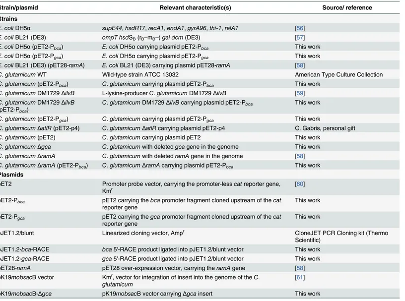
Table 1. Strains and plasmids used in this study and their relevant characteristics.
Strain/plasmid Relevant characteristic(s) Source/ reference Strains
E.coliDH5α supE44,hsdR17,recA1,endA1,gyrA96,thi-1,relA1 [56]
E.coliBL21 (DE3) ompT hsdSB(rB–mB–)gal dcm(DE3) [57]
E.coliDH5α(pET2-Pbca) E.coliDH5αcarrying plasmid pET2-Pbca This work
E.coliDH5α(pET2-Pgca) E.coliDH5αcarrying plasmid pET2-Pgca This work
E.coliBL21 (DE3) (pET28-ramA) E.coliBL21 (DE3) carrying plasmid pET28-ramA [58]
C.glutamicumWT Wild-type strain ATCC 13032 American Type Culture Collection C.glutamicum(pET2-Pbca) C.glutamicumcarrying plasmid pET2-Pbca This work
C.glutamicumDM1729ΔilvB L-lysine-producerC.glutamicumDM1729ΔilvB [59] C.glutamicumDM1729ΔilvB
(pET2-Pbca)
C.glutamicumDM1729ΔilvBcarrying plasmid pET2-Pbca This work
C.glutamicum(pET2-Pgca) C.glutamicumcarrying plasmid pET2-Pgca This work
C.glutamicumΔatlR(pET2-p4) C.glutamicumΔatlRcarrying plasmid pET2-p4 C. Gabris, personal gift C.glutamicum(pET2) C.glutamicumcarrying plasmid pET2 This work
C.glutamicumΔgca C.glutamicumwith deletedgcagene in the genome This work C.glutamicumΔramA C.glutamicumwith deletedramAgene in the genome [58] C.glutamicumΔramA(pET2-Pbca) C.glutamicumΔramAcarrying plasmid pET2-Pbca This work
Plasmids
pET2 Promoter probe vector, carrying the promoter-lesscatreporter gene,
Kmr [60]
pET2-Pbca pET2 carrying thebcapromoter fragment cloned upstream of thecat
reporter gene
This work
pET2-Pgca pET2 carrying thegcapromoter fragment cloned upstream of thecat
reporter gene
This work
pJET1.2/blunt Linearized cloning vector, Ampr CloneJET PCR Cloning kit (Thermo
Scientific) pJET1.2-bca-RACE bca5'-RACE product ligated into pJET1.2/blunt vector This work pJET1.2-gca-RACE gca5'-RACE product ligated into pJET1.2/blunt vector This work pET28-ramA pET28 over-expression vector, carrying theramAgene [58] pK19mobsacB vector Kmr, vector for integration of insert into the genome of theC.
glutamicum
[61]
pK19mobsacB-Δgca pK19mobsacB vector carryingΔgcainsert This work
Plasmids fromE.coliandC.glutamicumcells were isolated using the E.Z.N.A plasmid DNA Mini Kit (Omega Bio-tec Inc., Norcross, USA) or the method described in Green and Sambrook [62], respectively, and purified using the NucleoSpin Gel and PCR Clean-up Kit (Macherey-Nagel, Düren, Germany), according to the manufacturer’s instructions. Chromo-somal DNA was isolated fromC.glutamicum[64] and purified with phenol-chloroform purifi-cation method [62].
PCR experiments were performed in a Thermocycler (Biometra, Göttingen, Germany) using Phusion1
DNA orTaqDNA polymerase with oligonucleotides designed using the Clone Manager v.7 software and obtained from biomers.net (Ulm, Germany). All other reagents used for the PCR mix were obtained from Thermo Scientific. PCR products were puri-fied using the NucleoSpin Gel and PCR Clean-up Kit from Macherey-Nagel.
Plasmid transfer intoC.glutamicumwas carried out by electroporation with an Electropora-tor 2510 (Eppendorf, Hamburg, Germany), and the recombinant strains were selected on 2xTY medium [62] agar plates containing kanamycin (50 or 15μg/ml), as described by van der Rest et al. [65]. Electroporation ofE.coliwas carried out with competent cells according to the method of Dower et al. [66]. The success of the transformation was verified by plasmid prepa-ration and/or other analyses indicated below.
Cloning of the
bca
and
gca
promoter fragments
The promoter regions of thebcagene (position -500 to +20 with respect to the putative transla-tional start site ofbca) and of thegcagene (position -262 to +258 with respect to the putative translational start site of the upstream cg0154 gene) were amplified with primer pairsbca -pro-moter-fw/ -rev andgca-promoter-fw/ -rev, respectively. The two PCR products (i.e., thebca
andgcapromoter fragments) were separately ligated into the multiple cloning site of the pro-moter-probe vector pET2, upstream of the promoter-lesscatreporter gene, encoding chloram-phenicol acetyltransferase (CAT). The resulting plasmids pET2-Pbcaand pET2-Pgcawere
transformed intoE.coliDH5αcells, transformants were selected on 2xTY agar plates contain-ing kanamycin. The success of the transformation was verified by plasmid preparation, restric-tion analysis, and sequence analysis (GATC Biotech, Konstanz, Germany) of the insert(s) in the isolated and purified plasmids, using vector-specific primers namely cm4 and pET-rev. Subsequently, the pET2-Pbcaand pET2-Pgcaplasmids were transformed intoC.glutamicumby
electroporation.
RNA isolation and determination of the transcriptional start site
Total RNA was isolated fromC.glutamicumcarrying pET2-Pbcaor pET2-Pgcaplasmids, the
transcriptional start sites (TSSs) were determined by cDNA synthesis and 5’ “rapida mplifica-tion ofcDNA-ends”(5’-RACE) with PCR [67].
TheC.glutamicumstrains were grown in minimal medium with glucose 1% (w/v) as carbon source and harvested at the mid-exponential growth phase (OD600of about 5) by
centrifuga-tion (4500 rpm for 10 minutes at 4°C). The total RNA was isolated as described by Auchter et al. [64] and after DNase I treatment purified using the RNeasy Mini Kit (Qiagen) according to the manufacturer’s instructions.
Clean-up Kit and ligated into the pJET1.2/blunt vector of the CloneJetTMPCR Cloning Kit with blunt end ligation according to the manufacturer’s instructions, resulting in plasmids pJET1.2-bca-RACE and pJET1.2-gca-RACE. For both thebcaandgcapromoters, plasmids of three independent clones were sequenced using pJET1.2 vector-specific primers (pJET-fw and pJET-rev), sequence analysis was performed using the NCBI database and Clone Manager v.7 software.
Enzyme Assays
For determination of specific CAT enzyme activities in cell extracts,C.glutamicumcarrying pET2-Pbcaor pET2-Pgcaplasmid was grown in minimal medium containing glucose 1%, 2%
(w/v) and/or acetate 1% (w/v) as carbon source, to the mid-exponential growth phase (OD600
of about 5) and cultures were harvested by centrifugation (4500 rpm, 4°C, 10 minutes). For preparation of cell extracts, the cell pellets were dissolved in 1 ml of washing buffer (200 mM Tris/ HCl pH 7.8), added to screw cap tubes containing 250μl of glass-beads (diameter 0.1 mm) (Sigma Aldrich) and cell disruption was carried out in a Precellys 24 at speed 6.5 for 30 seconds three times with cooling on ice for 5 minutes each time. The glass-beads and cell debris were removed by centrifugation (14000 rpm for 30 minutes at 4°C). Protein quantification was performed using the Pierce BCA Protein Assay Kit (Thermo Scientific) in 96 well PS-Micro-plates, according to the manufacturer’s instructions. The specific CAT enzyme activities in the extracts were determined by the method described by Gerstmeir et al. [68].
Over-production and purification of His
6-RamA protein
The His6-RamA fusion protein was over-produced inE.coliBL21 (DE3) carrying
pET28-ramAplasmid [58]. The culture was grown in 500 ml of TB medium in a 2 L Erlenmeyer flask and over-production of His6-RamA fusion protein was induced by addition of Isopropyl-β
-D-thiogalactopyranoside (IPTG; 1 mM final concentration) after the culture reached an OD600of
0.6 and was grown further for 4 hours to an OD600of about 5. The over-produced His-tagged
RamA fusion protein was purified on an ÄKTATMpurifier (Amersham Biosciences, Freiburg, Germany) with a HisTrapTMHP column (GE Healthcare, Uppsala, Sweden) using loading
buffer (NNIG-20: 50 mM NaH2PO4, 300 mM NaCl, 20 mM imidazole, 5% glycerol (v/v), pH
8) and elution buffer (NNIG-500: 50 mM NaH2PO4,300 mM NaCl, 500 mM imidazole, 5%
glycerol (v/v), pH 7.8).
For identification and verification of the purified His6-RamA, the protein sample was
sepa-rated on a SDS-PAGE gel [69], the protein bands of interest were cut out of the gel (approxi-mately 5 x 1.5 x 1 mm in size), and MALDI-TOF (Matrix Assisted Laser Desorption/
Ionization—Time Of Flight) analysis was performed as described by Gerstmeir et al. [68]. The MALDI-TOF analysis was done at the Forschungszentrum Jülich (Germany), the data obtained were analyzed using Mascot (PMF) Peptide mass fingerprint (http://www. matrixscience.com).
Promoter binding assays with His-tagged RamA protein
The binding of purified His-tagged RamA protein with thebcapromoter fragment Pbcaand its
sub-fragments PF1, PF2 and PF3 was tested using an electrophoretic mobility shift assay (EMSA). The fragment 1b (described in [58]) was used as negative control and anaceA-aceB
intergenic fragment with known binding affinity for RamA [58] as positive control. Bovine serum albumin (BSA) was used as a negative protein control. The respective fragments were amplified by PCR with primersbca-promoter-fw and -rev, PF1-fw, PF2-fw, PF3-fw and
respectively. The products were purified using the NucleoSpin Gel and PCR Clean-up Kit. In the binding assays, about 70 ng of the fragments (each) were incubated for 20 minutes at room temperature with varying concentrations (0 to 2μg) of His-tagged RamA protein in a total of 20μl reaction mixture containing 10 mM Tris, 1 mM dithioerythritol, 1 mM EDTA, 1μg Poly [d (I-C)] in 10% (v/v) glycerol. Afterwards, the mixture was separated on a 2% agarose gel in 1x TAE buffer (200 mM Tris-HCl, 100 mM acetate, 5 mM EDTA, pH 7.5) at 70 volts and stained with ethidium bromide.
Construction of the
gca
deletion mutant in
C
.
glutamicum
To construct agcadeletion mutant ofC.glutamicum, the upper and lower regions (each 423 bp) ofgcawere generated by PCR using primer pairs Del-gca-upper-fw / -rev and Del-gca -lower-fw /–rev, respectively. The two products were purified using NucleoSpin Gel and PCR Clean-up Kit and subsequently combined in a cross-over PCR [70], using primer pair Del-gca -upper-fw / Del-gca-lower-rev, resulting in a truncated version of thegcagene with an intra-genic deletion of 422 bp. The truncatedgcagene then was ligated into the vector pK19mobsacB, resulting in plasmid pK19mobsacB-Δgca. This plasmid was subsequently transformed intoC.
glutamicum. The replacement of the nativegca(wild-type) gene with the truncated version in the genome ofC.glutamicumwas performed by homologous recombination (double cross-over) according to the protocol described by Schäfer et al. [61]. The deletion/truncation of the chromosomalgcagene in the resultingC.glutamicumstrainΔgcawas confirmed by colony PCR using primersgca-promoter-fw and Del-gca-lower-rev.
Results
Transcriptional start sites of the
bca
and
gca
genes in
C
.
glutamicum
The transcriptional start sites (TSSs) of thebcaandgcagenes were determined using 5'-RACE analysis. For this purpose, the promoter regions of both genes were amplified, ligated into pro-moter-probe vector pET2 in front of thecatreporter gene, and the resulting promoter-reporter fusion plasmids pET2-Pbcaand pET2-Pgca, were transformed intoC.glutamicum. The
transfor-mants were grown in minimal medium containing 1% (w/v) glucose and total RNA was iso-lated from cells harvested at the mid-exponential growth phase. cDNAs for thebca-catand the
gca-cattranscriptional fusions were synthesized, tailed with poly-(A), amplified and cloned into the pJET1.2/blunt cloning vector. For exact localization of thebcaandgcaTSSs, the ampli-fied products were sequenced and analyzed.
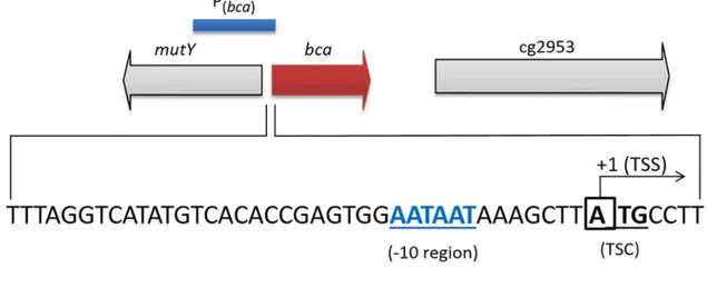
As indicated inFig 2A, thebcaTSS was found to be the first nucleotide“A”of the putative translational start codon (ATG), indicating that thebcagene codes for a leader-less transcript and thus, lacks a 5'-untranslated region. Centered 10 bp upstream of the TSS, an AATAAT motif was observed, which is very similar to the -10 consensus sequence forC.glutamicum
[71].
The TSS forgcawas identified to be an“A”residue at position—20 relative to the first nucleotide of the putative translational start codon (ATG) of cg0154, the gene located upstream of thegca(Fig 2B). In accordance, it has previously been shown that cg0154 and
gcagenes are co-transcribed [72]. Centered 10 bp upstream of the cg0154-gcaTSS, the motif TAGGCT was observed, which shows reasonable similarity to the -10 consensus [TA(C/T) AAT] sequence forC.glutamicum[71]. Six bp upstream of the putative cg0154 translational start, we observed an AGGAG motif, which represents an ideal ribosomal binding site forC.
Expression of the
bca
gene is subject to carbon source-dependent
regulation
Using theC.glutamicumstrains carrying plasmids pET2, pET2-Pbcaand pET2-Pgca, we
per-formed a comparative expression analysis of thebcaandgcagenes in cells grown in minimal medium with 1% (w/v) glucose or 1% (w/v) acetate with initial pH values at 6.8 and 6.3. Cultures were harvested at the mid-exponential phase of growth (OD600of about 5) and after cell
disrup-tion, the promoter activities were determined in the cell extracts by measuring the specific CAT activities. While the extracts from cells carrying the empty promoter-probe vector pET2 did not show any detectable CAT activity (<0.01 U/mg protein), the extracts of the strains carrying the bcaandgcapromoters within pET2 showed activity and expression ofbcaalso showed carbon source-dependent regulation (Table 2). Thebcapromoter activities were observed to be about 1.5- to 2-fold higher in extracts of glucose-grown cells as compared to that in extracts of acetate-grown cells. However, thegcapromoter activities were very low, i.e., about 20-fold lower than
Fig 2. Transcriptional organization of thebcaandgcagenes in the genome ofC.glutamicum.Genomic loci, promoter fragments used and transcriptional start sites (TSSs) of thebca(A)andgca(B)genes inC.glutamicum. The TSSs were identified by the 5'-RACE method. The putative -10 regions, the annotated translational start codon (TSC) ofbcaand of cg0154 and ribosome binding site (RBS) of cg0154 are indicated.
that of thebcapromoter in glucose- and in acetate-grown cells, indicating that expression of the
gcagene inC.glutamicumis very low under the given conditions. Furthermore, it was observed that activities of thebcapromoter were nearly the same on either glucose as carbon source on both pH values (6.3 and 6.8) or acetate as carbon source at both pH values (6.3 and 6.8)
(Table 2). These results indicate that expression of thebcagene on either carbon source as well as carbon source-dependent regulation is independent of the initial pH 6.3 or 6.8.
We also tested thebcapromoter activity in cell extracts of the L-lysine-producing strainC.
glutamicumDM1729ΔilvBtransformed with plasmid pET2-Pbcaand grown in minimal
medium containing 2% glucose. As can be seen inTable 2, the specific CAT activity was in the same range as in extracts ofC.glutamicumwild-type. In accordance, L-lysine production byC.
glutamicumDM1729ΔilvB(pET2-Pbca) was in the same range as previously reported [59] for
the parental strainC.glutamicumDM1729ΔilvB, i.e., a final L-lysine concentration of about 32.3 mM after 24 h of incubation.
Global regulator RamA negatively regulates expression of the
bca
gene
Based on the results of carbon source-dependent expression control of thebcagene inC. glu-tamicumwith glucose and acetate (see above), we speculated the regulator of acetate metabo-lism RamA [58] to be involved in this regulation. RamA is a LuxR-type global regulator, essential for growth on acetate or ethanol and is involved in expression control of a variety of genes in central carbon metabolism [73]. The involvement of RamA inbcaexpression was tested by comparativebcapromoter activity analysis with the wild-type and a RamA-defi-cient derivative ofC.glutamicum. For this purpose, plasmid pET2-Pbcawas transformed into
C.glutamicumΔramAand the specific CAT activities of the resulting transformant and ofC.
glutamicum(pET2-Pbca) were determined in cell extracts after growth of the cells in minimal
medium with either glucose 1% (w/v) or glucose plus acetate (1% each, w/v) and harvested at the mid-exponential growth phase (OD600of about 5). As shown inTable 2, the specific CAT
activity and thus, thebcapromoter activity in theΔramAmutant was about 1.6-fold higher in minimal medium with glucose and about four-fold higher with glucose plus acetate when
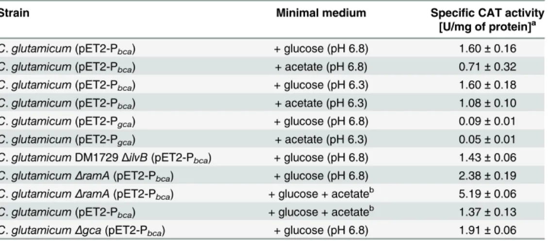
Table 2. Specific chloramphenicol acetyltransferase (CAT) activities of differentC.glutamicum
strains carrying plasmids pET2-Pbcaor pET2-Pgca, cultured in minimal medium containing 1% or 2%
(w/v) glucose and/or 1% (w/v) acetate with initial pH values of 6.3 or 6.8.
Strain Minimal medium Specific CAT activity [U/mg of protein]a
C.glutamicum(pET2-Pbca) + glucose (pH 6.8) 1.60±0.16
C.glutamicum(pET2-Pbca) + acetate (pH 6.8) 0.71±0.32
C.glutamicum(pET2-Pbca) + glucose (pH 6.3) 1.60±0.18
C.glutamicum(pET2-Pbca) + acetate (pH 6.3) 1.08±0.10
C.glutamicum(pET2-Pgca) + glucose (pH 6.8) 0.09±0.01
C.glutamicum(pET2-Pgca) + acetate (pH 6.3) 0.05±0.01
C.glutamicumDM1729ΔilvB(pET2-Pbca) + glucose (pH 6.8) 1.43±0.06
C.glutamicumΔramA(pET2-Pbca) + glucose (pH 6.8) 2.38±0.19
C.glutamicumΔramA(pET2-Pbca) + glucose + acetateb 5.19±0.06
C.glutamicum(pET2-Pbca) + glucose + acetateb 1.37±0.13
C.glutamicumΔgca(pET2-Pbca) + glucose (pH 6.8) 1.91±0.06
aThe values are means of at least three independent experiments. bThe initial pH values in these cultures were set to 6.3.
compared to that in wild-type cells ofC.glutamicum. The higher activity of thebcapromoter in the absence of a functional RamA protein indicates that RamA acts as a negative transcrip-tional regulator for the expression of thebcagene inC.glutamicum.
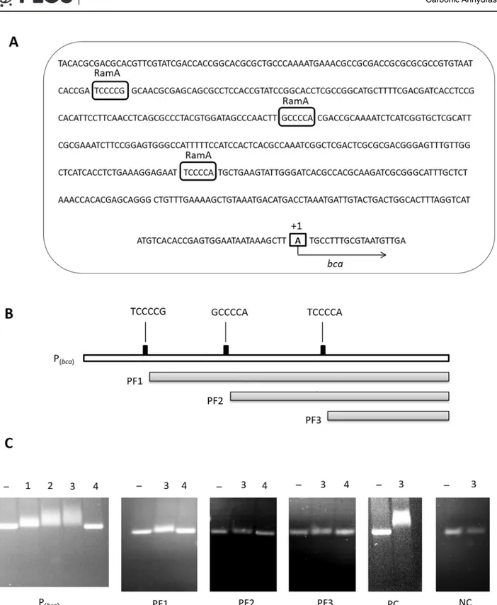
The most common RamA binding motifs have been identified as A/T/C-GGGG-N and A/ T/C-CCCC-N [73]. As shown inFig 3A, three such motifs were also observed in the sequence of thebcapromoter region and therefore, it was likely that the RamA protein binds to thebca
promoter region. To analyze the binding of RamA to thebcapromoter region, a His-tagged RamA protein was over-produced inE.coliBL21 (DE3) containing the pET28-ramAconstruct, identified by MALDI-TOF mass spectrometry, and used for EMSAs. For this purpose, thebca
promoter fragment (Pbca) and its sub-fragments with only two, one or no RamA binding motifs
(PF1, PF2 and PF3, respectively, as shown inFig 3B), were incubated with varying amounts (0–2μg) of purified His-tagged RamA protein and the assay mixture was separated on an aga-rose gel. AnaceA-aceBinter-genic fragment was used as positive control fragment for binding of the His-tagged RamA protein, as the RamA protein was already known to bind to this region [58], while fragment 1b having no binding affinity for RamA [58] was used as negative control fragment in the EMSA experiments. As shown inFig 3C, thebcapromoter fragment Pbcawith
all three RamA binding motifs was retarded by the RamA protein and the retardation was observed to be proportional to increasing concentration of the His-tagged RamA protein. The PF1 fragment showed less retardation with 2μg RamA, fragments PF2 and PF3 did not show significant retardation. These results show that RamA binds to thebcapromoter region and indicates that the two distal RamA binding motifs are functional.
Taken together, the higherbcapromoter activity inC.glutamicumΔramAcompared to that in the wild-type and binding of His-tagged RamA protein to thebcapromoter fragment in the EMSA experiments showed that RamA negatively regulates the expression of thebcagene in
C.glutamicum.
Expression of the
bca
gene not affected in the absence of a functional
Gca protein
To test for a possible effect of Gca on growth and on expression of thebcagene, agcadeletion mutant ofC.glutamicumwas generated, using the suicide vector pK19mobsacB and homolo-gous recombination. A mutant version ofgcagene was constructed by amplifying and con-densing thegcaupper and lower fragments (each 423 bp), resulting in a truncatedgcagene with an intragenic deletion of 422 bp, which was exchanged with the native chromosomal copy ofgca. In minimal medium containing glucose 4% (w/v), the resulting strainC.glutamicum Δgcawas observed to grow with the same growth rates and to the same final OD600as the
wild-typeC.glutamicum(data not shown), indicating that thegcagene is dispensable under the con-ditions tested. This result is consistent with the previous observation thatgcais not essential for growth ofC.glutamicumunder normal conditions [17].
Plasmid pET2-Pbcawas transformed intoC.glutamicumΔgcaand the activities of thebca
promoter were determined by analysis of the specific CAT activities in crude cell extracts of cultures grown in minimal medium with glucose 1% (w/v). As shown inTable 2, the specific CAT activities in extracts ofC.glutamicumΔgcawere observed to be nearly the same as in the respective wild type derivative. This result indicates that there is no significant effect of the absence of a functional Gca protein on expression of thebcagene inC.glutamicum.
Discussion
In nature, CO2is in chemical equilibrium with HCO3-, carbonic acid and carbonate. Of these,
Fig 3. Thebcapromoter sequence and EMSAs. (A):Sequence of thebcapromoter region (Pbca) with its potential RamA binding motifs (boxed and
indicated above the sequence) inC.glutamicum. The transcriptional start point is indicated by a box and“+1”.(B):Thebcapromoter (Pbca) and its
sub-fragments with exclusion of one, two and three RamA binding motifs in PF1, PF2 and PF3, respectively.(C):Representative EMSAs for binding assays using purified His- tagged RamA protein. TheaceA-aceBinter-genic region was used as a positive control fragment (shown as PC) [58], fragment 1b [58] as a negative control fragment (shown as NC) and bovine serum albumin (BSA) as negative protein control. Lane–shows the respective fragment without protein whereas lanes 1, 2 and 3 show EMSAs using 0.8, 1.5 and 2μg of His- tagged RamA, respectively, and lane 4 EMSAs using 2μg of BSA instead of RamA.
as they serve as substrate or product in carboxylating and decarboxylation reactions, are involved in ion transport and internal pH regulation, regulate virulence and toxin formation in pathogenic bacteria and recently have been shown to be regulatory triggers of global transcrip-tional regulation and of microbial and mammalian production processes [74,75,4]. In aerobic (micro)organisms, CO2is the product of respiration and as such in sufficient amounts present
within metabolically active cells. However, for anaplerotic (and also other) carboxylation reac-tions, the physiologically most important reactant is HCO3-[2,15,17]. As CO2(but not
HCO3-) can diffuse out of the cell (for a recent review see [76]), the intracellular conversion of
CO2to HCO3-is essential to retain CO2as genuine carboxylation substrate inside the cell. The
chemical inter-conversion of CO2and HCO3-is relatively slow at physiological pH [77] and
thus, nature has evolved enzymatic conversion by zinc-dependent CAs, catalyzing the revers-ible hydration of CO2with high turnover numbers and allowing the cells to maintain the
intra-cellular balance of CO2/HCO3-that is needed for cellular processes [2,78,79]. The role of CAs
has intensively been studied inE.coliand other microorganisms, in particular in relation to CO2/HCO3-balance in the intracellular environment under physiological conditions (for
refer-ences seeintroduction) and it has been observed that under atmospheric conditions, inactiva-tion of CA(s) is lethal or highly inhibitory unless the CO2content is increased to 5–10% [14–
18,80–82]. In spite of numerous studies on the physiological function of CAs in bacterial CO2/
HCO3-metabolism, there is much less information on the transcriptional organization and on
expression control of the respective CA genes.
The purpose of this work was to broaden our knowledge about the transcriptional organiza-tion of the CA genes as well as expression analysis in relaorganiza-tion to media composiorganiza-tion (carbon sources) and transcriptional regulation inC.glutamicum. This organism is an industrial work-horse widely used for the production of amino acids and a variety of other metabolites [31,32, 83,84,85,86]. The PEP-pyruvate-oxaloacetate node of this organism (seeFig 1), being an important branch-point of carbon flux distribution and having a role in anaplerosis, gluconeo-genesis and amino acid biosynthesis, involves several carboxylation/decarboxlation reactions and thus, a pivotal effect of the intracellular CO2/HCO3-balance on the overall physiology of
C.glutamicumcan be presumed. Two CA genes, namelybcaandgca, encodingβ- andγ-CA, respectively, have previously been identified andbcahas been shown to be essential under atmospheric conditions inC.glutamicum[17].
In this study, the TSS of thebcagene was identified to be the first nucleotide of its putative translational start codon (ATG). Thus,bcacodes for a leaderless transcript which lacks the 5ˊ -untranslated region. Leaderless mRNAs starting with an AUG start codon have been reported in bacteria, archaea, eukaryotes [87] and also inC.glutamicum[71]. In fact, a recent RNA sequence analysis (RNAseq) withC.glutamicumrevealed that about 33% of all mRNAs includ-ing that of thebcagene, in the cells are leaderless and that the translational start codon of these leaderless mRNAs generally is an AUG (about 79%) or GUG (about 21%) [72]. However, for leaderless mRNAs starting with the initiation codon AUG, no signals have been shown down-stream of the 5ˊ-terminal AUG for recruitment of ribosomes [87]. InE.coli, it has been shown that for translation initiation of leaderless mRNAs, the molar ratio of the initiation factors IF2 and IF3 plays a final role, indicating that the translation efficiency of these mRNAs can be altered, based on the availability of components of the translational machinery [87,88]. Homo-logues of genes encoding IF2 and IF3 have been found in the genome ofC.glutamicum
(cg1563 and cg2176, respectively; [89]), however, a role of these factors in translation of lead-erless transcripts remains to be investigated.
located upstream of thegca. Co-transcription of cg0154 andgcahas previously been shown based on RNAseq analysis ofC.glutamicumby Pfeifer-Sancar et al. [72].
To investigate a carbon source-dependent regulation of the CA genes inC.glutamicum, we investigated the expression ofbcaandgcain terms of their respective promoter activities in relation to glucose and acetate as carbon sources. This analysis revealed for both genes higher promoter activities when the cells were grown on glucose as compared to acetate. Furthermore, the activity of thebcapromoter was observed to be about 15-fold higher than that of thegca
promoter. The lower level ofgcaexpression as compared tobcaexpression is consistent with the results of Mitsuhashi et al. [17] who found in Northern blot analysis of growingC. glutami-cumcells thegcatranscript level below the detection limit, suggesting thatgcaexpression is either constantly very low or tightly regulated. Furthermore, it was also observed that expres-sion ofgcaunder control of thelacpromoter restored the growth ofbcamutant under normal environmental conditions [17]. However, our results suggest that thebcagene is subject to car-bon source-dependent regulation as is the case for a variety of genes encoding key enzymes in central metabolism inC.glutamicum[53,73,90,91].
The lowerbcapromoter activity and thus, the lowerbcaexpression in acetate-grown cells than in glucose-grown cells ofC.glutamicummight be attributed to a lower HCO3-demand
and reduced need of anaplerosis by pyruvate or PEP carboxylation and using the glyoxylate cycle for anaplerosis when growing on acetate instead of glucose. This hypothesis is in agree-ment with carbon flux analysis of the central metabolism ofC.glutamicumgrowing in minimal medium containing glucose and/or acetate [92] and with the previous finding that a PEP and pyruvate carboxylase-deficient double mutant ofC.glutamicumgrows on acetate but not on glucose [93]. However the crucial role ofbcaduring growth on glucose may not only be con-fined to replenishment of oxaloacetate or other TCA cycle intermediates as Mitsuhashi et al. [17] observed that addition of oxaloacetate, glutamate and succinate did not restore the growth ofC.glutamicumΔbca. It is, however, important to mention thatC.glutamicumwas found to be unable to take up and to grow on the TCA cycle intermediates fumarate, succinate and L-malate [94]. Therefore, a further potential role of CA inC.glutamicum, aside of replenishment of TCA cycle intermediates, has to be experimentally proven.
The transcriptional regulator RamA originally has been identified as the regulator of acetate metabolism inC.glutamicum[58]. Later it has been shown that RamA is functional as activator or as repressor in carbon metabolism of this organism and is involved in expression control of a variety of genes and operons encoding enzymes or pathways in the central metabolism ofC.
glutamicum(reviewed in [73,90,95]). Based on the observed carbon source-dependent regula-tion of thebcapromoter, we speculated RamA to be involved in expression control of thebca
Supporting Information
S1 Table. Oligonucleotides used in this study. (DOCX)
Acknowledgments
The German Academic Exchange Service (DAAD) and Higher Education Commission (HEC; Pakistan) award for AS is gratefully acknowledged. We thank C. Gabris for providing strainC.
glutamicumΔatlR(pET2-p4) and M. Brocker at the Forschungszentrum Jülich for MALDI-TOF analysis.
Author Contributions
Conceived and designed the experiments: AS BJE. Performed the experiments: AS. Analyzed the data: AS BJE. Wrote the paper: AS BJE.
References
1. Kaur S, Mishra MN, Tripathi AK. Regulation of expression and biochemical characterization of a β-class carbonic anhydrase from the plant growth-promoting rhizobacterium,Azospirillum brasilense Sp7. FEMS Microbiol Lett. 2009; 299:149–158. doi:10.1111/j.1574-6968.2009.01736.xPMID:
19694814
2. Smith KS, Ferry JG. Prokaryotic carbonic anhydrases. FEMS Microbiol Rev. 2000; 24:335–366. doi:
http://dx.doi.org/10.1111/j.1574-6976.2000.tb00546.xPMID:10978542
3. Emmett DH, Tashian RE. Functional diversity, conservation, and convergence in the evolution of the α-,β-, andγ-carbonic anhydrase gene families. Mol Phylogenet Evol. 1996; 5:50–77. PMID:8673298
4. Blombach B, Takors R. CO2–intrinsic product, essential substrate, and regulatory trigger of microbial and mammalian production processes. Front Bioeng Biotechnol. 2015; 3:108. doi:10.3389/fbioe.2015. 00108PMID:26284242
5. Supuran CT. Bacterial carbonic anhydrases as drug targets: toward novel antibiotics? Front Pharma-col. 2011; 2:34. doi:10.3389/fphar.2011.00034PMID:21779249
6. Zimmerman SA, Ferry JG. Theβandγclasses of carbonic anhydrase. Curr Pharm Des. 2008; 14:716–721. doi:10.2174/138161208783877929PMID:18336318
7. Smith KS, Jakubzick C, Whittam TS, Ferry JG. Carbonic anhydrase is an ancient enzyme widespread in prokaryotes. PNAS. 1999; 96:15184–15189. doi:10.1073/pnas.96.26.15184PMID:10611359
8. Kaur S, Mishra MN, Tripathi AK. Gene encodingγ-carbonic anhydrase is cotranscribed withargC and induced in response to stationary phase and high CO2inAzospirillum brasilenseSp7. BMC Microbiol. 2010; 10:184. doi:10.1186/1471-2180-10-184PMID:20598158
9. Lotlikar SR., Hnatusko S, Dickenson NE, Choudhari SP, Picking WL, Patrauchan MA. Three functional β-carbonic anhydrases inPseudomonas aeruginosaPAO1: role in survival in ambient air. Microbiology. 2013; 159:1748–1759. doi:10.1099/mic.0.066357–0PMID:23728627
10. Rowlett RS. Structure and catalytic mechanism of theβ-carbonic anhydrases. Biochimi Biophys Acta. 2010; 1804:362–373. doi:10.1016/j.bbapap.2009.08.002PMID:19679201
11. Guilloton MB, Lamblin AF, Kozliak EI, Gerami-Nejad M, Tu C, Silverman D, et al. A physiological role for cyanate-induced carbonic anhydrase inEscherichia coli. J Bacteriol. 1993; 175:1443–1451. PMID:
8444806
12. Nishimori I, Onishi S, Takeuchi H, Supuran CT. Theαandβclasses carbonic anhydrases from Helico-bacter pylorias novel drug targets. Curr Pharm Des. 2008; 14:622–630. doi:10.2174/
138161208783877875PMID:18336307
13. Kumar RSS, Hendrick W, Correll JB, Patterson AD, Melville SB, Ferry JG. Biochemistry and physiology of theβclass carbonic anhydrase (Cpb) from Clostridium perfringens strain 13. J Bacteriol. 2013; 195:2262–2269. doi:10.1128/JB.02288-12PMID:23475974
15. Merlin C, Masters M, McAteer S, Coulson A. Why is carbonic anhydrase essential toEscherichiacoli? J Bacteriol. 2003; 185:6415–6424. doi:10.1128/JB.185.21.6415–6424.2003PMID:14563877
16. Gai CS, Lu J, Brigham CJ, Bernardi AC, Sinskey AJ. Insights into bacterial CO2 metabolism revealed by the characterization of four carbonic anhydrases in Ralstonia eutropha H16. AMB Express. 2014; 4:2. doi:10.1186/2191-0855-4-2PMID:24410804
17. Mitsuhashi S, Ohnishi J, Hayashi M, Ikeda M. A gene homologous toβ-type carbonic anhydrase is essential for the growth ofCorynebacterium glutamicumunder atmospheric conditions. Appl Microbiol Biotechnol. 2004; 63:592–601. doi:10.1007/s00253-003-1402-8PMID:12937954
18. Ueda K, Nishida H, Beppu T. Dispensabilities of carbonic anhydrase in Proteobacteria. Int J Evol Biol. 2012; 2012:324549. doi:10.1155/2012/324549PMID:22675650
19. Kinoshita S, Udaka S, Shimono M. Studies on the amino acid fermentation. Part I. Production of L-glu-tamic acid by various microorganisms. J Gen Appl Microbiol. 1957; 3:193–205.
20. Liebl W. The genus Corynebacterium—nonmedical. In: Dworkin M, Falkow S, Rosenberg E, Schleifer KH, Stackebrandt E (eds) The Prokaryotes, 3rd ed, vol. 3, Springer, New York; 2006. pp. 796–818.
21. Arndt A, Auchter M, Ishige T, Wendisch VF, Eikmanns BJ. Ethanol catabolism inCorynebacterium glu-tamicum. J Mol Microbiol Biotechnol. 2008; 15:222–233. doi:10.1159/000107370PMID:17693703
22. Takors R, Bathe B, Rieping M, Hans S, Kelle R, Huthmacher K. Systems biology for industrial strains and fermentation processes—Example: Amino acids. J Biotechnol. 2007; 129:181–190. doi:10.1016/ j.jbiotec.2007.01.031PMID:17367886
23. Eggeling L, Bott M. A giant market and a powerful metabolism: L-lysine provided byCorynebacterium glutamicum. Appl Microbiol Biotechnol. 2015; 99:3387–3394. doi:10.1007/s00253-015-6508-2PMID:
25761623
24. Jensen JVK, Eberhardt D, Wendisch VF. Modular pathway engineering ofCorynebacterium glutami-cumfor production of the glutamate-derived compounds ornithine, proline, putrescine, citrulline, and arginine. J Biotechnol. 2015; 214:85–94. doi:10.1016/j.jbiotec.2015.09.017PMID:26393954
25. Park SH, Kim HU, Kim TY, Park JS, Kim SS, Lee SY. Metabolic engineering ofCorynebacterium gluta-micumfor L-arginine production. Nat Commun. 2014; 5:4618. doi:10.1038/ncomms5618PMID:
25091334
26. Blombach B, Schreiner ME, Bartek T, Oldiges M, Eikmanns BJ.Corynebacterium glutamicumtailored for high-yield L-valine production. Appl Microbiol Biotechnol. 2008; 79:471–479. doi: 10.1007/s00253-008-1444-zPMID:18379776
27. Hasegawa S, Suda M, Uematsu K, Natsuma Y, Hiraga K, Jojima T, et al. Engineering of Corynebacte-rium glutamicumfor high-yield L-valine production under oxygen deprivation conditions. Appl Environ Microbiol. 2013; 79:1250–1257. doi:10.1128/AEM.02806-12PMID:23241971
28. Oldiges M, Eikmanns BJ, Blombach B. Application of metabolic engineering for the biotechnological production of L-valine. Appl Microbiol Biotechnol. 2014; 98:5859–5870. doi: 10.1007/s00253-014-5782-8PMID:24816722
29. Cheng Y, Zhou Y, Yang L, Zhang C, Xu Q, Xie X, et al. Modification of histidine biosynthesis pathway genes and the impact on production of L-histidine inCorynebacterium glutamicum. Biotechnol Lett. 2013; 35:735–741. doi:10.1007/s10529-013-1138-1PMID:23355034
30. Kulis-Horn RK, Persicke M, Kalinowski J. Histidine biosynthesis, its regulation and biotechnological application in Corynebacterium glutamicum. Microb Biotechnol. 2014; 7:5–25. doi: 10.1111/1751-7915.12055PMID:23617600
31. Eikmanns BJ, Bott M. EngineeringCorynebacterium glutamicumfor the production of organic acids and alcohols. In:Corynebacterium glutamicum–From Systems Biology to Biotechnological Applica-tions, A. Burkowski (Ed.), Horizon Scientific Press and Caister Academic Press, Norwich, UK; 2015. pp. 111–137; ISBN: 978-1910190-05-0
32. Wieschalka S, Blombach B, Bott M, Eikmanns BJ. Bio-based production of organic acids with Coryne-bacterium glutamicum. Microb Biotechnol. 2013; 6:87–102. doi:10.1111/1751-7915.12013PMID:
23199277
33. Litsanov B, Kabus A, Brocker M, Bott M. Efficient aerobic succinate production from glucose in minimal medium with Corynebacterium glutamicum. Microb Biotechnol. 2012; 5:116–128. doi: 10.1111/j.1751-7915.2011.00310.xPMID:22018023
34. Okino S, Noburyu R, Suda M, Jojima T, Inui M, Yukawa H. An efficient succinic acid production process in a metabolically engineeredCorynebacterium glutamicumstrain. Appl Microbiol Biotechnol. 2008; 81:459–464. doi:10.1007/s00253-008-1668-yPMID:18777022
35. Hüser AT, Chassagnole C, Lindley ND, Merkamm M, Guyonvarch A, Elišáková V, et al. Rational design
flux analysis and genome-wide transcriptional profiling. Appl Environ Microbiol. 2005; 71:3255–3268. doi:10.1128/AEM.71.6.3255–3268.2005PMID:15933028
36. Mimitsuka T, Sawai H, Hatsu M, Yamada K. Metabolic engineering ofCorynebacterium glutamicumfor cadaverine fermentation. Biosci Biotechnol Biochem. 2007; 71:2130–2135. PMID:17895539
37. Schneider J, Wendisch VF. Putrescine production by engineeredCorynebacterium glutamicum. Appl Microbiol Biotechnol. 2010; 88:859–868. doi:10.1007/s00253-010-2778-xPMID:20661733
38. Schneider J, Wendisch VF. Biotechnological production of polyamines by bacteria: recent achieve-ments and future perspectives. Appl Microbiol Biotechnol. 2011; 91:17–30. doi: 10.1007/s00253-011-3252-0PMID:21552989
39. Kind S, Wittmann C. Bio-based production of the platform chemical 1,5 diaminopentane. Appl Microbiol Biotechnol. 2011; 5:1287–1296.
40. Nguyen AQD, Schneider J, Reddy GK, Wendisch VF. Fermentative production of the diamine putres-cine: System metabolic engineering ofCorynebacterium glutamicum. Metabolites. 2015; 5:211–231. doi:10.3390/metabo5020211PMID:25919117
41. Inui M, Kawaguchi H, Murakami S, Vertès AA, Yukawa H. Metabolic engineering ofCorynebacterium glutamicumfor fuel ethanol production under oxygen-deprivation conditions. J Mol Microbiol Biotech-nol. 2004; 8:243–254. doi:10.1159/000086705PMID:16179801
42. Blombach B, Riester T, Wieschalka S, Ziert C, Youn JW, Wendisch VF, et al.Corynebacterium glutami-cumtailored for efficient isobutanol production. Appl Environ Microbiol. 2011; 77:3300–3310. doi:10. 1128/AEM.02972-10PMID:21441331
43. Yamamoto S, Suda M, Niimi S, Inui M, Yukawa H. Strain optimization for efficient isobutanol production usingCorynebacterium glutamicumunder oxygen deprivation. Biotechnol Bioeng. 2013; 110:2938– 2948. doi:10.1002/bit.24961PMID:23737329
44. Siebert D, Wendisch VF. Metabolic pathway engineering for production of 1,2 propanediol and 1-propa-nol by Corynebacterium glutamicum. Biotech1-propa-nol Biofuels. 2015; 8:91. doi: 10.1186/s13068-015-0269-0PMID:26110019
45. Jojima T, Noburyu R, Sasaki M, Tajima T, Suda M, Yukawa H, et al. Metabolic engineering for improved production of ethanol byCorynebacterium glutamicum. Appl Microbiol Biotechnol. 2015; 99:1165– 1172. doi:10.1007/s00253-014-6223-4PMID:25421564
46. Krause FS, Blombach B, Eikmanns BJ. Metabolic Engineering ofCorynebacterium glutamicumfor 2-Ketoisovalerate Production. Appl Environ Microbiol. 2010; 76:8053–8061. doi: 10.1128/AEM.01710-10PMID:20935122
47. Buchholz J, Schwentner A, Brunnenkan B, Gabris C, Grimm S, Gerstmeir R, et al. Platform engineering ofCorynebacterium glutamicumwith reduced pyruvate dehydrogenase complex activity for improved production of L-Lysine, L-Valine, and 2 Ketoisovalerate. Appl Environ Microbiol. 2013; 79:5566–5575. doi:10.1128/AEM.01741-13PMID:23835179
48. Bückle-Vallant V, Krause FS, Messerschmidt S, Eikmanns BJ. Metabolic engineering of Corynebacte-rium glutamicumfor 2-ketoisocaproate production. Appl Microbiol Biotechnol. 2014; 98:297–311. doi:
10.1007/s00253-013-5310-2PMID:24169948
49. Vogt M, Haas S, Polen T, Ooyen JV, Bott M. Production of 2-ketoisocaproate with Corynebacterium glutamicum strains devoid of plasmids and heterologous genes. Microb Biotechnol. 2015; 8:351–360. doi:10.1111/1751-7915.12237PMID:25488800
50. Heider SAE, Peters-Wendisch P, Wendisch VF. Carotenoid biosynthesis and overproduction in Cory-nebacterium glutamicum. BMC Microbiol. 2012; 12:98. doi:10.1186/1471-2180-12-198PMID:
22963379
51. Jo SJ, Maeda M, Ooi T, Taguchi S. Production system for biodegradable polyester polyhydroxybutyrate byCorynebacterium glutamicum. J Biosci Bioeng. 2006; 102:233–236. doi:10.1263/jbb.102.233
PMID:17046539
52. Song Y, Matsumoto K, Yamada M, Gohda A, Brigham CJ, Sinskey AJ, et al. Engineered Corynebacte-rium glutamicumas an endotoxin-free platform strain for lactate-based polyester production. Appl Microbiol Biotechnol. 2012; 93:1917–1925. doi:10.1007/s00253-011-3718-0PMID:22127753
53. Bott M & Eikmanns B.J. TCA cycle and glyoxylate shunt of Corynebacterium glutamicum. In: Coryne-bacterium glutamicum: Biology and Biotechnology (Microbiology Monographs), Yukawa H. & Inui M. (eds), Springer Verlag Berlin Heidelberg; 2012. pp. 281–314
54. Sauer U, Eikmanns BJ. The PEP–pyruvate–oxaloacetate node as the switch point for carbon flux distri-bution in bacteria. FEMS Microbiol Rev. 2005; 29:765–794. doi:http://dx.doi.org/10.1016/j.femsre. 2004.11.002PMID:16102602
response to the availability of sulphide acceptor molecules. BMC Genomics. 2008; 9:483. doi:10. 1186/1471-2164-9-483PMID:18854009
56. Hanahan D. Studies on transformation ofEscherichia coliwith plasmids. J Mol Biol. 1983; 166:557– 580. doi:10.1016/S0022-2836(83)80284-8PMID:6345791
57. Studier FW, Moffatt BA. Use of bacteriophage T7 RNA polymerase to direct selective high-level expres-sion of cloned genes. J Mol Biol. 1986; 189:113–130. doi:10.1016/0022-2836(86)90385-2PMID:
3537305
58. Cramer A, Gerstmeir R, Schaffer S, Bott M, Eikmanns BJ. Identification of RamA, a novel LuxR-type transcriptional regulator of genes involved in acetate metabolism ofCorynebacterium glutamicum. J Bacteriol. 2006; 188:2554–2567. doi:10.1128/JB.188.7.2554–2567.2006PMID:16547043
59. Blombach B, Hans S, Bathe B, Eikmanns BJ. Acetohydroxyacid synthase, a novel target for improve-ment of L-Lysine production byCorynebacterium glutamicum. Appl Environ Microbiol. 2009; 75:419– 427. doi:10.1128/AEM.01844-08PMID:19047397
60. Vasicová P, Abrhámová Z, Nesvera J, Pátek M, Sahm H, Eikmanns B. Integrative and autonomously replicating vectors for analysis of promoters inCorynebacterium glutamicum. Biotechnology Tech-niques. 1998; 12:743–746.
61. Schäfer A, Tauch A, Jäger W, Kalinowski J, Thierbach G, Pühler A. Small mobilizable multi-purpose cloning vectors derived from theEscherichia coliplasmids pK18 and pK19: selection of defined dele-tions in the chromosome ofCorynebacterium glutamicum. Gene. 1994; 145:69–73. PMID:8045426
62. Green MR, Sambrook J. Molecular cloning, a laboratory manual. 4th ed. Cold Spring Harbor Labora-tory Press, Cold Spring Harbor New York, USA; 2012.
63. Eikmanns BJ, Metzger M, Reinscheid D, Kircher M, Sahm H. Amplification of three threonine biosyn-thesis genes inCorynebacterium glutamicumand its influence on carbon flux in different strains. Appl Microbiol Biotechnol. 1991; 34:617–622. PMID:1369320
64. Auchter M, Laslo T, Fleischer C, Schiller L. Arndt A, Gaigalat L, et al. Control ofadhAandsucR expres-sion by the SucR regulator inCorynebacterium glutamicum. J Biotechnol. 2011; 152:77–86. doi:10. 1016/j.jbiotec.2011.02.003PMID:21320555
65. van der Rest ME, Lange C, Molenaar D. A heat shock following electroporation induces highly efficient transformation ofCorynebacterium glutamicumwith xenogeneic plasmid DNA. Appl Microbiol Biotech-nol. 1999; 52:541–545. PMID:10570802
66. Dower WJ, Miller JF, Ragsdale CW. High efficiency transformation ofE.coliby high voltage electropora-tion. Nucleic Acids Res. 1988; 13:6127–6145.
67. Frohman MA, Dush MK. Martin GR. Rapid production of full-length cDNAs from rare transcripts: amplifi-cation using a single gene-specific oligonucleotide primer. PNAS. 1988; 85:8998–9002. PMID:
2461560
68. Gerstmeir R, Cramer A, Dangel P, Schaffer S, Eikmanns BJ. RamB, a novel transcriptional regulator of genes involved in acetate metabolism ofCorynebacterium glutamicum. J Bacteriol. 2004; 186:2798– 2809. doi:10.1128/JB.186.9.2798–2809.2004PMID:15090522
69. Laemmli UK. 1970. Cleavage of structural proteins during the assembly of the head of bacteriophage T4. Nature. 227:680–685. PMID:5432063
70. Ho SN, Hunt HD, Horton RM, Pullen JK, Pease LR. Site-directed mutagenesis by overlap extension using the polymerase chain reaction. Gene. 1989; 77:51–59. doi:10.1016/0378-1119(89)90358-2
PMID:2744487
71. Patek M, Nesvera J, Guyonvarch A, Reyes O, Leblon G. Promoters ofCorynebacterium glutamicum. J Biotechnol. 2003; 104:311–323. doi:10.1016/S0168-1656(03)00155-XPMID:12948648
72. Pfeifer-Sancar K, Mentz A, Rückert C, Kalinowski J. Comprehensive analysis of theCorynebacterium glutamicumtranscriptome using an improved RNAseq technique. BMC Genomics. 2013; 14:888. doi:
10.1186/1471-2164-14-888PMID:24341750
73. Auchter M, Cramer A, Huser A, Rückert C, Emer D, Schwarz P, et al. RamA and RamB are global tran-scriptional regulators inCorynebacterium glutamicumand control genes for enzymes of the central metabolism. J Biotechnol. 2011; 154:126–139. doi:10.1016/j.jbiotec.2010.07.001PMID:20620178
74. Blombach B, Buchholz J, Busche T, Kalinowski J, Takors R. Impact of different CO2/HCO3−levels on
metabolism and regulation inCorynebacterium glutamicum. J Biotechnol. 2013; 168:331–340. doi:10. 1016/j.jbiotec.2013.10.005PMID:24140290
75. Follonier S, Escapa IF, Fonseca PM, Henes B, Panke S, Zinn M, Prieto MA. New insights on the reorga-nization of gene transcription in Pseudomonas putida KT2440 at elevated pressure. Microb Cell Fact. 2013; 12:30. doi:10.1186/1475-2859-12-30PMID:23537069
77. Kern DM. The hydration of carbon dioxide. J. Chem. Educ. 1960; 37. doi:10.1021/ed037p14
78. Tashian RE. The carbonic anhydrases: widening perspectives on their evolution, expression and func-tion. Bioessays. 1989; 10:186–192. PMID:2500929
79. Tripp BC, Smith K. Ferry JG. Carbonic anhydrase: new insights for an ancient enzyme. J Biol Chem. 2001; 276:48615–48618. PMID:11696553
80. Götz R, Gnann A, Zimmermann FK. Deletion of the carbonic anhydrase-like geneNCE103of the yeast Saccharomyces cerevisiaecauses an oxygen-sensitive growth defect. Yeast. 1999; 15:855–864. doi:
10.1002/(SICI)1097-0061(199907)15:10A<855::AID-YEA425>3.0.CO;2-CPMID:10407265
81. Kusian B, Sültemeyer D, Bowien B. Carbonic anhydrase is essential for growth ofRalstonia eutrophaat ambient CO2concentrations. J Bacteriol. 2002; 184:5018–5026. doi:10.1128/JB.184.18.5018–5026. 2002PMID:12193617
82. Cottier F, Raymond M, Kurzai O, Bolstad M, Leewattanapasuk W, et al. The bZIP transcription factor Rca1p is a central regulator of a novel CO2sensing pathway in yeast. PLoS Pathog. 2012; 8: e1002485. doi:10.1371/journal.ppat.1002485PMID:22253597
83. Eikmanns BJ. Blombach B. Isobutanol. In: Bioprocessing of renewable resources to commodity biopro-ducts. V.S. Bisaria & A. Kondo (Eds.), John Wiley and Sons, Inc; 2014. pp. 327–352, ISBN: 978-1-118-17583-5
84. Vertès AA, Inui M, Yukawa H. Postgenomic approaches to using Corynebacteria as Biocatalysts. Annu
Rev Microbiol. 2012; 66:521–50. doi:10.1146/annurev-micro-010312-105506PMID:22803796
85. Wendisch VF. Molecular Biotechnology: From enzymes and metabolically engineered microbes to superior and sustainable products and processes. J Biotechnol. 2015; 201:1 doi:10.1016/j.jbiotec. 2015.02.010PMID:25683385
86. Heider SA, Wendisch VF. Engineering microbial cell factories: Metabolic engineering of Corynebacte-rium glutamicumwith a focus on non-natural products. Biotechnol J. 2015; 10:1170–1184. doi:10. 1002/biot.201400590PMID:26216246
87. Moll I, Grill S, Gualerzi CO, Bläsi U. Leaderless mRNAs in bacteria: surprises in ribosomal recruitment and translational control. Mol Microbiol. 2002; 43:239–246. doi:10.1046/j.1365-2958.2002.02739.x
PMID:11849551
88. Grill S, Moll I, Hasenöhrl D, Gualerzi CO, Bläsi U. Modulation of ribosomal recruitment to 5ˊ- terminal start codons by translation initiation factors IF2 and IF3. FEBS Lett. 2001; 495:167–171. doi:10.1016/ S0014-5793(01)02378-XPMID:11334885
89. Kalinowski J, Bathe B, Bartels D, Bischoff N, Bott M, Burkovski A, et al. The completeCorynebacterium glutamicumATCC 13032 genome sequence and its impact on the production of L-aspartate-derived amino acids and vitamins. J Biotechnol. 2003; 104:5–25. doi:10.1016/S0168-1656(03)00154-8PMID:
12948626
90. Toyoda K, Inui M. Regulons of global transcription factors inCorynebacterium glutamicum. Appl Micro-biol Biotechnol. 2015; 100:45–60. doi:10.1007/s00253-015-7074-3PMID:26496920
91. Toyoda K, Teramoto H, Gunji W, Inui M, Yukawa H. Involvement of regulatory interactions among global regulators GlxR, SugR, and RamA in expression oframAinCorynebacterium glutamicum. J Bacteriol. 2013; 195:1718–1726. doi:10.1128/JB.00016-13PMID:23396909
92. Wendisch VF, de Graaf A, Sahm H, Eikmanns BJ. Quantitative determination of metabolic fluxes during coutilization of two carbon sources: Comparative analyses withCorynebacterium glutamicumduring growth on acetate and/or glucose. J Bacteriol. 2000, 182:3088–3096.
93. Peters-Wendisch PG, Kreutzer C, Kalinowski J, Pátek M, Sahm H, Eikmanns BJ. Pyruvate carboxylase fromCorynebacterium glutamicum: characterization, expression and inactivation of thepycgene. Microbiology. 1998; 144:915–927. doi:10.1099/00221287-144-4-915PMID:9579065
94. Youn JW, Jolkver E, Krämer R, Marin K, Wendisch VF. Identification and characterization of the dicar-boxylate uptake system DccT inCorynebacterium glutamicum. J Bacteriol. 2008; 190: 6458–6466. doi:10.1128/JB.00780-08PMID:18658264