110
CLINICS 2010;65(1):110-3
LETTER TO THE EDITOR
I General Surgery, Afyon Kocatepe University, Medical Faculty -
Afyon-karahisar, Turkey.
II Department of Pathology, Afyon Kocatepe University, Medical Faculty -
Afyonkarahisar, Turkey.
Email: [email protected] Tel: +90 505 266 45 00
A GIANT MESENTERIC FIBROMATOSIS CASE
PRESENTING WITH MECHANICAL INTESTINAL
OBSTRUCTION AND SUCCESSFULLY RESECTED
WITH PARTIAL DUODENO-JEJUNECTOMY AND
RIGHT HEMICOLECTOMY
doi: 10.1590/S1807-59322010000100017
Coskun Polat,I Fatma Aktepe,II Serkan Turel, I Burc Yazicioglu,I Taner Ozkececi, I Yuksel ArıkanI
The term “desmoid” was coined by Muller in 1838 and is derived from the Greek word “desmos”, meaning band or tendon.1 Mesenteric ibromatosis accounts for approximately
0.03% of all malignant and benign neoplasms.2 It often
arises from the abdominal wall or the extremities of parous women. It can also originate, though rarely, from the mesentery.3 It may be locally aggressive but does
not metastasize. Its biological behavior is similar to that of ibrous lesions and ibrosarcoma. The most common symptoms are abdominal pain, nausea, vomiting, abdominal mass on palpation, weight loss, and fever. The tumor may lead to complications such as small bowel or ureteric obstruction, intestinal perforation, enterocutaneous istula, and intestinal hemorrhagia.4,5
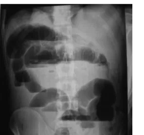
A 57-year-old male presented to our hospital with complaints of abdominal pain, vomiting, distension, and swelling. The patient had been suffering abdominal discomfort for five years and had undergone two colonoscopic examinations in those five years, but no disorders had been found. Abdominal examination revealed distension, a metallic sound, muscular rigidity, and tenderness. X-ray examination revealed multiple air-luid levels (Figure 1). An abdominal ultrasound showed a large solid mass in the right upper quadrant of the abdominal region. Since the intravesical pressure was measured as 25 cm H2O, the patient was diagnosed with mechanical intestinal obstruction, and laparotomy was performed
following luid and electrolyte replacement and antibiotic treatment. Abdominal exploration revealed an elastic hard tumor of 8x10 cm in diameter that appeared to originate from the jejunal mesentery, involving the third and fourth parts of the duodenum, the proximal jejunum and the ascending colon. The ascending colon, the proximal jejunum and the anterior face of the involved parts of the duodenum were surrounded by the mass. Thus, by right hemicolectomy, the irst 60 cm massive small intestine and partial duodenum were exised to resect the mass en bloc. The proximal small bowel segment was anastomosed to the transverse colon in an end-to-side fashion, and the duodenum was sutured transversely. The postoperative course was uneventful, and the patient was allowed to go home on the eighth postoperative day.
111
CLINICS 2010;65(1):110-3 A giant mesenteric ibromatosis case presenting with mechanical intestinal obstruction Polat C et al.
cells had pale eosinophilic cytoplasm and were embedded in a collagen network interrupted by ibrotic sections, but mitosis was not seen (Figure 3). Immunohistochemical analysis showed that the tumor cells expressed vimentin and actin (Figure 4) but not S100, desmin, CD34 or CD117. Macroscopic examination, microscopic examination and immunohistochemical features were suggestive of an intraabdominal desmoid tumor.
Desmoid tumors are the most common primary tumors of the mesentery and constitute about 3.5% of all ibrous tissue tumors3,4 The difference in sex distribution is statistically
insigniicant, but there is a slightly higher incidence of this tumor in women than in men.7 There are two forms:
the sporadic or primary form and the secondary form. The sporadic or primary form is extremely rare and is a variant of a benign stromal neoplasm of ibroblast-myoibroblast origin. It is usually secondary to trauma or hormonal stimulation or associated with familial polyposis coli or Gardner’s syndrome. Desmoid tumors have been seen in about 10% of familial adenomatous polyposis cases.8 At
present, desmoid tumors are clinicopathologically classiied into three types: abdominal, extra-abdominal and intra-abdominal.9 Desmoids of the abdominal wall are proliferative
ibrous tumors, and extra-abdominal desmoids (desmoids outside the abdominal wall) are histologically the same as abdominal desmoids. Extra-abdominal desmoids are different in that they are usually invasive and spread even to areas surrounding deep blood vessels and nerves; in addition, they are dificult to remove completely and are likely to recur after surgery. Trauma may be a major factor precipitating the onset of this type of tumor. A deinite episode of trauma has been noted in a high percentage of cases (19-63%).10 Accidental blunt injuries, lacerations,
intramuscular injections, fractures, and different endocrine or genetic factors have also often been implicated.11 In the
present case, symptoms were sporadic, and the patient had no previous history of any of the above conditions. Imaging techniques such as abdominal USG and CT are the most useful means for determining the exact localization of the tumor. In CT scans, a solid lesion is observed as a mass with soft tissue density.12
Most mesenteric tumors are large and appear in women in their reproductive years, often during or after pregnancy.6 Regression of these tumors has been associated
with menopause and menarche.13,14 Lim et al. investigated
the incidence and binding characteristics of the cytosol estrogen receptor and found that estrogen receptors were present in 33% of the desmoid tumors assayed.15 The signs
and symptoms of mesenteric fibromatosis are insidious and usually manifest when there is a large palpable tumor resulting in abdominal discomfort or pain. Weight loss and symptoms of ureteral obstruction, mesenteric ischemia, or intestinal obstruction were observed in this patient. Mechanical intestinal obstruction is extremely rare, and we have seen only one previous case in the literature. Gastrointestinal stromal tumors, lymphomas, carcinoid tumors, ibrosarcomas or inlammatory ibroid polyps should be considered in the differential diagnosis.16
Histologically, desmoid tumors are composed of long Figure 2 - Tumoral tissue iniltrating the adipose tissue (H&Ex20)
Figure 3 - Microscopic image showing cell groups with fusiform nucleus that is forming bundles (H&Ex100)
112
CLINICS 2010;65(1):110-3 A giant mesenteric ibromatosis case presenting with mechanical intestinal obstruction
Polat C et al.
sweeping fascicles of differentiated ibroblastic cells with ill-deined cytoplasmic borders, delicately staining nucleoli, and rare mitosis. Unfortunately in some cases, the differential diagnosis between fibromatosis and well-differentiated ibrosarcoma (Grade I) is dificult.17 A variety of treatments,
including wide surgical excision, non-steroidal anti-inflammatory drugs, antiestrogens, radiotherapy and cytotoxic chemotherapy have been attempted, but the efficacy of most of these is unpredictable. Treatment modalities other than surgical excision are controversial. Surgery should be performed by radical resection with wide margins, but these tumors are often unresectable because of massive involvement of adjacent vital structures. Wide excision is recommended, as these tumors have a tendency toward local recurrence.1,3-7,10,12,16 The principal
chemotherapy drugs used are vinblastine, methotrexate, doxorubicin, dacarbazine and carboplatin. In addition, it has been shown that tamoxifen, either alone or in combination with indomethacin, has produced a good response.1,18 Despite
radical surgery and adjuvant radiotherapy, recurrence rates in most studies can be in the range of 25-50%.19 However,
recurrent disease can be resected, and patients may live for extended periods with recurrent disease, especially in cases of sporadic desmoids. In addition, Kollevold et al. have reported that a desmoid tumor temporarily regressed after salpingo-oophorectomy in a patient with breast cancer.20
Moreover, Baliski et al. have proposed that neoadjuvant treatment with doxorubicin and radiotherapy with delayed
surgery is a better option than surgery alone.19 To the best
of our knowledge, this is the first documented case of successful margin-negative resection of jejunal mesenteric intraabdominal ibromatosis by combined partial duodeno-jejunectomy and right hemicolectomy. The effects of radiation therapy on treatment are not obvious, but several reports have advocated that complete regression may be achieved using dose levels greater than 50 Gy. Furthermore, Nuyttens et al. have also reported that radiotherapy or surgery with radiotherapy results in a signiicantly lower local recurrence rate.12,21 Objective response was seen in
52% of patients with desmoid tumors treated with endocrine therapy.16 These tumors infiltrate the surrounding tissue
and lead to severe morbidity and death. The frequency of local recurrence following excision is high, ranging from 10-90%. The signiicance of margins is a very controversial topic. Some studies suggest that margins are signiicant for predicting recurrence, while others claim that they are of no prognostic value.22,23 The patient presented in this report had
a small bowel obstruction caused by mesenteric ibromatosis and underwent only an aggressive surgical operation without any postoperative adjuvant therapy. The patient has shown no signs of recurrence at present, 30 months after surgery. As a result, we may say that intra-abdominal desmoids are very rare and benign tumors but are very aggressive and should be considered in the differential diagnosis of mechanical intestinal obstruction.
REFERENCES
1. Tauro LF, Sathyamoorthy PA, Hegde RB, Ravikrishnan HI, Singla MM. Giant desmoid tumor in the posterior abdominal wall/retroperitoneum. Indian J Surg. 2007;69:105-7.
2. McAdam WF, Goligher JC. The occurence of desmoids in patients with Familial Polyposis Coli. Br J Surg. 1970;57:618-31.
3. Yannopoulos K, Stout AP. Primary solid tumors of the mesentery. Cancer. 1963;16:914-27.
4. Cole RP. Peritonitis due to perforeted mesenteric ibroma: A hormonal etiology? Postgrad Med J. 1988;64:971-2.
5. Murayama T, Imoto S, Ito M, Matsushita K, Matozaki S, Nakagawa T, et al. Mesenteric ibromatosis presenting as fever of unknown origin. Am J Gastroenterol. 1992;87:1503-5.
6. Hayry P, Scheinin TM. The desmoid (Reitamo) syndrome: etiology, manifestations, pathogenesis, and treatment. Curr Probl Surg. 1988;25:225-30.
7. Clark SK, Neale KF, Landgrebe JC, Phillips RK. Desmoid tumours complicating familial adenomatous polyposis. Br J Surg. 1999;86:1185-9. 8. Clark SK, Phillips RKS. Desmoids in familial adenomatous polyposis.
Br J Surg. 1996;83:1494-504.
9. Caldwell EH. Desmoid tumor. Musculoaponeurotic ibrosis of the abdominal wall. Surgery. 1976;79;104-6.
10. Rock MG, Pritchard DJ, Reiman HM, Soule EH, Brewster RC. Extra-abdominal desmoid tumors. J Bone Joint Surg. 1984;66:1369-74. 11. Shimizu J, Kawaura Y, Tatsuzawa Y, Maeda K, Oda M, Kawashima A.
Desmoid tumor of the chest wall follwing chest surgery: report of a case. Surg Today. 1999;29:945-7.
12. Kato Y, Tsuyuki A, Kikuchi K, Kurihara N, Fujishiro Y. Mesenteric fibromatosis succesfully resected with düodeno-jejunectomy and Neprectomy. Hepato-Gastroenterology. 2005;52:1730-3.
13. Strode JE. Desmoid tumors particulary as related to their surgical removal. Ann Surg. 1954;139:335-40.
14. Dahn I, Jonsson J, Lundh G. Desmoid tumors: A series of 33 cases. Acta Chir Scand. 1963;126:305-14.
15. Lim CL, Walker MJ, Mehta RR, Das Gupta TK. Estrogen and antiestrogen binding sites in desmoid tumors. Eur J Cancer Clin Oncol. 1986;22:583-7.
113
CLINICS 2010;65(1):110-3 A giant mesenteric ibromatosis case presenting with mechanical intestinal obstruction Polat C et al.
17. Miettinen M, Weiss SW. Soft tissue tomours. In: Damjanov I, Linder J, eds. Anderson’s Pathology. 10th edition. St Louis: Mosby,1996:2488-9.
18. William JG. Abdominal wall, omentum, mesentery, and retroperitoneum-desmoid tumours. Morris PJ, Malt RA. Oxford Textbook of Surgery. International Ed, Vol. 1, Oxford University Press: New York; 1994, p. 1325-9.
19. Baliski CR, Temple WJ, Arthur K, Schachar NS. Desmoid tumors: A Novel approach for local control. J Surg Oncol. 2002;80:96-9. 20. Kollevold T. Desmoid tumours and carcinoma mamma in the same
patient. Acta Chir Scand. 1973;139:573-6.
21. Plukker JT, van Oort I, Vermey A, Molenaar I, Hoekstra HJ, Panders AK, et al. Aggressive ibromatosis (non-familial desmoid tumor): therapeutic problems and the role of adjuvant radiotherapy. Br J Surg. 1995;82:510-4.
22. Brodsky JT, Gordon MS, Hajdu SI, Burt M. Desmoid tumors of the chest wall. A locally recurrent problem. J Thorac Cardiovasc Surg. 1992;104:900-3.