ON L I N E AR T I C L E*
Dental Press J Orthod 42 2010 Sept-Oct;15(5):42-3
* Access www.dentalpress.com.br/journal to read the full article.
Editor’s summary
The voxel size, the smallest unit of a Cone-Beam Computed Tomography (CBCT) image, is related to the definition of tomographic image.
The question raised by the authors of this study is whether voxel size can affect radiation dose dur-ing CT scanndur-ing. Measurement of dose-area prod-uct (DAP) and entrance skin dose (ESD) when
Evaluation of referential dosages
obtained by Cone-Beam Computed
Tomography examinations acquired
with different voxel sizes
Marianna Guanaes Gomes Torres**, Paulo Sérgio Flores Campos***, Nilson Pena Neto Segundo****, Marlos Ribeiro*****, Marcus Navarro******, Iêda Crusoé-Rebello*******
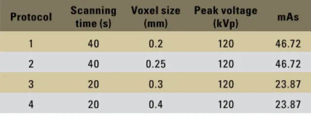
Objectives: The aim of this study was to evaluate the dose–area product (DAP) and the entrance skin dose (ESD), using protocols with different voxel sizes, obtained with i-CAT Cone-Beam Computed Tomography (CBCT), to determine the best parameters based on radioprotection principles. Methods: A pencil-type ionization chamber was used to measure the ESD and a PTW device was used to measure the DAP. Four protocols were tested: (1) 40s, 0.2 mm voxel and 46.72 mAs; (2) 40s, 0.25 mm voxel and 46.72 mAs; (3) 20s, 0.3 mm voxel and 23.87 mAs; (4) 20s, 0.4 mm voxel and 23.87 mAs. The kilovolt-age remained constant (120 kVp). Results: A significant statistical difference (p<0.001) was found among the four protocols for both methods of radiation dosage evaluation (DAP and ESD). For DAP evaluation, protocols 2 and 3 presented a statistically significant difference, and it was not possible to detect which of the protocols for ESD evaluation promoted this result. Conclusions: DAP and ESD are evaluation methods for radiation dose for Cone-Beam Computed Tomography, and more studies are necessary to explain such result. The voxel size alone does not affect the radiation dose in CBCT (i-CAT) ex-aminations. The radiation dose for CBCT (i-CAT) examinations is directly related to the exposure time and milliamperes.
Abstract
Keywords: Cone-Beam Computed Tomography. Radiation. Voxel.
** MSc in Dentistry, Federal University of Bahia (UFBA). Specialist in Dental Radiology and Imaging. *** Associate Professor, UFBA.
**** PhD in Dental Radiology, Campinas State University (UNICAMP). ***** Undergraduate Research Internship - PET, School of Dentistry, UFBA.
Torres MGG, Campos PSF, Pena N Neto Segundo, Ribeiro M, Navarro M, Crusoé-Rebello I
obtaining CBCT images with an i-CAT (Imaging Sciences International, Hatfield, PA, USA) was performed according to the protocols specified in Table 1. In all protocols, the field of view (collima-tion) of the scan was equivalent to 6 cm. The tests were repeated four times for each protocol.
The median DAP and ESD values found for the four protocols are shown in Table 2. A significant difference (p <0.001) was found among the four protocols for the two radiation dose assessment methods. The size of the voxel by itself did not in-fluence the exposed radiation dose. When the ex-posure factors (TE, kVp and mAs) are maintained, simply changing the voxel size does not influence the radiation dose significantly. However, the proto-cols correlate the use of smaller voxels with greater milliamperage exposure times, which invariably in-creases the exposure dose.
Contact address
Marianna Guanaes Gomes Torres Rua Araújo Pinho, 62, Canela CEP: 40.110-150 - Salvador / BA, Brazil E-mail: [email protected]
Questions to the authors
1) Which of the image acquisition protocols you tested is the most cost-effective? Why?
Not only this but other studies have shown that the protocol using a 0.3 mm voxel offers a combination of good resolution and reduced radiation dose. It is therefore the most cost-effective.
2) Does the size of the field of view (FOV) used in Cone-Beam CT examinations influence the radiation dose?
Yes. Especially when it comes to kerma area product (KAP), which increases the probabil-ity of stochastic effects. However, in our study, no influence was observed because we used the same FOV in all incidences and measurements. But, for example, in CBCT scans with a reduced FOV or restricted to measurement levels by sex-tants, the dose received is significantly reduced, implying very specific indications.
3) Do studies of radiation dose with Cone-Beam CT pose any difficulties or limitations?
Yes, researchers are still seeking a dosimet-ric quantity and/or a methodology that allows CBCT exposures to be assessed in order to esti-mate stochastic effects and compare exposures with other technologies. This is only made pos-sible thanks to the volumetric acquisition and advanced technology of CBCT equipment. TABLE 2 - Mean values of radiation doses (ESD and DAP) for the four
protocols.
Protocol
Entrance Skin Dose - ESD Dose Area Product-DAP
(mGy) (mGy m2)
1 3.77 44.92
2 3.78 45.30
3 2.00 24.43
4 2.00 24.98
(p = 0.00083) (p = 0.000145)
Protocol Scanning time (s) Voxel size (mm) Peak voltage (kVp) mAs
1 40 0.2 120 46.72
2 40 0.25 120 46.72
3 20 0.3 120 23.87
4 20 0.4 120 23.87
OR I G I N A L AR T I C L E
Dental Press J Orthod 1 2010 Sept-Oct;15(5):42.e1-4
Marianna Guanaes Gomes Torres*, Paulo Sérgio Flores Campos**, Nilson Pena Neto Segundo***, Marlos Ribeiro****, Marcus Navarro*****, Iêda Crusoé-Rebello******
intROduCtiOn
Successful dental treatment must be based on full planning and that includes the use of images to help with diagnosis. Computed tomography (CT) provides important
three-dimensional images and its use is increasing. However, the radiation dose accumulated in head and neck structures and its high cost are
major disadvantages of this technique.1-8
A new CT technology, Cone-Beam
Com-Evaluation of referential dosages
obtained by Cone-Beam Computed
Tomography examinations acquired
with different voxel sizes
Objectives: The aim of this study was to evaluate the dose–area product (DAP) and the entrance skin dose (ESD), using protocols with different voxel sizes, obtained with i-CAT Cone-Beam Computed Tomography (CBCT), to determine the best parameters based on radioprotection principles. Methods: A pencil-type ionization chamber was used to measure the ESD and a PTW device was used to measure the DAP. Four protocols were tested: (1) 40s, 0.2 mm voxel and 46.72 mAs; (2) 40s, 0.25 mm voxel and 46.72 mAs; (3) 20s, 0.3 mm voxel and 23.87 mAs; (4) 20s, 0.4 mm voxel and 23.87 mAs. The kilovolt-age remained constant (120 kVp). Results: A significant statistical difference (p<0.001) was found among the four protocols for both methods of radiation dosage evaluation (DAP and ESD). For DAP evaluation, protocols 2 and 3 presented a statistically significant difference, and it was not possible to detect which of the protocols for ESD evaluation promoted this result. Conclusions: DAP and ESD are evaluation methods for radiation dose for Cone-Beam Computed Tomography, and more studies are necessary to explain such result. The voxel size alone does not affect the radiation dose in CBCT (i-CAT) ex-aminations. The radiation dose for CBCT (i-CAT) examinations is directly related to the exposure time and milliamperes.
Abstract
Keywords: Cone-Beam Computed Tomography. Radiation. Voxel.
* MSc in Dentistry, Federal University of Bahia (UFBA). Specialist in Dental Radiology and Imaging. ** Associate Professor, UFBA.
*** PhD in Dental Radiology, Campinas State University (UNICAMP). **** Undergraduate Research Internship - PET, School of Dentistry, UFBA.
Evaluation of referential dosages obtained by Cone-Beam Computed Tomography examinations acquired with different voxel sizes
puted Tomography (CBCT), has recently be-come available. This technology was specifically developed for the head and neck region and provides three-dimensional volumetric images similar to medical tomographic images, at low cost and with reduction of patient exposure to radiation, because its field of vision (FOV) is limited to the axial dimension.2,5,7,9-12 The
voxel size is lower on CBCT compared with conventional CT. On the i-CAT device, for ex-ample, the voxel size can vary from 0.12 to 0.4 mm for the acquisition of images from the mandible, whereas on conventional CT the voxel size is normally 0.5–1 mm.6,13 Generally,
the smaller the voxel size and the longer the scanning time, the better the resolution and the details. However, a smaller voxel size is associated with a longer scanning time, which has some disadvantages such as greater possi-bility of patient movement during the exami-nation, elevated radiation doses and longer re-construction time.14,15
The aim of this study was to evaluate the dosage area product (DAP) and entrance skin dose (ESD), using protocols with different vox-el sizes, using the i-CAT CBCT device, to deter-mine better parameters based on radioprotec-tion principles.
MAteRiAls And MethOds
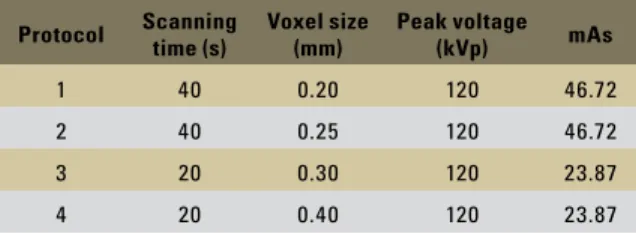
The DAP and ESD measurements using CBCT images from the i-CAT device (Imag-ing Sciences International, Hatfield, PA) were performed according to the protocols in Table 1. The scan height (collimation) was 6 cm for all protocols. The examinations were repeated four times for each protocol.
The RADCAL 9095 dose meter (Radcal. Corp., Monrovia, CA, USA) and the PTW DAP meter (PTW, Freiburg, Germany) were used. All equipment was calibrated in laboratories within the Brazilian Metrology Network (Rede Brasilei-ra de Metrologia-RBM). A pencil-type
ioniza-tion chamber (100 mm) was fixed on one end of the tomograph, coupled to an eletrometer, so that it was possible to measure the doses given while the images were obtained (ESD). A multi-plicative factor calculation was performed based on the distance between the x-ray beam output and the sensor, to compensate for the distance from the center of the device to the position of the ionization chamber. For the DAP measure-ment, a PTW device was coupled to the other end of the device.
The Kruskal-Wallis and Dunn tests were used to assess the data; p<0.001 was considered statistically significant.
Results
The median values for ESD and DAP for the four protocols are shown in Table 2. Sta-tistically significant differences (p<0.001) were found among the four protocols for both radia-tion dose evaluaradia-tion methods.
Dunn’s test showed that in the DAP evalu-ation, protocols 2 and 3 showed a statistically significant difference, and it was not possible to detect which of the protocols were significantly difference in the ESD evaluation.
disCussiOn
CBCT is a new technology and adequate knowledge is necessary to measure the radiation dose. We believe that the proposed method, us-ing the ESD and DAP, can be considered for dose measurements in this type of examination.
Protocol Scanning time (s) Voxel size (mm) Peak voltage (kVp) mAs
1 40 0.20 120 46.72
2 40 0.25 120 46.72
3 20 0.30 120 23.87
4 20 0.40 120 23.87
Torres MGG, Campos PSF, Pena N Neto Segundo, Ribeiro M, Navarro M, Crusoé-Rebello I
Dental Press J Orthod 3 2010 Sept-Oct;15(5):42.e1-4 Protocols 1 and 2 showed very similar ESD
and DAP values, and even though the voxel sizes were different, the exposure time (ET), the kilovoltage (kVp) and the milliamperage x exposure time (mAs) remained constant. The same applies to protocols 3 and 4 (Tables 1 and 2). This shows that the voxel size does not in-fluence the radiation dose; that is, when the ex-posure factors (ET, kVp and mAs) are the same, a single alteration of the voxel size does not alter the radiation dose significantly. However, the protocols couple the use of smaller voxels with greater exposure time and milliamperage, which invariably cause an increase in the ex-posure dose. Completely pre-established
proto-cols are provided by the i-CAT manufacturer.15
A greater voxel size, associated with a low
mAs and reduced ET, is able to reduce the dose
by as much as 50%.16 In our study, whereas the
ET and mAs practically doubled from proto-cols 3 and 4 to protoproto-cols 1 and 2, the radiation doses (ESD and DAP) behaved similarly for all protocols, being approximately doubled in pro-tocols 1 and 2 compared with propro-tocols 3 and 4 (Tables 1 and 2).
The limitation of the Dunn test in present-ing significant difference among the proto-cols and in evaluating ESD occurred because of the small sample. But, despite the small sample, protocols 2 and 3 showed a signifi-cant difference between (p=0.0065) for the DAP; this was only possible because of the extremely relevant difference that exists be-tween these protocols.
In conclusion, DAP and ESD are presented as evaluation methods for radiation doses in CBCT, and more studies are necessary to further elucidate such findings. The voxel size alone does not affect the radiation dose in CBCT (i-CAT) examinations. The radiation dose for CBCT (i-CAT) examinations is directly related to the exposure time and milliamperage.
ACKnOwledgMents
The authors express sincere gratitude to CAPES (Coordination of Improvement of Higher Education), IFBA (Federal Institute of Technological of Bahia) and Clinica Odonto-bioimagem, for supporting our projects. TABLE 2 - Mean values of radiation doses (ESD and DAP) for the four
protocols.
Protocol
Entrance Skin Dose - ESD Dose Area Product-DAP
(mGy) (mGy m2)
1 3.77 44.92
2 3.78 45.30
3 2.00 24.43
4 2.00 24.98
Evaluation of referential dosages obtained by Cone-Beam Computed Tomography examinations acquired with different voxel sizes
1. Lam EW, Ruprecht A, Yang J. Comparison of two-dimensional orthoradially reformatted computed tomography and panoramic radiography for dental implant treatment planning. J Prosthet Dent. 1995 Jul;74(1):42-6.
2. Mah JK, Danforth RA, Bumann A, Hatcher D. Radiation absorbed in maxillofacial imaging with a new dental computed tomography device. Oral Surg Oral Med Oral Pathol Oral Radiol Endod. 2003 Oct;96(4):508-13.
3. Kobayashi K, Shimoda S, Nakagawa Y, Yamamomto A. Accuracy in measurements of distance using limited cone-beam computerized tomography. Int J Oral Maxillofac Implants. 2004;19:228-31.
4. Tsiklakis K, Donta C, Gavala S, Karayianni K, Kamenopoulou V, Hourdakis CJ. Dose reduction in maxillofacial imaging using low dose cone beam CT. Eur J Radiol. 2005 Dec;56(3):413-7. 5. Guerrero ME, Jacobs R, Loubele M, Schutyser F, Suetens P, van
Steenberghe D. State-of-the-art on cone beam CT imaging for preoperative planning of implant placement. Clin Oral Investig. 2006 Mar;10(1):1-7.
6. Pinsky HM, Dyda S, Pinsky RW, Misch KA, Sarment DP. Accuracy of three-dimensional measurements using cone beam CT. Dentomaxillofac Radiol. 2006;35:410-6.
7. Van Assche N, van Steenberghe D, Guerrero ME, Hirsch E, Schutyser F, Quirynen M et al. Accuracy of implant placement based on pre-surgical planning of three-dimensional cone-beam images: a pilot study. J Clin Periodontol. 2007 Sep;34(9):816-21.
8. Hirsch E, Wolf U, Heinicke F, Silva MAG. Dosimetry of the cone beam computed tomography Veraviewepocs 3D
compared with the 3D Accuitomo in different ields of view.
Dentomaxillofac Radiol. 2008;37:268-73.
9. Loubele M, Maes F, Schutyser F, Marchal G, Jacobs R, Suetens P. Assessment of bone segmentation quality of cone beam CT versus multislice spiral CT: a pilot study. Oral Surg Oral Med Oral Pathol Oral Radiol Endod. 2006 Aug;102(2):225-34.
RefeRenCes
10. Loubele M, Van Assche N, Carpentier K, Maes F, Jacobs R, van Steenberghe D, et al. Comparative localized linear accuracy of
small-ield cone-beam CT and multislice CT for alveolar bone
measurements. Oral Surg Oral Med Oral Pathol Oral Radiol Endod. 2008 Apr;105(4):512-8.
11. Suomalainen A, Vehmas T, Kortesniemi M, Robinson S, Peltola J. Accuracy of linear measurements using dental cone beam and conventional multislice computed tomography. Dentomaxillofac Radiol. 2008 Jan;37(1):10-7.
12. Lascala CA, Panella J, Marques MM. Analysis of the accuracy of linear measurements obtained by cone beam computed tomography (CBCT-NewTom). Dentomaxillofac Radiol. 2004 Sep;33(5):291-4.
13. Mischkowski RA, Pulsfort R, Ritter L, Neugebauer J, Brochhagen HG, Keeve E, et al. Geometric accuracy of a newly developed cone-beam device for maxillofacial imaging. Oral Surg Oral Med Oral Pathol Oral Radiol Endod. 2007 Oct;104(4):551-9.
14. Stratemann SA, Huang JC, Maki K, Miller AJ, Hatcher DC. Comparison of cone beam computed tomography imaging with physical measures. Dentomaxillofac Radiol. 2008 Feb;37(2):80-93.
15. Ludlow JB, Davies-Ludlow LE, Brooks SL, Howerton WB. Dosimetry of 3 CBCT devices for oral and maxillofacial radiology: CB Mercuray, NewTom 3G and i-CAT. Dentomaxillofac Radiol. 2006 Jul;35(4):219-26. 16. Mozzo P, Procacci C, Tacconi A, Martini PT, Andreis IA. A
new volumetric CT machine for dental imaging based on the cone-beam technique: preliminary results. Eur Radiol. 1998;8(9):1558-64.
Contact address
Marianna Guanaes Gomes Torres Rua Araújo Pinho, 62, Canela CEP: 40.110-150 - Salvador / BA, Brazil E-mail: [email protected]
Submitted: July 2010