Original Article
Size Effects of Gold and Iron Nanoparticles on Radiation Dose Enhancement in
Brachytherapy and Teletherapy: A Monte Carlo Study
Ahad Ollah Ezzati1*, Seyed Rabi Mahdavi2, Hossein Mousavie Anijdan3
Abstract
Introduction
In this study, we aimed to calculate dose enhancement factor (DEF) for gold (Au) and iron (Fe) nanoparticles (NPs) in brachytherapy and teletherapy, using Monte Carlo (MC) method.
Materials and Methods
In this study, a new algorithm was introduced to calculate dose enhancement by AuNPs and FeNPs for Iridium-192 (Ir-192) brachytherapy and Cobalt-60 (Co-60) teletherapy sources, using the MC method. In this algorithm, the semi-random distribution of NPs was used instead of the regular distribution. Diameters were assumed to be 15, 30, and 100 nm in brachytherapy and 15 and 30 nm in teletherapy. Monte Carlo MCNP4C code was used for simulations, and NP density values were 0.107 mg/ml and 0.112 mg/ml in brachytherapy and teletherapy, respectively.
Results
AuNPs significantly enhanced the radiation dose in brachytherapy (approximately 60%), and 100 nm diameter NPs showed the most uniform dose distribution. AuNPs had an insignificant effect on teletherapy radiation field, with a dose enhancement ratio of 3% (about the calculation uncertainty) or less. In addition, FeNPs had an insignificant effect on both brachytherapy and teletherapy radiation fields. FeNPs dose enhancement was 3% in brachytherapy and 6% (about the calculation uncertainty) or less in teletherapy.
Conclusion
It can be concluded that AuNPs can significantly increase the absorbed dose in brachytherapy; however, FeNPs do not have a noticeable effect on the absorbed dose. Overall, AuNPs and FeNPs cannot increase the absorbed dose for the Co-60 teletherapy source.
Keywords: Brachytherapy, Dose Enhancement Factor, Monte Carlo, Nanoparticle.
1- Faculty of Physics, University of Tabriz, Tabriz, Iran
*Corresponding Author: Tel: +984113393349; Fax: +984113341424; E-Mail: [email protected]
2- Department of Medical Physics, Iran University of Medical Sciences, Tehran, Iran
1. Introduction
Medical application of nanoparticles (NPs) has grown rapidly in recent years. NPs have been studied for their potential use in thermal therapy, diagnostic imaging, drug delivery, and genetic research [1-8]. Laboratory studies have shown that gold nanoparticles (AuNPs) can enhance the radiation absorbed dose in radiotherapy [9-14].
AuNPs can be distributed in tumors, given their size. In addition, they can be tumor-selective by labeling with ligands or molecules [15]. Dose enhancement factor (DEF) for NPs can be calculated by Monte Carlo (MC) method or measured by experimental setups [15-22]. The ratio of AuNPs to tissue in MC studies was based on previous studiy by Hainfeld et al., in which 1.9 nm AuNPs were injected into mice and no toxicity was observed [5].
Iridium-192 (Ir-192) brachytherapy source has been widely used in DEF calculations for AuNPs, due to its unique properties such as low photon energy. Cho et al. simulated gold
tissue mixture by using the
BEAMnrc/DOSXYZnrc code to estimate the DEF. According to their study, the DEF calculated for AuNPs was approximately 31% at 7 mg/ml Au concentration [5].
According to Lechtman et al., in order to double the absorbed dose, 20 mg/ml concentration of AuNPs is required for the Ir-192 brachytherapy source [23]. Bahreyni Toossi et al. and Ghorbani et al. reported 1.16 DEF for 100 nm AuNPs at 30 mg/ml concentration [16,17]. According to the performed studies, different authors reported different DEFs by 100 nm AuNPs for the Ir-192 brachytherapy source [16, 17, 24-26]. Different DEFs for the same size of AuNPs show that DEF of AuNPs varies significantly in terms of concentration and simulation setup. Recent MC studies have used heavy element tissue mixtures or regular distribution of NPs [16, 17, 24-26]. The regular distribution of NPs is very different from their random distribution in the tissue. In the regular distribution, unlike the random distribution, NP distances from the neighbors are constant.
In the regular distribution, photon interactions decrease within the tumor, since some photons do not interact with any NPs in their trajectories.
In this study, a new algorithm was introduced for generating the semi-random distribution of NPs within the tissue. DEF for AuNPs and FeNPs of different sizes was calculated for two different photon sources (Ir-192 and Co-60). The calculated DEFs were compared between different NPs; the DEFs were also compared with the recently published data.
2. Materials and Methods
In this study, semi-randomly distributed NPs were used. In this distribution, a new algorithm was proposed to simulate the random distribution of NPs within the tissue. To model this arrangement, a computer program was developed using MATLAB 7.04 software, in which a specific number of NPs (900 NPs) were randomly placed in a cubic lattice of length L, and then the tumor volume was filled with the cubic lattice. MCNP4C code was used to calculate DEF for the NPs, and the absorbed dose was calculated using *f8 energy deposition tally.
A Theratron cobalt therapy unit was simulated in MCNP4C for obtaining percent depth dose (PDD) curve. To benchmark the MC simulations, the obtained PDD was compared with the measured data. Then, a point source was used instead of cobalt teletherapy unit to speed up MC calculations.
Dose distributions were calculated with and without NPs, and DEFs were calculated using equation 1. Dose enhancements by AuNPs and FeNPs were calculated for the Co-60 source with a field size of 2 cm diameter on a phantom surface, at 80 cm source-to-surface distance (SSD) (Fig. 1).
100 1
AbsorbeddosewithNP Absorbeddosewithout NP DEF
Absorbeddosewithout NP
-= ´
be 112 mg/ml within the tumor, and NPs, with two sizes of 15 and 30 nm, were simulated in teletherapy (Co-60 source) irradiation field. High dose rate (HDR) brachytherapy with an Ir-192 source was simulated in spherical water phantom to calculate DEF for NPs. The active cylindrical Ir-192 core was 3.5 mm in length and 0.6 mm in diameter. The source capsule was assumed to be stainless steel with 8.02 g/cm3 density (Fig. 2). The Ir-192 source was placed in the center of phantom with a diameter of 8 cm. The tumor was assumed as a cube with 1 cm side length; the bottom of this cube was placed 1 cm above the Ir-192 source. The concentration of AuNPs and FeNPs in the tumor area was 107 mg/ml, and three sizes of 15, 30, and 100 nm were simulated to obtain DEF for NPs. This configuration is shown schematically in Figure 3. The calculated DEFs and dose distributions for Ir-192 source
were benchmarked against the study results of Zhang et al. for 100 nm AuNPs [25].
Figure 1. Schematic representation of teletherapy simulation setup
Figure 2. Geometry of Ir-192 HDR brachytherapy source[25]
Figure 3. Schematic representation of brachytherapy simulation setup
80 cm
30 cm
31 cm 31
cm
Cobalt-60 source
3. Results
Theratron cobalt therapy unit with 10x10 cm2 field size at 100 cm SSD was simulated with MCNP4C code, and the obtained PDD was compared with the ion chamber measurement. As it can be seen in Figure 4, measurement and simulation are in good agreement within 1% of the accepted level.
Figure 4. Comparison of measured and simulated PDDs for Co-60 source with a 10x10 cm2 field size at 100 cm SSD
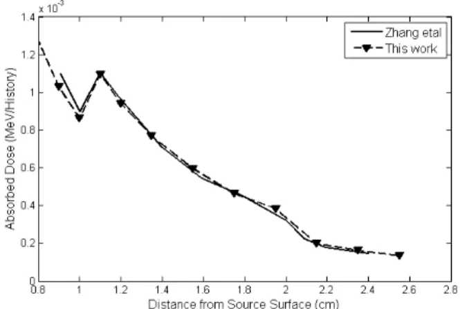
For benchmarking the brachytherapy calculations, the setup of the study by Zhang et al. [25] was exactly simulated, and the results were compared in Figure 5. As it can be seen, the two simulations are in agreement within 2% of the accepted level.
Figure 5. Comparison between dose distributions of 100 nm AuNPs in the current research and the study by Zhang et al.
Figure 6a shows the effect of AuNPs on the absorbed dose for Ir-192 brachytherapy source. As it can be seen from this figure, 100 nm AuNPs have maximum effect on absorbed dose distribution. In addition, 100 nm AuNPs improve the uniformity of the absorbed dose distribution more than the smaller sizes. Figure 6b shows the effects of FeNPs on the absorbed dose for Ir-192 brachytherapy source.
Average DEF due to AuNPs with different sizes are shown in Table 1. As it can be seen from this table, 100 nm AuNPs enhance the average absorbed dose in the tumor area about 55%, which is 1% less than gold-water mixture.
Figure 6. The effect of NPs with different sizes on absorbed dose for Ir-192 brachytherapy source; a) AuNPs, b) FeNPs
Table 1. Average DEF for different sizes of AuNPs for Ir-192 brachytherapy source
NP size (diameter)
15 nm
30 nm
100 nm
Gold-water mixture Average DEF
(%) 33 54 55 56
The effects of different sizes of FeNPs on the average absorbed dose are shown in Table 2. As it can be seen, FeNPs can decrease or increase the absorbed dose, depending on their sizes.
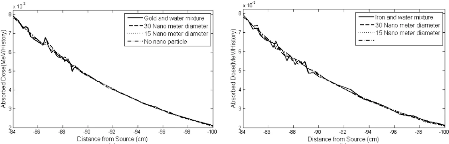
Figure 7 shows the effects of AuNPs and FeNPs on the absorbed dose for teletherapy source (Co-60) and different sizes of NPs. The PDDs were obtained for a small field size (2 cm diameter on a phantom surface) at 80 cm SSD. Figure 7b shows that the effect of FeNPs on the absorbed dose is negligible and these NPs decrease the absorbed dose by approximately 2% (order of calculation uncertainty) in the tumor area.
Table 2. Effects of different sizes of FeNPs on the average DEF for Ir-192 brachytherapy source
NP size (diameter)
15 nm
30 nm
100 nm
Iron-water mixture Average DEF
(%) -6 -0.4 1.6 0.6
Table 3. Effects of different sizes of AuNPs on the average DEF for teletherapy source (Co-60)
NP size
(diameter)
15 nm 30 nm Gold-water mixture Average DEF
(%)
0 1.7 1.5
Table 4. Effects of different sizes of FeNPs on the average DEF for teletherapy source (Co-60)
NP size
(diameter)
15 nm 30 nm Gold-water mixture Average DEF
(%)
-0.1 -2.2 -1.5
Average DEFs of different sizes of AuNPs and FeNPs for teletherapy source (Co-60) are shown in tables 3 and 4, respectively. As it can be seen from Table 3, AuNPs cannot increase the absorbed dose more than 2%. Table 4 shows that FeNPs decrease the average absorbed dose by approximately 2% in the tumor area.
Figure 7. The effects of NPs with different sizes on the absorbed dose for Co-60 teletherapy source; a) AuNPs, b) FeNPs
4. Discussion
Recent MC studies have used heavy element tissue mixture or regular distribution of NPs for calculating their DEFs [16, 17, 24-26]. Cho et al. used gold tissue mixture to calculate DEF for AuNPs. According to their study, DEF for AuNPs was approximately 31%, with
Bahreyni Toossi et al. and Ghorbani et al. also used regular distributions of AuNPs and reported 16% DEF for 100 nm AuNPs with 30mg/ml Au concentration [16-17]. According to these published studies [16, 17, 24-26], different researchers reported different DEFs for 100 nm AuNPs and Ir-192 brachytherapy source. Different DEFs for the same size of AuNPs show that DEFs for AuNPs vary significantly according to concentration and simulation setup.
Regular distribution of NPs is very different from their random distribution within the tissue. In this study, we used the semi-random distribution of NPs to evaluate the effects of AuNPs and FeNPs (with sizes smaller than 100 nm) on the absorbed dose, using MC method for Ir-192 brachytherapy and Co-60 teletherapy sources. According to the results, 100 nm AuNPs could increase the absorbed dose by approximately 55% in the tumor area, and these NPs enhanced the uniformity of absorbed dose distribution more than the smaller sizes (Fig. 6a and Table 1). Also, the FeNPs could not increase the absorbed dose as much as AuNPs for Ir-192 brachtherapy source, because of the low photoelectric cross cross-section and low electron density of the iron, with respect to the gold.
Roeske et al. used a gold-tissue mixture model and reported that the dose enhancement effect of AuNPs for Co-60 source ranged between 1.004 and 1.006 [26]. Moreover, Bahreyni Toossi et al. used the regular distribution of AuNPs and reported that the dose enhancement effect of AuNPs for Co-60 source was 2-4% for different distances from
the source [16]. In the current study, semi-random distribution of AuNPs was used, and as it can be seen from Table 3, DEFs of 15 nm and 30 nm AuNPs were zero and 2%, respectively.
Our simulations also showed that DEFs for AuNPs and FeNPs are negligible for the Co-60 source (Fig.7, Tables 3 & 4). The low photoelectric cross-section of gold and iron for Co-60 photons is the main reason for the small DEFs. The results of the current study were in agreement with those of previous research, and showed that the application of AuNPs could increase the absorbed dose of low-energy brachytherapy sources and decrease treatment duration.
5. Conclusion
It can be concluded that AuNPs can significantly increase the absorbed dose in brachytherapy; however, FeNPs do not have a noticeable effect on the absorbed dose. Overall, AuNPs and FeNPs cannot increase the absorbed dose for the Co-60 teletherapy source. It also can be concluded that the simple model of gold and- tissue mixture can be used in MC calculations of AuNPs with the size of 100 nm. However, for sizes smaller than 100 nm, the gold and water mixture simple model cannot could not be applied.
Acknowledgment
This work was financially supported by the University of Tabriz.
References
1. Bullis K. Remotely activated nanoparticles destroy cancer, Technology Review 2007. Available at: http://www.technologyreview.com/Nanotech/17956/.Published January 2, 2007. Accessed 25 Nov, 2007 2. Krishnan S, Diagaradjane P, Cho SH. Nanoparticle-mediated thermal therapy: evolving strategies for prostate
cancer therapy. Int J Hyperthermia. 2010;26(8):775-89.
3. Shen H, You J, Zhang G, Ziemys A, Li Q, Bai L, et al. Cooperative, nanoparticle-enabled thermal therapy of breast cancer. Adv Healthc Mater. 2012 Jan 11;1(1):84-9.
4. Gobin AM, Lee MH, Halas NJ, James WD, Drezek RA, West JL. Near-infrared resonant nanoshells for combined optical imaging and photothermal cancer therapy. Nano Lett. 2007 Jul;7(7):1929-34.
5. Hainfeld JF, Slatkin DN, Focella TM, Smilowitz HM. Gold nanoparticles: a new X-ray contrast agent. Br J Radiol. 2006 Mar;79(939):248-53.
7. Panyam J, Labhasetwar V. Biodegradable nanoparticles for drug and gene delivery to cells and tissue. Adv Drug Deliv Rev. 2003 Feb 24;55(3):329-47.
8. Mehta A, Ghaghada K, Mukundan S, Jr. Future Clinical Applications of Molecular Imaging: Nanoparticles, Cellular Probes, and Imaging of Gene Expression. In: Pillai JJ, editor. Functional Brain Tumor Imaging: Springer New York; 2014. p. 225-37.
9. Herold DM, Das IJ, Stobbe CC, Iyer RV, Chapman JD. Gold microspheres: a selective technique for producing biologically effective dose enhancement. Int J Radiat Biol. 2000 Oct;76(10):1357-64.
10. Hainfeld JF, Slatkin DN, Smilowitz HM. The use of gold nanoparticles to enhance radiotherapy in mice. Phys Med Biol. 2004 Sep 21;49(18):N309-15.
11. Chithrani DB, Jelveh S, Jalali F, van Prooijen M, Allen C, Bristow RG, et al. Gold nanoparticles as radiation sensitizers in cancer therapy. Radiat Res. 2010 Jun;173(6):719-28.
12. Jain S, Hirst DG, O'Sullivan JM. Gold nanoparticles as novel agents for cancer therapy. Br J Radiol. 2012 Feb;85(1010):101-13.
13. Ngwa W, Makrigiorgos GM, Berbeco RI. Gold nanoparticle-aided brachytherapy with vascular dose painting: estimation of dose enhancement to the tumor endothelial cell nucleus. Med Phys. 2012 Jan;39(1):392-8. 14. Hainfeld JF, Smilowitz HM, O'Connor MJ, Dilmanian FA, Slatkin DN. Gold nanoparticle imaging and
radiotherapy of brain tumors in mice. Nanomedicine (Lond). 2013 Oct;8(10):1601-9.
15. Loo C, Lowery A, Halas N, West J, Drezek R. Immunotargeted nanoshells for integrated cancer imaging and therapy. Nano Lett. 2005 Apr;5(4):709-11.
16. Bahreyni Toossi MT, Ghorbani M, Mehrpouyan M, Akbari F, Sobhkhiz Sabet L, Soleimani Meigooni A. A Monte Carlo study on tissue dose enhancement in brachytherapy: a comparison between gadolinium and gold nanoparticles. Australas Phys Eng Sci Med. 2012 Jun;35(2):177-85.
17. Ghorbani M, Pakravan D, Bakhshabadi M, Meigooni AS. Dose enhancement in brachytherapy in the presence of gold nanoparticles: a Monte Carlo study on the size of gold nanoparticles and method of modelling. Nukleonika. 2012;57:401-6.
18. Pakravan D, Ghorbani M, Momennezhad M. Tumor dose enhancement by gold nanoparticles in a 6 MV photon beam: a Monte Carlo study on the size effect of nanoparticles. Nukleonika. 2013;58.
19. Ghorbani M, Bakhshabadi M, Golshan A, Knaup C. Dose enhancement by various nanoparticles in prostate brachytherapy. Australasian Physical & Engineering Sciences in Medicine. 2013;36(4):431-40.
20. Khadem-Abolfazli M, Mahdavi M, Mahdavi S, Ataei G. Dose enhancement effect of gold nanoparticles on MAGICA polymer gel in mega voltage radiation therapy. Int J Radiat Res. 2013;11(1):55-61.
21. Anijdan SM, Shirazi A, Mahdavi S, Ezzati A, Mofid B, Khoei S, et al. Megavoltage dose enhancement of gold nanoparticles for different geometric set-ups: Measurements and Monte Carlo simulation. Iran J Radiat Res. 2012;10(3-4):183-6.
22. Mousavie Anijdan SH, Mahdavi SR, Shirazi A, Zarrinfard MA, Hajati J. Megavoltage X-ray Dose Enhancement with Gold Nanoparticles in Tumor Bearing Mice. Int J Mol Cell Med. 2013 Summer;2(3):118-23.
23. Lechtman E, Chattopadhyay N, Cai Z, Mashouf S, Reilly R, Pignol JP. Implications on clinical scenario of gold nanoparticle radiosensitization in regards to photon energy, nanoparticle size, concentration and location. Phys Med Biol. 2011 Aug 7;56(15):4631-47.
24. Cho SH, Jones BL, Krishnan S. The dosimetric feasibility of gold nanoparticle-aided radiation therapy (GNRT) via brachytherapy using low-energy gamma-/x-ray sources. Phys Med Biol. 2009 Aug 21;54(16):4889-905.
25. Zhang SX, Gao J, Buchholz TA, Wang Z, Salehpour MR, Drezek RA, et al. Quantifying tumor-selective radiation dose enhancements using gold nanoparticles: a monte carlo simulation study. Biomed Microdevices. 2009 Aug;11(4):925-33.
![Figure 2. Geometry of Ir-192 HDR brachytherapy source[25]](https://thumb-eu.123doks.com/thumbv2/123dok_br/18341349.352023/3.892.99.787.557.739/figure-geometry-ir-hdr-brachytherapy-source.webp)