The impact of topically applied preservation
solu-tions on the respiratory epithelium of tracheal grafts
submitted to cold ischemia: functional and
morpho-logical analysis
Artur Eugeˆnio de Azevedo-Pereira,IJuliana Akemi Saka,IKarina Andrighetti de Oliveira-Braga,IRoge´rio Pazetti,IMauro Canzian,IIPaulo Manuel Peˆgo-Fernandes,IFabio Biscegli JateneI
IInstituto do Corac¸a˜o (InCor), Faculdade de Medicina da Universidade de Sa˜o Paulo, Laboratory of Research on Thoracic Surgery (LIM-61), Sa˜o Paulo/SP,
Brazil.IIInstituto do Corac¸a˜o (InCor), Faculdade de Medicina da Universidade de Sa˜o Paulo, Laboratory of Pathology, Sa˜o Paulo/SP, Brazil.
OBJECTIVE: Advances in graft reepithelialization and revascularization have renewed interest in airway transplantation. This study aims to determine whether topically applied preservation solutions can ameliorate ischemic injury to tracheal grafts. We analyzed 1) the effects of cold ischemia on the mucociliary clearance of tracheal grafts and 2) the impact of topically applied preservation solutions on the effects of cold ischemia on mucociliary clearance.
METHOD:Tracheal segments (n = 217) from 109 male Wistar rats were harvested, submerged in low-potassium-dextran-glucose, histidine-tryptophan-ketoglutarate, or saline solution (saline group), and stored at 4
˚
C for 6, 10, 16, or 24 hours. A control group (not submerged) was analyzed immediately after harvesting. In situ mucociliary transport and ciliary beating frequency were measured using a stroboscope. Epithelial integrity, cellular infiltration, and mucus storage were quantified by light microscopy and image analysis software, along with transmission electron microscopy.RESULTS: 1) The effects of cold ischemia: in situ mucociliary transport and ciliary beating frequency were greater in the control group than after cold ischemia. Microscopic analysis results were similar between groups. 2) The effects of preservation solutions: there was no difference between the low-potassium-dextran-glucose, histidine-tryptophan-ketoglutarate, and saline groups in functional or light microscopy analysis. The saline group presented stronger signs of ischemic injury with transmission electron microscopy.
CONCLUSIONS: Cold ischemia diminished the mucociliary clearance of the tracheal respiratory epithelium. Topically applied preservation solutions did not ameliorate the injury caused by cold ischemia to the tracheal respiratory epithelium.
KEYWORDS: Trachea; Mucociliary Clearance; Organ Preservation Solutions.
Azevedo-Pereira AE, Saka JA, Oliveira-Braga KA, Pazetti R, Canzian M, Peˆgo-Fernandes PM, et al. The impact of topically applied preservation solutions on the respiratory epithelium of tracheal grafts submitted to cold ischemia: functional and morphological analysis. Clinics. 2013;68(5):702-709.
Received for publication onOctober 10, 2012;First review completed onNovember 4, 2012;Accepted for publication onJanuary 14, 2013 E-mail: [email protected]
Tel.: 55 11 2661-5248
& INTRODUCTION
Tracheal transplantation remains a challenge (1). A large number of surgical techniques and implantable devices have been tested, with disappointing results (1,2). In spite of that, some recent findings on tracheal revascularization and
graft reepithelialization have renewed interest in tracheal transplantation (2–5). However, for tracheal transplantation to succeed, the effects of ischemic injury secondary to harvesting and storage on tracheal tissues need to be better understood.
Because of the particular segmental pattern of tracheal vascularization, with no major tracheal vessels, the intra-vascular administration of preservation solutions into tracheal grafts is far from useful in a clinical scenario (2,6,7). Therefore, we previously investigated whether preservation solutions were effective when applied topi-cally. Due to the small sample size, preliminary data were inconclusive in both measuring the effects of cold ischemia on the tracheal respiratory epithelium and defining whether Copyrightß2013CLINICS– This is an Open Access article distributed under
the terms of the Creative Commons Attribution Non-Commercial License (http:// creativecommons.org/licenses/by-nc/3.0/) which permits unrestricted non-commercial use, distribution, and reproduction in any medium, provided the original work is properly cited.
No potential conflict of interest was reported.
topically applied preservation solutions could ameliorate those effects (8).
The trachea acts as both a mechanical conduit of environmental air to and from the lower respiratory tract and a defense organ through mucociliary clearance. Mucociliary clearance is a delicate mechanism through which mucus is propelled from the lower to the upper airways, carrying with it noxious organic and inorganic particles to be swallowed or expelled by coughing (9). Based on its delicate mechanism, we consider mucociliary clear-ance, especially mucociliary transportation, an excellent criterion by which to verify tracheal graft preservation. Our aims were 1) to analyze the effects of cold ischemia on the tracheal respiratory epithelium and 2) to analyze the effects of topically applied preservation solutions on tracheal respiratory epithelia submitted to cold ischemia.
& MATERIALS AND METHODS
This research was approved by the Ethical Committee of our Institution. All animals received humane care in compliance with the ‘Principles of Laboratory Animal Care’ formulated by the National Society for Medical Research and the ‘Guide for the Care and Use of Laboratory Animals’ prepared by the Institute of Laboratory Animal Resources and published by the National Institutes of Health (NIH Publication No. 86–23, revised 1996).
We obtained 217 tracheal segments from 109 Wistar male rats (weight, 300¡50 g). The rats were anesthetized with intraperitoneal pentobarbital (50 mg/kg) and euthanized by exsanguination. A median cervicosternotomy was per-formed, followed by tracheal harvesting. The trachea was sectioned at the midpoint between the cricoid cartilage and the carina, providing two tracheal segments from each rat. Immediately after harvesting, each tracheal segment was randomly allocated to be submerged in one of three solutions: saline solution (saline group); low-potassium-dextran-glucose (LPD-glucose) (Vitrolife AB, Sweden) (LPD group); or histidine-tryptophan-ketoglutarate (HTK) (Dr F. Ko¨hler Chemie, Germany) (HTK group). Each tracheal segment was submerged in the solution within a sterile test tube. The submerged tracheal segments were immediately allocated to be stored at 4
˚
C for 6, 10, 16, or 24 hours. Each tracheal segment was submitted to only one period of cold ischemia. At the end of the ischemic time (6, 10, 16, or 24 hours), the tracheal segments were incubated at room temperature for 20 minutes and submitted to the functional analyses of mucociliary clearance as described below. After functional analyses, the segments were prepared for morphological analyses as described below. The control group was composed of tracheal segments that were not submerged in any solution or submitted to cold ischemia. The tracheal segments of the control group were submitted to functional analyses of mucociliary clearance immediately after harvesting. After functional analyses, the control group samples were prepared for morphological analyses similar to the other groups.Functional analysis
After the specified ischemic time and before undergoing the functional analyses, all tracheal segments were incu-bated at room temperature for 20 minutes. The functional analyses were performed at room temperature and in room
air. Mucociliary clearance was analyzed through in situ
mucociliary transport and the ciliary beating frequency, as described previously (10,11). Briefly, the ventral wall of each tracheal segment was opened to expose the ciliated epithelium. The tracheal segment was placed under the 100x objective of a light microscope (BX50, Olympus, Japan) that was connected to a video camera (Trinitron 3CCD, Sony, Japan).In situmucociliary transport was measured by timing the movement of mucous particles across the tracheal surface with the aid of a reticulated eyepiece. In situ mucociliary transport was expressed in millimeters/ minute. Subsequently, under the same microscope, a stroboscope (Machine Vision Strobe, Cedar-Hurst, USA) was placed in front of the tracheal segment, and the ciliary beating frequency was measured by synchronization between cilia movement and the stroboscope flashlight. The ciliary beating frequency was expressed in hertz. The researchers performing the functional analyses were blinded to the groupings.
Morphological analysis
Morphological analysis was performed using light micro-scopy and transmission electron micromicro-scopy. After func-tional analysis, tracheal segments were fixed in formalin and embedded in paraffin. Serial sections (0.5mm thick)
were cut with a microtome and stained with hematoxylin-eosin, alcian blue, and periodic acid-Schiff (PAS). A small sample of each tracheal segment was prepared for transmis-sion electron microscopy.
Light microscopy
The hematoxylin-eosin-stained slides were used to deter-mine the ischemic injury on the tracheal grafts. We analyzed ischemic injury through epithelial integrity and cellular infiltration. The analysis was performed using modified semi-quantitative scales for epithelial integrity and cellular infiltration based on a previous work on tracheal transplan-tation (12). Epithelial integrity was quantified with a semi-quantitative scale rated from 1 to 3: 1 (low integrity), epithelial lining up to 1/3 of the tracheal surface; 2 (moderate integrity), epithelial lining from 1/3 to 2/3 of the tracheal surface; and 3 (high integrity), epithelial lining of more than 2/3 of the tracheal surface. Cellular infiltration was quantified with a semi-quantitative scale rated from 1 to 3: 1 (mild infiltration), scattered and diffuse cellular infiltration, without islets of dense infiltrate; 2 (moderate infiltration), more intense cellular infiltration, with up to two islets of dense infiltrate; and 3 (severe infiltration), a thick layer of dense infiltrate or more than two islets of dense infiltrate. Each slide was distinctly quantified by three observers blinded to the groupings, and the final score was achieved with the concordance of at least two observers.
quantification was expressed as the percentage of the respiratory epithelial area stained by alcian blue and PAS.
Transmission electron microscopy
We obtained one sample from each tracheal segment. The samples were fixed in 2% glutaraldehyde and 0.1% tannic acid for 2 hours, followed by fixation in 1% osmium tetroxide for 1 hour. After dehydration in a series of ethanol solutions for 1 hour, the samples were embedded in acetone and resin for 3 hours. The samples were baked for 24 hours to be embedded in pure resin and stored. Only samples from the control group and the more affected tracheal segments, as indicated by functional and light microscopy analysis, were stained and inspected. Thick sections (0.5mm) were stained with toluidine blue to determine the
area of interest. Then, ultrathin sections (90 nm) were prepared and stained with uranyl acetate and lead citrate. The specimens were inspected and photographed under a transmission electron microscope (1200EXII, Jeol, Japan).
Data presentation and statistical analysis
The data obtained were analyzed from two perspectives according to the objectives of the study. To analyze the effects of cold ischemia on the tracheal respiratory epithe-lium (our first objective), we grouped the tracheal segments according to the duration of ischemia and performed the first statistical analysis. Therefore, we had five groups: control group (not submitted to ischemia), 6 hours, 10 hours, 16 hours, and 24 hours. The type of preservation solution used was not considered in this first analysis. Subsequently, we focused on the effects of the topically applied preservation solutions on the ischemic tracheal respiratory epithelium (our second objective). The second statistical analysis compared the data from the different preservation solutions (control group, LPD group, HTK group, and saline group), including all ischemic times in each preservation solution group but not including the ischemic time itself.
The distribution of the variables was evaluated using the Kolmogorov-Smirnov test. For variables following a normal distribution, the data were presented as the mean ¡-standard deviation and compared with an analysis of variance (ANOVA) with Bonferroni’s post-test. Despite analyzing two factors (ischemic time and preservation solutions), we did not perform a two-way ANOVA because we focused on the two objectives separately. For the non-normally distributed variables, the data were presented as the median and interquartile range and compared with the Kruskal-Wallis test. For comparisons among groups of qualitative variables, we used the chi-square test. Statistical significance was considered at p,0.05. The
statistical analysis was performed with SPSS 13.0 software (SPSS Inc., Chicago, IL, USA).
& RESULTS
Effects of cold ischemia on the tracheal respiratory epithelium
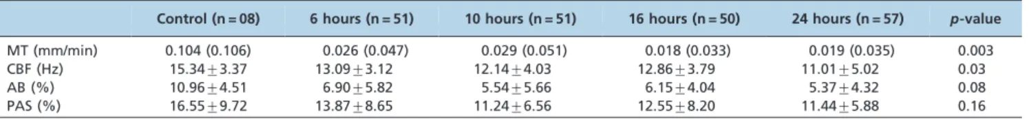
As shown in Table 1, there was a statistically significant difference between the control group and the groups submitted to cold ischemia in terms ofin situ mucociliary transport (p,0.003) (Figure 1A) and ciliary beating fre-quency (p,0.03) (Figure 1B). In the morphological analysis, there was no statistically significant difference among the groups in either epithelial integrity or cellular infiltration (data not shown). Figure 2 illustrates how the semi-quantitative scores were calculated for some histological slides. Indeed, there was no statistically significant differ-ence in the quantification of the area stained by alcian blue and PAS among the groups (Figure 1C and 1D).
Effects of topically applied preservation solutions on the tracheal respiratory epithelium
As shown in Table 2, in situ mucociliary transport was higher in the control group compared to the other groups (p,0.0001) (Figure 3A). In situ mucociliary transport was also higher in the HTK group than the saline group (p,0.006). However, there was no difference in mucociliary transport between the other groups in which tracheal segments were submerged in preservation solutions. The ciliary beating frequency in the control group was different compared to the LPD, HTK, and saline groups (p,0.02). However, there was no difference among the LPD, HTK, and saline groups in the same variable (Figure 3B). In the morphological analysis, there was no statistically significant difference among the groups in either epithelial integrity or cellular infiltration (data not shown). There was no statistically significant difference in the quantification of the area stained by alcian blue and PAS among the groups (Figure 3C and 3D).
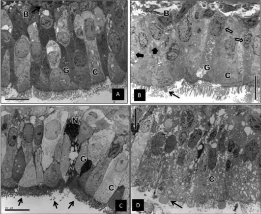
The transmission electron microscopy findings are shown in Figure 4. The LPD group showed nuclear shrinkage, most likely related to osmotic differences between the respiratory cells and the LPD-glucose solution. Some signs of cytoplas-mic disruption and vacuolization were found (Figure 4B). The HTK group showed electron-dense crystals throughout the respiratory cells. The cilia coincided with those crystals in all fields. Some necrotic cells were found, which contained a large amount of those crystals. However, the ultrastructural architecture of the respiratory cells was preserved (Figure 4C). The saline group showed marked signs of loss of cellular integrity, cytoplasmic disruption, and intense vacuolization (Figure 4D).
Table 1 -Effects of cold ischemia on mucociliary clearance.
Control (n = 08) 6 hours (n = 51) 10 hours (n = 51) 16 hours (n = 50) 24 hours (n = 57) p-value
MT (mm/min) 0.104 (0.106) 0.026 (0.047) 0.029 (0.051) 0.018 (0.033) 0.019 (0.035) 0.003
CBF (Hz) 15.34¡3.37 13.09¡3.12 12.14¡4.03 12.86¡3.79 11.01¡5.02 0.03
AB (%) 10.96¡4.51 6.90¡5.82 5.54¡5.66 6.15¡4.04 5.37¡4.32 0.08
PAS (%) 16.55¡9.72 13.87¡8.65 11.24¡6.56 12.55¡8.20 11.44¡5.88 0.16
& DISCUSSION
Some advances, such as the use of angiogenic factors for tracheal graft revascularization and improved knowledge on the reepithelialization phenomenon after tracheal allo-transplantation, have renewed interest in clinical airway transplantation (3,4). More recently, the use of tissue-engineering techniques has produced promising results (5). However, although some successful case reports have been published, there is no universally accepted clinical approach for tracheal transplantation. We believe that the poor knowledge on the effects of ischemia on tracheal grafts is one of the most important obstacles for performing tracheal transplantation in a clinical setting. Research on tracheal graft ischemia is scarce, and the principles of tracheal graft preservation are based on extrapolations from research on the preservation of other solid organs, primarily the lung. To our knowledge, there are no data regarding the impact of cold ischemia on the mucociliary clearance of tracheal grafts.
Mucociliary clearance is the most important innate defense mechanism of the respiratory system (9). Mucociliary clearance relies on the production and secretion of mucus by the goblet cells of the respiratory epithelium and submucosal glands in conjunction with the propulsion
of that mucus by the ciliated cells. The trachea plays an essential role in mucociliary clearance by transporting potentially dangerous organic and inorganic particles adhered to the mucus from the lower to the upper airways to be swallowed or expelled by coughing (13). In fact, the trachea acts as a defense organ by effecting mucociliary clearance. Therefore, the maintenance of tracheal mucocili-ary clearance appears to be an important prerequisite for tracheal graft preservation. Indeed, the adequacy of tracheal mucociliary clearance could mirror the adequacy of tracheal graft preservation.
small sample size and proposed that another study with a larger sample size be performed to clarify this issue (8).
The present data show that cold ischemia significantly impairs the mucociliary clearance of tracheal grafts. Bothin situ mucociliary transportation and ciliary beating fre-quency were adversely affected by cold ischemia. As stated above, we previously hypothesized that the longer the ischemic time, the stronger the impairment of tracheal mucociliary clearance. However, despite a trend toward worse functional results after longer ischemic times, the difference between mucociliary clearance in the shorter (6 hours) and longer (24 hours) ischemic groups was not statistically significant (Figure 1). This finding could be partially explained by the method of functional analysis of mucociliary clearance employed in this work. When submerging the tracheal grafts, the preservation solution remained in contact with the mucus of the respiratory epithelium for several hours. This contact could have affected the rheological properties of the mucus and influenced the in situ mucociliary transport and ciliary beating frequency measurements, especially in the groups with longer ischemic times. For instance, the contact between the mucus and the preservation solution could
diminish the viscosity of the mucus, which could facilitate ciliary movement and compensate for the ciliary dysfunc-tion caused by the ischemic injury.
The morphological analysis demonstrated that mucous storage in the respiratory epithelium was not affected by cold ischemia up to 24 hours. Mucus production and secretion are two important events of mucociliary clearance. Organic and inorganic noxious agents adhere to mucous and are expelled by ciliary beating. A number of factors can diminish the mucous storage of airway epithelium by inducing goblet cell secretion, e.g., irritant gases, inflammatory mediators, oxy-gen metabolites, and pH variations (15). In other solid organs, such as the liver and lung, cold ischemia has some effects that modify the intracellular environment, such as the production of reactive oxygen species (oxygen metabolites) and the induction of intracellular calcium overload through calcium release from intracellular depots (16). We hypothesized that the release of mucus could be affected by the cellular modifications in the goblet cells caused by cold ischemia. However, our data did not show any difference in mucous storage after up to 24 hours of cold ischemia.
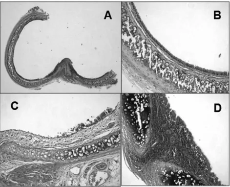
The trachea has a simple anatomic and physiologic structure compared to other solid organs. The trachea is a Figure 2 -Examples of tracheal histological examination (hematoxylin-eosin stain). A: High-integrity epithelium (25x). B: High-integrity epithelium and mild cellular infiltration (200x). C: Low-integrity epithelium and moderate cellular infiltration (200x). D: Severe cellular infiltration (200x).
Table 2 -Effects of topically applied preservation solutions on mucociliary clearance.
Control group (n = 08) LPD group (n = 86) HTK group (n = 81) Saline group (n = 42) p-value
MT (mm/min) 0.104 (0.106) 0.023 (0.039) 0.028 (0.045) 0.013 (0.018) 0.0001
CBF (Hz) 15.34¡3.37 12.16¡4.54 12.50¡4.14 11.90¡3.22 0.02
AB (%) 10.96¡4.51 5.94¡5.02 5.86¡4.71 6.11¡5.59 0.14
PAS (%) 16.55¡9.72 12.30¡6.55 12.03¡7.94 12.27¡7.75 0.56
semi-rigid tube, approximately 3 mm thick, whose lumen remains open even after extraction (6). Conversely, the tracheal arterial supply has a segmental pattern, without a major tracheal artery or vein. In the clinical environment, this anatomical arrangement makes the intravascular administration of preservation solutions into the trachea exceedingly difficult (2,7).
Based on the tracheal anatomy of both human and Wistar rats and on previous studies, we did not attempt to deliver preservation solutions through any intravascular route to fulfill the objectives of this study (17,18). Therefore, we tried to obviate the need for the intravascular administration of preservation solutions to tracheal grafts. The respiratory mucosa is highly permeable by water but not by larger molecules under normal conditions. However, the mucosal absorptive ability is greatly increased by some strong stimuli that disrupt the epithelial lining (19). LPD-glucose is a preservation solution specially developed for lung preservation. The advantages of LPD-glucose are the low potassium concentration and the presence of dextran-40, a polysaccharide implicated in the enhancement of micro-circulation in lung grafts (20). HTK is a low-potassium and low-sodium preservation solution used for liver, kidney, and pancreatic transplantation. The advantages of HTK are
its strong histidine buffer activity and low viscosity (21). We tested whether topically applied preservation solutions could penetrate the tracheal layers after cold ischemia and contribute to the preservation of the tracheal respiratory epithelium. Our data demonstrate that neither topically applied LPD-glucose nor HTK not affect mucociliary clearance after cold ischemia. In fact,in situmucociliary transportation and ciliary beating frequency were not significantly different in the LPD, HTK, and saline groups (Figure 3). Regarding the morphological preservation of the respiratory epithelium, a significant difference among the groups was still absent.
Transmission electron microscopy was the only analysis that showed a marked difference among the groups. In the LPD group, the nuclear shrinkage was most likely caused by osmosis. Because the respiratory epithelial cell has a water-permeable apical membrane, the free water could move from the cells toward the sodium-rich lumen provided by the composition of the LPD-glucose solution (19,20). Nuclear shrinkage was not observed in the HTK group, most likely because of the low-sodium and low-potassium composition of HTK (21).
or the cartilage, which are also important for tracheal function. Another limitation is the sample size of the control group, which was smaller than the other groups. Although this difference could lead to bias when interpreting the statistical analysis, this bias did not occur in this study. Despite the larger dispersion measures in some variables of the control group, significant differences among the groups could be clearly observed. As another limitation, morphological analysis through transmission electron microscopy was performed in only a small proportion of samples. Therefore, only a qualitative analysis, with not statistical analysis, could be performed, despite the large differences among the groups.
Our conclusions are the following: 1) cold ischemia diminished the mucociliary clearance of the tracheal respiratory epithelium, and 2) topically applied preserva-tion solupreserva-tions did not ameliorate the ischemic injury caused by cold ischemia to the tracheal respiratory epithelium. Further studies that address the function and morphology of connective and muscular tissues after ischemia could elucidate whether those effects occur in other tracheal cells.
& ACKNOWLEDGMENTS
This work was supported by Fundac¸a˜o de Amparo a` Pesquisa do Estado de Sa˜o Paulo (FAPESP).
& AUTHOR CONTRIBUTIONS
Azevedo-Pereira AE designed the study, collected the data, and wrote the manuscript. Saka JA collected the data. Oliveira-Braga KA and Pazetti R
designed the study and collected the data. Canzian M designed the study and collected the data. Peˆgo-Fernandes PM designed the study and revised the manuscript. Jatene FB revised the manuscript.
& REFERENCES
1. Grillo HC. Tracheal replacement: a critical review. Ann Thorac Surg. 2002;73(6):1995-2004, http://dx.doi.org/10.1016/S0003-4975(02)03564-6. 2. Pego-Fernandes PM, Azevedo-Pereira AE. Tracheal transplantation: is
there lumen at the end of the tunnel? Sao Paulo Med J. 2009;127(5):249-50. 3. Sung SW, Won T. Effects of basic fibroblast growth factor on early revascularization and epithelial regeneration in rabbit tracheal ortho-topic transplantation. Eur J Cardiothoracic Surg. 2001;19(1):14-8, http:// dx.doi.org/10.1016/S1010-7940(00)00624-2.
4. Genden EM, Iskander AJ, Bromberg JS, Mayer L. Orthotopic tracheal allografts undergo reepithelialization with recipient-derived epithelium. Arch Otolaryngol Head Neck Surg. 2003;129(1):118-23, http://dx.doi. org/10.1001/archotol.129.1.118.
5. Macchiarini P, Jungebluth P, Go T, Asnaghi MA, Rees LE, Cogan TA, et al. Clinical transplantation of a tissue-engineered airway. Lancet. 2008; 372(9662):2023-30, http://dx.doi.org/10.1016/S0140-6736(08)61598-6. 6. Grillo HC. Anatomy of the trachea. In: Grillo HC. Surgery of the trachea
and bronchi. London: BC Decker, 2004:51-8.
7. Macchiarini P, Mazmanian GM, de Montpre´ville VT, Dulmet EM, Chapelier AR, Dartevelle PG. Maximal preservation time of tracheal allografts. Ann Thorac Surg. 1995;60(6):1597-604, http://dx.doi.org/10. 1016/0003-4975(95)00811-X.
8. Azevedo-Pereira AE, Saka JA, Oliveira KA, Pazetti R, Peˆgo-Fernandes PM, Jatene FB. Impact of topically-applied LPD-glucose on tracheal mucociliary clearance after warm and cold ischemia: short communica-tion. Clinics. 2011;66(2):347-9, http://dx.doi.org/10.1590/S1807-5932201 1000200027.
9. Randell SH, Boucher RC. Effective mucus clearance is essential for respiratory health. Am J Respir Cell Mol Biol. 2006;35(1):20-8. 10. Rivero DHRF, Lorenzi-Filho G, Pazetti R, Jatene BF, Saldiva PHN. Effects
of bronchial transection and reanastomosis on mucociliary system. Chest. 2001;119(5):1510-5, http://dx.doi.org/10.1378/chest.119.5.1510.
11. Pazetti R, Peˆgo-Fernandes PM, Lorenzi-Filho G, Saldiva PHN, Moreira LFP, Jatene FB. Effects of Cyclosporine A and bronchial transection on mucociliary transport in rats. Ann Thorac Surg. 2008;85(6):1925-9, http://dx.doi.org/10.1016/j.athoracsur.2008.02.084.
12. Qu N, de Vos P, Schelfhorst M, de Haan A, Timens W, Prop J. Integrity of airway epithelium is essential against obliterative airway disease in transplanted rat tracheas. J Heart Lung Transplant. 2005;24(7):882-90, http://dx.doi.org/10.1016/j.healun.2004.04.020.
13. Knowles MR, Boucher RC. Mucus clearance as a primary innate defense mechanism for mammalian airways. J Clin Invest. 2002;109(5):571-7. 14. Wagner EM, Foster WM. Importance of airway blood flow on particle
clearance from the lung. J Appl Physiol. 1996;81(5):1878-83.
15. Rogers DF. Airway goblet cells: responsive and adaptable front-line defenders. Eur Respir J. 1998;7(9):1690-706.
16. de Perrot M, Liu M, Waddell TK, Keshavjee S. Ischemia-reperfusion-induced lung injury. Am J Respir Crit Care Med. 2003;167(4):490-511, http://dx.doi.org/10.1164/rccm.200207-670SO.
17. Ferreira PG, Silva AC, A´ guas AP, Pereira AS, Grande NR. Detailed arrangement of the bronchial arteries in the Wistar rat: a study using vascular injections and scanning electron microscopy. Eur J Anat. 2001;5(2):67-76. 18. Macchiarini P, Mazmanian GM, Montpre´ville V, Dulmet E, Fattal M,
Lenot B, et al. Experimental tracheal and tracheoesophageal allotrans-plantation. Paris-sud university lung transplantation group. J Thorac Cardiovasc Surg. 1995;110(4 Pt 1):1037-46.
19. Persson CGA. Airway mucosal exudation of plasma. In: Takishima T, Shimura S (eds). Airway secretion. Physiological bases for the control of mucous hypersecretion. New York: Marcel Dekker, 1994:451-65. 20. Keshavjee SH, Yamazaki F, Yokomise H, Cardoso PF, Mullen JBM,
Slutsky AS, et al. The role of dextran 40 and potassium in extended hypothermic lung preservation for transplantation. J Thorac Cardiovasc Surg. 1992;103(2):314-25.