Rogério Lacerda dos Santos1, Matheus Melo Pithon2, Fernanda Otaviano Martins3, Maria Teresa Villela Romanos4
Cytotoxicity of separation orthodontic elastics.
original article
Objective: To test the hypothesis that there is no difference in cytotoxicity between separating elastics of different manufacturers.
Methods: The present article compared latex elastics (4.0 mm, 4.4 mm and 4.8 mm) of four different manufactur-ers. The sample was allocated to seven groups of 9 elastics: Group A (American Orthodontics, green color, mod-ules), Groups M1 and M2 (Morelli, blue color, modules and free in pack respectively), Groups M3 and M4 (Morelli, green color, modules and free in pack respectively), Group U (Uniden, blue color, free in pack) and Group T (Tec-nident, blue color, free in pack) regarding their possible cytotoxic effects on oral tissues. Cytotoxicity assays were performed using cell culture medium containing epithelioid-type cells (Hep-2 line) derived from human laryngeal carcinoma and submitted to the methods for evaluating the cytotoxicity by the “dye-uptake” test, at time intervals 24, 48, 72 and 168 h. Data were compared by analysis of variance (ANOVA) and Tukey’s test (p < 0.05).
Results: Results showed statistically significant difference (p < 0.05) between group U and all the other Groups (A, M1, M2, M3, M 4 and T) at 24 and 48 hours.
Conclusions: Uniden elastics evoked more cell lysis at 24 and 48 h, although, all brands showed biocompatibility from 72 h onwards.
Keywords: Cytotoxicity. Elastics. Biocompatibility. Orthodontics.
How to cite this article: Santos RL, Pithon MM, Martins FO, Romanos MTV. Cy-totoxicity of separation orthodontic elastics.. Dental Press J Orthod. 2012 July-Aug;17(4):110-4.
Submitted: February 02, 2009 - Revised and accepted: August 16, 2009 » The authors report no commercial, proprietary or financial interest in the products or companies described in this article.
Contact address: Rogério Lacerda dos Santos Universidade Federal de Campina Grande – UFCG
Centro de Saúde e Tecnologia Rural (CSTR) – Av. dos Universitários, S/N, Rodovia Patos–Teixeira, Km 1 – Santa Cecília – Patos/PB – Brazil CEP: 58.700-970 – E-mail: [email protected] 1 Specialist in Orthodontics, Federal University of Alfenas. MSc and PhD in
Orthodontics, Federal University of Rio de Janeiro. Adjunct Professor of Orthodontics, Federal University of Campina Grande.
2 Specialist in Orthodontics, Federal University of Alfenas. MSc and PhD in Orthodontics, Federal University of Rio de Janeiro. Assistant Professor of Orthodontics, State University of Southwest of Bahia.
3 Graduated in Biological Sciences, Federal University of Rio de Janeiro. 4 MSc and PhD in Microbiology, Federal University of Rio de Janeiro. Adjunct
Rogério Lacerda dos Santos1, Matheus Melo Pithon2, Fernanda Otaviano Martins3, Maria Teresa Villela Romanos4
Citotoxicidade de elásticos ortodônticos de separação
Objetivo: o propósito do presente estudo foi testar a hipótese que não existe diferença de citotoxicidade entre
elásticos de diferentes marcas.
Métodos: foram comparadas entre si 4 marcas de elásticos de separação (4,0mm, 4,4mm e 4,8mm) intrabucais
de látex quanto ao possível efeito citotóxico nos tecidos bucais, divididos em 7 grupos de 9 elásticos cada: grupo A (cor verde – modular, American Orthodontics), grupos M1 e M2, (cor azul – modular e a granel, respectivamente, Morelli), grupos M3 e M4 (cor verde – modular e a granel, respectivamente, Morelli), grupo U (cor azul – a gra-nel, Uniden) e grupo T (cor azul – a gragra-nel, Tecnident). O ensaio de citotoxicidade foi realizado utilizando-se cul-tura de células da linhagem HEp-2 (do tipo epitelióide, que tem origem em carcinoma de laringe humana), sendo submetido o material ao teste para células viáveis em vermelho neutro (“dye-uptake”), no tempo de 24, 48, 72 e 168 horas. A análise de variância e comparação múltipla (ANOVA) e o teste de Tukey foram utilizados (p<0,05).
Resultados: os resultados evidenciaram diferença estatisticamente significativa dos grupos A, M1, M2, M3, M4 e T
com o grupo U nos tempos de 24 e 48h (p<0,05).
Conclusão: pôde-se evidenciar que os elásticos da marca Uniden causaram alta quantidade de lise celular em 24 e
48h, porém, todas as marcas mostraram-se biocompatíveis a partir de 72h.
Palavras-chave: Teste de materiais. Látex. Ortodontia. Toxicidade.
Como citar este artigo: Santos RL, Pithon MM, Martins FO, Romanos MTV. Cy-totoxicity of separation orthodontic elastics. Dental Press J Orthod. 2012 July--Aug;17(4):110-4.
Enviado em: 02 de fevereiro de 2009 - Revisado e aceito: 16 de agosto de 2009 » Os pacientes que aparecem no presente artigo autorizaram previamente a publica-ção de suas fotografias faciais e intrabucais.
» Os autores declaram não ter interesses associativos, comerciais, de propriedade ou financeiros, que representem conflito de interesse nos produtos e companhias des-critos nesse artigo.
Endereço para correspondência: Rogério Lacerda dos Santos Universidade Federal de Campina Grande – UFCG
Centro de Saúde e Tecnologia Rural (CSTR) – Av. dos Universitários, S/N, Rodovia Patos–Teixeira, Km 1 – Santa Cecília – Patos/PB – CEP: 58.700-970 E-mail: [email protected]
1 Especialista em Ortodontia, Universidade Federal de Alfenas. Mestre e Doutor
em Ortodontia, Universidade Federal do Rio de Janeiro. Professor Adjunto de Ortodontia, Universidade Federal de Campina Grande.
2 Especialista em Ortodontia, Universidade Federal de Alfenas. Mestre e Doutor
em Ortodontia, Universidade Federal do Rio de Janeiro. Professor Assistente de Ortodontia, Universidade Estadual do Sudoeste da Bahia.
3 Graduada em Ciências Biológicas, Universidade Federal do Rio de Janeiro. 4 Doutora em Microbiologia e Professora Adjunta de Microbiologia, Universidade
Santos RL, Pithon MM, Martins FO, Romanos MTV
INTRODUCTION
Orthodontic elastics are widely used in orthodon-tic pracorthodon-tice with the purpose of helping orthodonorthodon-tic treatment, and therefore need to be inert to oral tis-sues. Elastics in contact with the oral mucosa for sev-eral hours a day is a situation that may continue for months. Therefore, the question arises about the pos-sibility of toxic substances being released by elasto-mers, which may be capable of harming the cells.
Latex is constituted of chains of cis-1, 4-poly-iso-prene. After obtaining the liquid from latex, it is pre-served by the addition of conservants (usually ammo-nia). When it is manufactured, various substances are added in order to achieve the final properties.22
There are variations in the composition of latex elastics, and consequently, there are differences in their properties. This may be one of the reasons why companies produce various sizes to compensate for the variations in physical properties.1 Depending on
how latex is stored, alterations may occur in its com-position, as its major limitation is sensitivity to ozone or other systems generating free radicals, such as sun-light which weakens the latex polymer chain.22
Pre-vulcanized latex production involves the mixture of latex from the purest and highest molecular weight natural rubber with stabilizers, such as zinc oxide and vulcanized chemical products. The mixture is heated to a temperature of 70°C.14 Zinc is known to be
neu-rotoxic.8 Although the zinc released from orthodontic
elastics may be swallowed, the results suggest that the use of latex elastics in orthodontics is appropriate.5
Natural latex is not in the category of materials generally considered safe.6,16 Allergy caused by
pro-teins from latex has been well documented,13 and
may present immediate hypersensitivity reactions.21
Among the reactions caused by orthodontic elastics, there have been reports of the development of stoma-titis with swelling, erythematous oral lesions, in addi-tion to respiratory, and systemic reacaddi-tions, and in ex-treme cases, anaphylactic shock.3,18 The prevalence of
latex allergy is between 3% and 17%.20
Cell culture tests for the evaluation of dental mate-rial toxicity are a valid method to enable understand-ing of their biologic behavior.16 The aim of the present
study was to test the hypothesis that there is no dif-ference in cytotoxicity between separating elastics of different brands.
MATERIAL AND METHODS
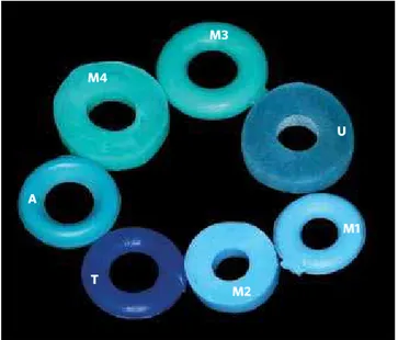
Samples of intraoral latex separating elastics (4.0 mm, 4.4 mm and 4.8 mm) (Fig 1) of 4 different brands and colors were selected (Table 1), and divided into 7 groups containing 9 elastics each: Group A (Green color- modular, American Orthodontics, Sheboygan, Wisconsin, USA), Group M1 (Blue color- modular, Morelli, Sorocaba, Brazil), Group M2 (Blue color- in bulk, Morelli, Sorocaba, Brazil), Group M3 (Green color- modular, Morelli, Sorocaba, Brazil), Group M4 (Green color- in bulk, Morelli, Sorocaba, Bra-zil), Group U (Blue color- in bulk, Uniden, Sorocaba, Brazil) and Group T (Blue color- in bulk, Tecnident, São Carlos, Brazil) with regard to the possible cyto-toxic effect on oral tissues.The elastics used in this research were from the same production lot for each tested color. Copper amalgam was used as positive control (Vigodent, Rio de Janeiro, Brazil), standard-ized by size and weight, and as negative control, stainless steel wire (American Orthodontics, She-boygan, Wisconsin, USA) (Table 1).
To conduct this study, HEp-2 (human carcinoma of the larynx) cell culture was used, maintained in Eagle’s Minimum Essential Medium (MEM-Eagle) (Cultilab, Campinas, Brazil) with the addition of 0.03 mg/ml glu-tamine (Sigma, St. Louis, Missouri, USA), 50 µg/ml
ga-ramicine (Schering Plough, Kenilworth, New Jersey, USA), 2.5 mg/ml fungizone (Bristol-Myers-Squibb,
Figure 1 - Separating elastics evaluated in this study: A (green modular, American Orthodontics), M1 (blue, modular, Morelli), M2 (blue, in bulk, Morelli), M3 (green, modular, Morelli), M4 (green, in bulk, Morelli), U (blue, in bulk, Uniden) and T (blue, in bulk, Tecnident).
M4
A
T
M2
New York, USA), 0.25% sodium bicarbonate solution (Merck, Darmstadt, Germany), 10 mM HEPES (Sigma, St. Louis, Missouri) and 10% fetal bovine serum (Cul-tilab, Campinas, Brazil) (growth medium) or without fetal bovine serum (maintenance medium) and incu-bated at 37ºC for 48 hours.
The elastics were previously sterilized by ultra-violet radiation (Labconco, Kansas, Missouri) for 30 minutes on each surface of the elastic. To determine the cytotoxicity of the orthodontic elastics, the tech-nique denominated “dye-uptake” was used,11 which is
based on the incorporation of neutral red dye by live cells. The time intervals of 24 h, 48 h, 72 h and 168 h were used, as these elastics are usually maintained in the oral cavity for up to 168 h (7 days) to separate the teeth. This time represents maintenance of the elastic in the cell culture medium for 24 h, 48 h, 72 h, 168 h, and later removal.
Dye-uptake
Volumes of 100 µl of HEp-2 cell suspension were distributed into 96-well microplates. After 48 hours, the growth medium was replaced by 100 µl of culture
medium (MEM-Eagle) obtained after incubation with different elastics for time intervals of 24 h, 48 h, 72 h and 168 h. As positive and negative controls the culture media obtained after contact with amalgam and stainless steel wire respectively were used. The experiment was conducted in quadruplicate.
After 24 hours of incubation, 100l of 0.01% neu-tral red (Sigma, St. Louis, Missouri, USA), were add-ed to culture madd-edium in each well of the miniplate, and these were incubated at 37o C for 3 hours for the
dye to penetrate into the live cells. After the elapse of this time interval, and after disregarding the dye, 100µl of 4% formaldehyde solution (Vetec, Rio de
Ja-neiro, Brazil) was added to PBS (NaCl 130 mM; KCl 2 mM; Na2HPO4 2H2O 6 mM; K2HPO4 1mM, pH7.2) for 5 minutes, to promote cell fixation to the plates. Next, in order to extract the dye, 100µl of 1% acetic
acid solution (Vetec, Rio de Janeiro, Brazil) with 50% methanol was added (Vetec, Rio de Janeiro, Brazil). After 20 minutes the readout was taken in a spectro-photometer (BioTek, Winooski, Vermont, USA) at a wavelength of 492nm (λ = 492 nm).
Data were compared by analysis of variance (ANOVA) and afterwards by Tukey’s test for evalu-ation among groups, with reliability at a level of sig-nificance of 0.05.
RESULTS
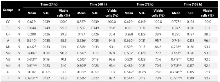
The results showed statistically significant differ-ence among Groups A (Green color- modular, Ameri-can Orthodontics), M1 (Blue color- modular, Mo-relli), M2 (Blue color- in bulk, MoMo-relli), M3 (Green color- modular, Morelli), M4 (Green color- in bulk, Morelli) and T (Blue color- in bulk, Tecnident) with Group U (Blue color- in bulk, Uniden) at the time intervals of 24 h and 48 h (p<0.05) (Table 2). There was no statistically significant difference between Groups M2, M3, U and T at 72 h (p>0.05) (Table 2). At 72 h, the Morelli and Tecnident brands of elastics caused a larger quantity of cell lysis in comparison with the time intervals of 24, 48 and 168 h. This may mean greater release of toxic substances by these elastics in 3 days.
Discussion
In this study, the option was taken to use copper amalgam as positive control and stainless steel wire as negative con-trol (Table 1) as they have been proved to be adequate for this test.9,12 The cytotoxic
potential of dental amalgam comes from the presence of mercury, however, the amalgam contains other substances that
Groups Brand Color Diameter (mm) Reference
A A. Orthodontics Green 4.44 854-251
M1 Morelli Blue 4.00 60.04.201
M2 Morelli Blue 4.00 60.04.200
M3 Morelli Green 4.80 60.04.401
M4 Morelli Green 4.80 60.04.400
U Uniden Blue 4.80 000-1320
T Tecnident Blue 4.40 A-007
Positive Control Copper Amalgam. Pratic NG 2. Vigodent
Negative Control Stainless steel wire American Orthodontics. 0.019 x 0.025-in
Santos RL, Pithon MM, Martins FO, Romanos MTV
may also be neurotoxic, depending on its composi-tion and manufacturer.8
Sterilization is a pre-requisite for the cytotoxic-ity test. Autoclave sterilization may be used, however, elastics have been shown to darken and harden after this type of sterilization due to the heat liberated,23
which may cause degradation and the release of sub-stances that are toxic to cells. In this study, steriliza-tion by ultraviolet radiasteriliza-tion was used15 for 30 minutes
on each side of the elastic. In this study the elastics were shown to have the same aspects of color and mal-leability after UV light sterilization.
With the increasing use of rubber latex as dental material, many cytotoxic factors have been reported.7
Sulphur and zinc oxide, as conservants, exhibit cy-totoxicity, and dithiocarborates, N-nitrosodibutyl-amine, and N-nitrosopiperidine, which act as anti-oxidants, are also known to be cytotoxic substances.4
Holmes and cols6 verified whether coloring agents
used in colored latex manufacture may have any toxic effect. Their results showed that they have low toxic-ity. Clinically therefore, this effect is harmless.
Although case reports on allergy to latex do not ap-pear frequently, allergic reactions have become some-what more prevalent with the increase in latex-based products. The majority of allergic reactions17 have
been related to the use of latex gloves, but only 2 cases
were related to the use of orthodontic elastics.10 In the
cases related to orthodontic elastics, the presence of small vesicles or acute edema occurred, and the pa-tients complained of burning and itching.
Allergy to natural latex occurs because it contains many types of proteins, and the powder present in the coating of orthodontic elastics function as a vehicle for these proteins. Therefore, from a clinical point of view, the development of elastics without latex is in-creasingly important.
In this study, the talc was removed before the in vitro studies were conducted, and it is not known whether the talcum powder would have made any difference.
According to Schmalz,16 the great danger with the
use of intraoral elastics with cytotoxic potential would be the fact that the substances released by these would be ingested by the patient, and over the course of time, cause diseases resulting from the cumulative effect of toxic substances. It is known that latex is not a com-pletely biocompatible substance. It may cause aller-gic reactions20,22 and generate cross-reactions with
foods2,20 and medications.19
As they are widely used materials in the orthodon-tic clinic, one must be concerned about the cytotox-icity of elastics, particularly the intraoral type that comes into intimate contact with the mucosa, and opt
Groups n
Time (24 h) Time (48 h) Time (72 h) Time (168 h)
Mean S.D. Viable
cells (%) Mean S.D.
Viable
cells (%) Mean S.D.
Viable
cells (%) Mean S.D.
Viable cells (%)
CC 9 0.672a 0.139 100.0 0.557a 0.139 100.0 0.690a 0.149 100.0 0.778ª 0.124 100.0
C- 9 0.644 0.149 95.9 0.539 0.149 96.8 0.681 0.131 98.8 0.747 0.120 96.1
C+ 9 0.200 0.126 29.8 0.197 0.126 35.4 0.268 0.129 38.9 0.295 0.127 38.0
A 9 0.640b 0.135 95.3 0.526b 0.135 94.5 0.660b 0.131 95.7 0.749b 0.131 96.4
M1 9 0.617cb 0.133 91.9 0.518b 0.133 93.1 0.598c 0.113 86.8 0.728cb 0.130 93.7
M2 9 0.606dc 0.116 90.2 0.517b 0.116 92.9 0.532d 0.126 77.2 0.729dbc 0.120 93.8
M3 9 0.612ecd 0.119 91.1 0.515b 0.119 92.6 0.521d 0.128 75.6 0.719ecd 0.112 92.5
M4 9 0.611fcde 0.123 91.0 0.509b 0.123 91.4 0.489e 0.121 70.9 0.718fcde 0.117 92.4
U 9 0.114g 0.096 17.1 0.068c 0.096 12.3 0.542d 0.089 78.6 0.724gcdef 0.115 93.1
T 9 0.620bcdef 0.122 92.3 0.516b 0.122 92.7 0.544d 0.112 78.9 0.721cdefg 0.119 92.7
Table 2 - Dye-uptake technique. Statistical description for optical density of elastic evaluated.
1. Bishara SE, Andreasen GF. A comparison of time related forces between plastic alastiks and latex elastics. Angle Orthod. 1970 Oct;40(4):319-28.
2. Carey AB, Cornish K, Schrank P, Ward B, Simon R. Cross-reactivity of alternate plant sources of latex in subjects with systemic IgE-mediated sensitivity to Hevea brasiliensis latex. Ann Allergy Asthma Immunol. 1995 Apr;74(4):317-20. 3. Everett FG, Hice TL. Contact stomatitis resulting from the use of orthodontic
rubber elastics: report of case. J Am Dent Assoc. 1974 May;88(5):1030-1.
4. Fiddler W, Pensabene J, Sphon J, Andrzejewski D. Nitrosamines in rubber bands
used for orthodontic purposes. Food Chem Toxicol. 1992 Apr;30(4):325-6. 5. Hanson M, Lobner D. In vitro neuronal cytotoxicity of latex and nonlatex
orthodontic elastics. Am J Orthod Dentofacial Orthop. 2004 Jul;126(1):65-70. 6. Holmes J, Barker MK, Walley EK, Tuncay OC. Cytotoxicity of orthodontic elastics.
Am J Orthod Dentofacial Orthop. 1993 Aug;104(2):188-91.
7. Hwang CJ, Cha JY. Mechanical and biological comparison of latex and silicone
rubber bands. Am J Orthod Dentofacial Orthop. 2003 Oct;124(4):379-86. 8. Lobner D, Asrari M. Neurotoxicity of dental amalgam is mediated by zinc. J Dent
Res. 2003 Mar;82(3):243-6.
9. Moreira TC, Quintão CAC, Menezes LM, Wigg MD, Chevitarese O. Elásticos
plásticos: avaliação da citotoxicidade após esterilização. Rev SBO. 1998;3(2):172-7. 10. Neiburger EJ. A case of possible latex allergy. J Clin Orthod. 1991 Sep;25(9):559-60. 11. Neyndorff HC, Bartel DL, Tufaro F, Levy JG. Development of a model to
demonstrate photosensitizer-mediated viral inactivation in blood. Transfusion. 1990 Jul-Aug;30(6):485-90.
12. Pacheco MC, Wigg MD, Chevitarese O. Biocompatibilidade das soldagens
ortodônticas. Rev SBO. 1995 Jun;2(3):233-8.
REFERENCES
13. Palosuo T, Alenius H, Turjanmaa K. Quantitation of latex allergens. Methods. 2002 May;27(1):52-8. Review.
14. Perrella FW, Gaspari AA. Natural rubber latex protein reduction with an emphasis on enzyme treatment. Methods. 2002 May;27(1):77-86.
15. dos Santos RL, Pithon MM, Mendes GS, Romanos MTV, Ruellas ACO. Cytotoxicity
of intraoral orthodontic elastics. J Appl Oral Sci. 2009 Jul-Aug;17(4):326-9. 16. Schmalz G. Use of cell cultures for toxicity testing of dental materials—advantages
and limitations. J Dent. 1994;22 Suppl 2:S6-11.
17. Snyder HA, Settle S. The rise in latex allergy: implications for the dentist. J Am Dent Assoc. 1994 Aug;125(8):1089-97.
18. Tomazic VJ, Withrow TJ, Fisher BR, Dillard SF. Latex-associated allergies and anaphylactic reactions. Clin Immunol Immunopathol. 1992 Aug;64(2):89-97. Review.
19. Towse A, O’Brien M, Twarog FJ, Braimon J, Moses AC. Local reaction secondary
to insulin injection. A potential role for latex antigens in insulin vials and syringes. Diabetes Care. 1995 Aug;18(8):1195-7.
20. Turjanmaa K, Alenius H, Mäkinen-Kiljunen S, Reunala T, Palosuo T. Natural rubber latex allergy. Allergy. 1996 Sep;51(9):593-602.
21. Wakelin SH, White IR. Natural rubber latex allergy. Clin Exp Dermatol. 1999 Jul;24(4):245-8.
22. Weiss ME, Hirshman CA. Latex allergy. Can J Anaesth. 1992 Jul;39(6):528-32. 23. Wigg MD, Menezes LM, Quintão CCA, Moreira TC, Chevitarese O. Elásticos
extra-orais: avaliação da citotoxicidade. Ortod Gaúcha. 1997 Jul;1(2):151-7.
for materials that have been proved to be biocompat-ible from this aspect. Previous studies on the toxicity of latex orthodontic elastics used for separating the teeth, both clear and neon, have been shown to be cy-totoxic to gingival fibroblasts.6
The cytotoxic nature was evidenced after the elastics were exposed to the culture medium. The Uniden brand of separating elastics caused the greatest quantity of cell death in comparison with the other brands evaluated in the time intervals of 24 and 48 h, which suggests the release of toxic in-gredients in the first 48 h for this elastic, however, as from the 3rd day, all the elastics demonstrated a
less cytotoxic nature.
The percentage of viable cells was obtained by means of comparison of the mean optic density (OD) of the control cells (without coming into contact with the elastics) with the means of OD obtained from the
supernatant of the cell cultures that were placed in contact with the elastics, and toxicity was calculated for 50% of the cell cultures (CC50) (Table 2).
Variations occur in the composition of the latex elastics and this may explain the difference in the results obtained among the brands. Although the in vitro evaluation does not simulate the oral medium, it is necessary to consider that the elastics are not clinically inert.
CONCLUSION
It could be concluded that:
1) The separating elastics of American Orthodon-tics, Morelli and Tecnident brands caused a small quantity of cell lysis.