Angle Class I malocclusion, with anterior
open bite, treated with extraction of
permanent teeth*
Mírian Aiko Nakane Matsumoto**
Open bite is an anomaly with distinct characteristics which, in addition to involving complex, multiple etiologic factors, entails aesthetic and functional consequences. Many alternative approaches have been employed to treat open bite, including palatal crib, orthopedic forces, occlusal adjustment, camouflage with or without extractions, mini-implants or mini-plates, and orthognathic surgery. By determining accurate diagnosis and etiology professionals can set the goals and ideal treatment plan for this malocclu-sion. This report, describing the two stages treatment of Angle Class I malocclusion with Class II skeletal pattern and anterior open bite, was presented to the Brazilian Board of Orthodontics and Dentofacial Orthopedics (BBO), representative of category 2, as partial fulfillment of the requirements for obtaining the title of BBO diplomate.
Abstract
Keywords: Angle Class I malocclusion. Open bite. Corrective orthodontics. Tooth extraction.
** Associate Professor, Department of Child Dentistry, Preventive and Social Dentistry, Ribeirão Preto School of Dentistry, São Paulo Univer-sity. PhD in Orthodontics, School of Dentistry, Rio de Janeiro Federal UniverUniver-sity. Diplomate of the Brazilian Board of Orthodontics. introduction
Patient was a 12-year-old Caucasian girl re-ferred by a speech therapist for orthodontic treatment and presented with a chief complaint of “lack of contact between the anterior teeth and altered position of canines.” During the in-terview, she reported having had a pacifier suck-ing habit until 5 years of age and havsuck-ing under-gone adenotonsillectomy at age four. She was in good general health, with no history of serious illnesses or trauma.
diAGnoSiS
Clinical examination revealed an increased low-er face, lip incompetence, a slightly convex profile, obtuse nasolabial angle and good cervical-mandib-ular line (Fig 1).
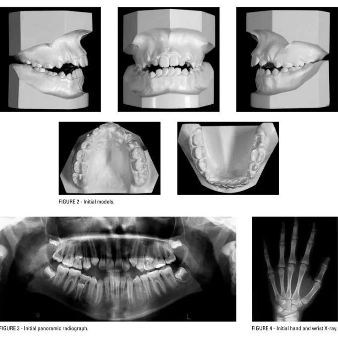
Intraoral evaluation disclosed low risk for caries, healthy gums, molars and canines in normal occlu-sion, open bite that extended to the premolar region, maxillary atresia, 6 mm overjet, 2 mm lower midline deviation to the right side, and upper midline coin-ciding with the mid-palatine raphe (Figs 1 and 2).
From a functional standpoint, she exhibited mixed breathing, predominantly oral, tongue thrusting and adapted swallowing and speech.
Panoramic radiography showed all permanent teeth, third molar crowns in formation, maxillary primary second molars in exfoliation phase and lower right second deciduous molar with ankylosis (Fig 3). It was also noted that the patient was in the stage of maximum pubertal growth spurt (Fig 4).
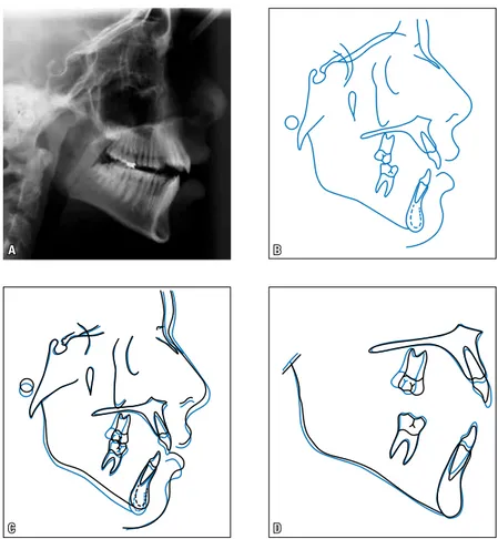
Cephalometric analysis was performed and vealed that both the maxilla and mandible were re-truded in relation to the skull base. She had a Class II skeletal pattern (ANB = 6º), predominance of vertical growth (SN.GoGn = 43º), protruding up-per and lower incisors (1-NA = 5.5 mm and 1-NB = 6.0 mm), with decreased axial inclination (1.NA = 19.5º and 1.NB = 22.5º) (Fig 5 and Table 1).
trEAtMEnt GoALS (phASE 1)
Initially, the goal was to eliminate the orofa-cial myofunctional disorder (adapted swallowing and speech), redirect facial growth by stimulat-ing mandibular rotation in the counterclockwise direction to counter a growth tendency noted in the lower face and correct the anterior open bite.
trEAtMEnt pLAn (phASE 1)
Planning for the first phase consisted in redi-recting facial growth, corredi-recting the Class II skel-etal pattern and growth tendency in the lower face, using a modified Thurow appliance. This appliance was intended to prevent further verti-cal alveolar growth and stimulate mandibular ro-tation in the counterclockwise direction. The in-stallation of a palatal crib was also planned, with
A B FIGURE 2 - Initial models.
FIGURE 3 - Initial panoramic radiograph.
FIGURE 5 - Initial lateral cephalogram (A) and cephalometric tracing (B).
FIGURE 6 - Intermediate facial and intraoral photographs.
referral of the patient to a speech therapist to intercept the tongue thrusting habit. Addition-ally, her lower right second deciduous molar was extracted as it showed an abnormal root resorp-tion pattern and ankylosis.
trEAtMEnt proGrESS
A modified Thurow appliance was installed, combined with a palatal crib from premolar to premolar to control maxillary vertical growth and prevent the tongue from being placed in the anterior region. In addition, speech thera-py was started to treat the orofacial myofunc-tional disorder. The patient was followed up on a monthly basis during the treatment period, which lasted for twelve months.
The use of a modified Thurow appliance was dis-continued when all permanent teeth had erupted.
The Class III molar relationship was evident due to a distalization component in the Thurow pliance, and a slight posterior crossbite which ap-peared over time.
rESuLtS (phASE 1)
A B FIGURE 7 - Intermediate models.
A B
C D
FIGURE 10 - Lateral cephalogram (A) and cephalometric tracing (B). Total (C) and partial (D) super-impositions of initial (black) and intermediate (blue) cephalometric tracings.
trEAtMEnt GoALS (phASE 2)
In the second phase, the goal was to correct the mild crossbite that resulted from wearing the modified Thurow appliance, control the clockwise rotational tendency in the mandible and balance the lower face, eliminating the orofacial myofunc-tional disorder (adapted swallowing and speech) to establish correct overbite and overjet, normal molar and canine occlusion, correct the Class II skeletal pattern, as well as align and level all the teeth, thereby correcting the lower midline.
trEAtMEnt pLAn (phASE 2)
At this treatment stage, correction of the slight crossbite was planned by installing a Haas expand-er. Mandibular growth control was achieved with
an anterior vertical-pull chin cup, while speech therapy was maintained to correct the orofacial myofunctional disorder. Concurrently, fixed orth-odontic appliances were set up on both arches to perform corrective treatment with extraction of the first upper and lower premolars.
trEAtMEnt proGrESS
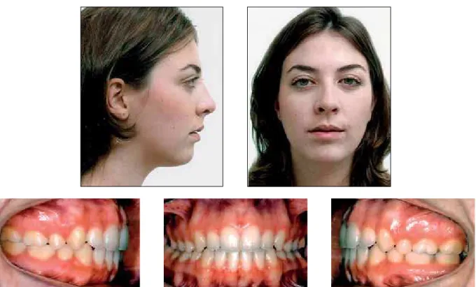
FIGURE 11 - Final facial and intraoral photographs.
the first upper and lower molars and brackets bond-ed to the other teeth, except for the first upper and lower premolars, which were extracted.
Alignment and leveling were performed as well as torque correction using nickel-titanium 0.012-in and 0.014-0.012-in wires, and sta0.012-inless steel 0.016-0.012-in to 0.020-in wires. From the moment that round 0.020-in wires began to be utilized, elastic chains were inserted for closure of extraction spaces, with anchorage loss. Next, stainless steel 0.019x 0.025-in archwires were fabricated for 0.025-incisor and can0.025-ine retraction, with posterior anchorage loss. At this stage, intermaxillary elastics were used to improve intercuspation and finishing. After completion and verification that the main treatment goals had been achieved, the fixed orthodontic appliance was re-moved. A removable maxillary retainer was installed
with a wraparound-type archwire, in addition to a fixed lingual canine-to-canine retainer made with 0.032-in stainless steel wire. The patient was in-structed to wear the upper retainer 24/7 during the first year, and at night during the second year. The mandibular bonded retainer was kept indefinitely.
trEAtMEnt rESuLtS
A B
A B
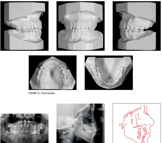
FIGURE 12 - Final models.
FIGURE 14 - Final lateral cephalogram (A) and cephalometric tracing (B).
A B
A B
FIGURE 19 - Control lateral cephalogram (A) and cephalometric tracing (B), two years and three months after treatment completion.
FIGURE 18 - Control panoramic radiograph two years and three months after treat-ment completion.
FIGURE 20 - Total (A) and partial (B) superimpositions of cephalometric tracings: initial (black), final (red) and two years and three months after treatment completion (green).
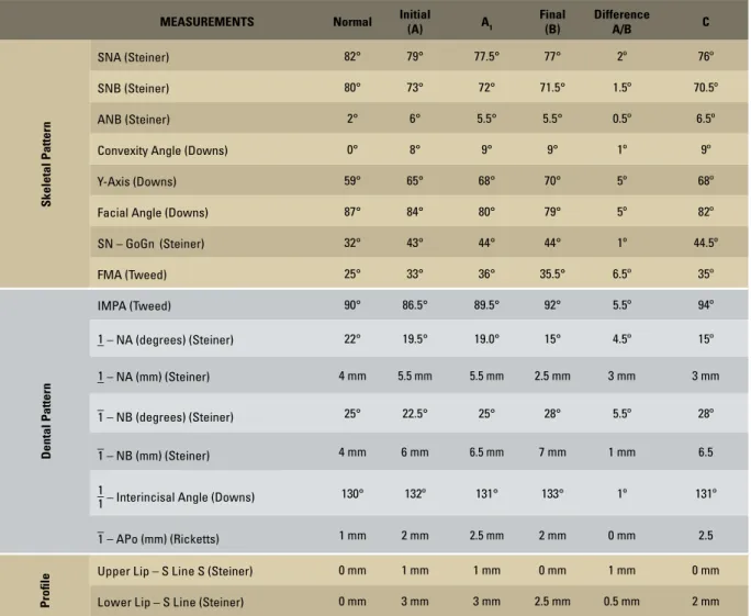
form and no undesirable effects to the periodon-tium. Cephalometric measurements did not ex-perience major changes as ANB remained at 5.5º, SN.GoGn remained at 44º and Y-axis increased from 68º to 70º (Fig 14 and Table 1). Two years and three months after the end of treatment,
MEASUREMENTS Normal Initial
(A) A1
Final (B)
Difference
A/B C
Skeletal Pattern
SNA (Steiner) 82° 79° 77.5° 77° 2º 76º
SNB (Steiner) 80° 73° 72° 71.5° 1.5º 70.5º
ANB (Steiner) 2° 6° 5.5° 5.5° 0.5º 6.5º
Convexity Angle (Downs) 0° 8° 9° 9° 1º 9º
Y-Axis (Downs) 59° 65° 68° 70° 5º 68º
Facial Angle (Downs) 87° 84° 80° 79° 5º 82º
SN – GoGn (Steiner) 32° 43° 44° 44° 1º 44.5º
FMA (Tweed) 25° 33° 36° 35.5° 6.5º 35º
Dental Pattern
IMPA (Tweed) 90° 86.5° 89.5° 92° 5.5º 94º
–
1 – NA (degrees) (Steiner) 22° 19.5° 19.0° 15° 4.5º 15º
–
1 – NA (mm) (Steiner) 4 mm 5.5 mm 5.5 mm 2.5 mm 3 mm 3 mm
–
1 – NB (degrees) (Steiner) 25° 22.5° 25° 28° 5.5º 28º
–
1 – NB (mm) (Steiner) 4 mm 6 mm 6.5 mm 7 mm 1 mm 6.5
–1
1 – Interincisal Angle (Downs) 130° 132º 131° 133° 1º 131º
–
1 – APo (mm) (Ricketts) 1 mm 2 mm 2.5 mm 2 mm 0 mm 2.5
Proile
Upper Lip – S Line S (Steiner) 0 mm 1 mm 1 mm 0 mm 1 mm 0 mm
Lower Lip – S Line (Steiner) 0 mm 3 mm 3 mm 2.5 mm 0.5 mm 2 mm TABLE 1 - Summary of cephalometric measurements.
FinAL conSidErAtionS
Open bite is an anomaly with distinct, easily recognizable features that can be found in 25% to 38% of orthodontic patients.1-4 Several etiologi-cal factors are involved in this type of malocclu-sion, such as: Facial growth pattern, sucking hab-its, tongue posture, mouth breathing, enlarged adenoids, syndromes, occlusal and eruption forces, dental ankylosis and mandibular posture imbal-ance. Other factors such as case severity and tim-ing of treatment initiation can render correction harder and produce unstable results.5,6,7 Palatal crib5-8, bite-block,9,10 modified Thurow appliance,11
orthodontic camouflage,12,13,14 magnets15, mini-im-plants16, mini-plates17,18 and orthognathic surgery.19 To ensure that the most appropriate therapy is em-ployed, it is necessary to establish a correct diagno-sis and treatment plan.14,20,21
patient compliance. Some authors, like Subtelny and Sakuda,5 and Epker and Fish22 argue that palatal cribs are unable to correct open bite, with the exception of cases with a favorable growth pattern and Class I malocclusion. In this case report, tongue thrusting was treated with a palatal crib combined with a modified Thu-row appliance and speech therapy. The modified Thurow appliance has the function of control-ling maxillary vertical growth and, consequently, displacing the mandible in the counterclockwise direction. After twelve months, it was observed that the mechanics employed in this case was not effective in closing the bite and was not able to promote cephalometric changes (Table 1), probably due to lack of patient cooperation and her excessive vertical growth.
In general, stability is the most important crite-rion in choosing the method for treating open bite as this malocclusion can prove difficult to control. Authors such as Goto et al.23 argue that treat-ments with extractions do not show stability since retraction of anterior teeth can encroach upon the tongue area. On the other hand, Janson et al.3 and Vaden24 claim that treatments with extractions allow greater stability since retraction, associated
with anchorage loss, promotes bite closure, there-by decreasing the need for vertical elastics and the need to perform correction by extruding anterior teeth. In addition, extractions can often help in achieving lip seal14 as they allow retraction of the upper and lower incisors.23
contact address
Mírian Aiko Nakane Matsumoto
Faculdade de Odontologia de Ribeirão Preto Av. do Café, s/n / Monte Alegre
CEP: 14.040-904 – Ribeirão Preto / SP, Brazil E-mail: [email protected]
1. Lopez-Gavito G, Wallen TR, Little RM, Joondeph DR. Anterior open-bite malocclusion: a longitudinal 10-year postretention evaluation of orthodontically treated patients. Am J Orthod. 1985 Mar;87(3):175-86.
2. Katsaros C, Berg R. Anterior open bite malocclusion: a follow-up study of orthodontic treatment effects. Eur J Orthod. 1993 Aug;15(4):273-80.
3. Janson GRP, Valarelli FP, Beltrão RTS, Freitas MR, Henriques JFC. Stability of anterior open bite nonextraction treatment in the permanent dentition. Am J Orthod Dentofacial Orthop. 2003 Sep;124(3):265-76.
4. Espeland L, Dowling PA, Mobarak KA, Stenvik A. Three-year stability of open-bite correction by 1-piece maxillary osteotomy. Am J Orthod Dentofacial Orthop. 2008 Aug;134(1):60-6.
5. Subtenly JD, Sakuda M. Openbite: diagnosis and treatment. Am J Orthod. 1964 May;50(5):337-58.
6. Nahoum HI. Vertical proportions and the palatal plane in anterior openbite. Am J Orthod. 1971 Mar;59(3):273-82. 7. Spyroulus MN, Askarieh M. Vertical control: a multifatorial
problem and its clinical implications. Am J Orthod. 1986 Jul; 70(1):70-80.
8. Haryett RD, Hansen FC, Davidson PO, Sandilands ML. Chronic thumb-sucking. The psycologic effects and relative effectiveness of various methods of treatment. Am J Orthod Dentofacial Orthop. 1967 Aug;53(8):569-85.
9. McNamara JA. An experimental study of increased vertical dimension in the growing face. Am J Orthod. 1977 Apr;71(4):382-95.
10. Sehgal V, Chahdna A, Saini M. Use of semiixed posterior
bite blocks to open a deep bite. J Clin Orthod. 2008 Jun;42(6):358-60.
11. Stuani MBS, Stuani AS. Modiied Thurow appliance. A
clinical alternative for correcting skeletal open bite. Am J Orthod Dentofacial Orthod. 2005 Jul;128(1):118-25. 12. Kim YH. Anterior openbite and its treatment with multiloop
edgewise archwire. Angle Orthod. 1987 Oct;57(4):290-321. 13. Küçükkeles N, Acar A, Dermikaya AA, Evrenoi B, Enacar A. Cephalometric evaluation of open bite treatment with NiTi arch wires and anterior elastics. Am J Orthod Dentofacial Orthop. 1999 Nov;116(5):555-62.
rEFErEncES
14. Denny JM, Weiskircher MA, Dorminey JC. Anterior open
bite and overjet treated with camoulage therapy. Am J
Orthod Dentofacial Orthop. 2007 May;131(5):670-8. 15. Melsen B, McNamara JA Jr, Hoenie DC. The effect of
bite blocks with and without repelling magnets studied histomorphometrically in the rhesus monkey (macaca mullata). Am J Orthod Dentofacial Orthop. 1995 Nov;108(5):500-9.
16. Park YC, Lee HA, Choi NC, Kim DH. Open bite correction by intrusion of posterior teeth with miniscrews. Angle Orthod. 2008 Jul;78(4):699-710.
17. Chung KR, Kim YS, Linton JL, Lee YJ. The miniplate with tube for skeletal anchorage. J Clin Orthod. 2002 Jul;36(7):407-12.
18. Sugawara J, Baik UB, Umemori M, Takahashi J, Kawamura H, Mitani H. Treatment and posttreatment dentoalvelar changes following intrusion of mandibular molars with application of a skeletal anchorage system (SAS) for open bite correction. Int J Adult Orthodon Orthognath Surg. 2002 Winter;17(4):243-53.
19. Denison TF, Kokich VG, Shapiro PA. Stability of maxillary surgery in open bite versus nonopen bite malocclusions. Angle Orthod. 1989 Mar;59(1):5-10.
20. Nielsen IL. Vertical malocclusions: etiology, development, diagnosis and some aspects of treatment. Angle Orthod. 1991 Dec;61(4):247-60.
21. Vaden JL, Pearson LE. Diagnosis of the vertical dimension. Semin Orthod. 2002 Dec;8(4):120-9.
22. Epker BN, Fish LC. Surgical orthodontic corrections of openbite deformity. Am J Orthod. 1977 Mar;71(3):278-99. 23. Goto S, Boyd RL, Iizuka T. Case report: nonsurgical
treatment of an adult with severe anterior open bite. Angle Orthod. 1994;64(4):311-8.
24. Vaden JL. The vertical dimension: The “low-angle” patient. World J Orthod. 2005 Summer;6(2):115-24.
Submitted: October 2010