The roles of Tenascin C and Fibronectin 1 in adhesive
capsulitis: a pilot gene expression study
Carina Cohen,I ,#Mariana Ferreira Leal,I,II ,*,# Paulo Santoro Belangero,IEduardo Antoˆnio Figueiredo,I Marı´lia Cardoso Smith,IICarlos Vicente Andreoli,IAlberto de Castro Pochini,I Moises Cohen,I
Benno Ejnisman,IFla´vio FaloppaI
IUniversidade Federal de Sa˜o Paulo, Departamento de Ortopedia e Traumatologia, IIDepartamento de Morfologia e Gene´tica, Disciplina de Gene´tica,
Sa˜o Paulo/SP, Brazil.
OBJECTIVES: We evaluated mRNA expression levels of genes that encode TGF-b1; the TGF-b1 receptor; the collagen-modifying enzymes LOX, PLOD1, and PLOD2; and the extracellular matrix proteins COMP, FN1, TNC and TNXB in synovial/capsule specimens from patients with idiopathic adhesive capsulitis. Possible associations between the measured mRNA levels and clinical parameters were also investigated.
METHODS: We obtained glenohumeral joint synovium/capsule specimens from 9 patients with idiopathic adhesive capsulitis who had not shown improvement in symptoms after 5 months of physiotherapy. Adhesive capsulitis was confirmed in all patients by magnetic resonance imaging. We also obtained specimens from 8 control patients who had underwent surgery for acute acromioclavicular joint dislocation and who had radiological indication of glenohumeral capsule alteration based on arthroscopic evaluation. mRNA expression in the synovium/capsule specimens was analyzed by quantitative reverse transcription PCR. TheB2MandHPRT1
genes were used as references to normalize target gene expression in the shoulder tissue samples.
RESULTS:The synovium/capsule samples from the patients with adhesive capsulitis had significantly higherTNC
and FN1 expression than those from the controls. Additionally, symptom duration directly correlated with expression ofTGFb1 receptor I.
CONCLUSION:Elevated levels ofTNCandFN1expression may be a marker of capsule injury. Upregulation of
TGFb1 receptor Iseems to be dependent on symptom duration; therefore,TGFbsignaling may be involved in adhesive capsulitis. As such, TNC, FN1 and TGFb1 receptor I may also play roles in adhesive capsulitis by contributing to capsule inflammation and fibrosis.
KEYWORDS: Adhesive Capsulitis; Glenohumeral Capsule; Gene Expression; Extracellular Matrix; TGFb1
Signaling.
Cohen C, Leal MF, Belangero PS, Figueiredo EA, Smith MC, Andreoli CV, et al. The roles of Tenascin C and Fibronectin 1 in adhesive capsulitis: a pilot gene expression study. Clinics. 2016;71(6):325-331
Received for publication onDecember 15, 2015;First review completed onFebruary 5, 2016;Accepted for publication onMarch 21, 2016
*Corresponding author. E-mail: [email protected]
#contributed equally to this work
’ INTRODUCTION
Adhesive capsulitis, or frozen shoulder, is a debilitating condition in which patients present limited active and passive glenohumeral motion. Adhesive capsulitis occurs in 3%-5% of the general population (1) and the main cause of the painful restriction of movement is inflammatory
contracture of the joint capsule. The initial inflammation seems to lead to capsular fibrosis, stiffness and pain (2). Therefore, it has been hypothesized that similarities exist between adhesive capsulitis and the fibrous contractures that occur in Dupuytren disease (3,4). However, the molecular mechanism responsible for the underlying glenohumeral capsule inflammation and fibrosis is poorly understood.
Rodeo et al. suggested that cytokines, such as transform-ing growth factor beta (TGFb), may be involved in the inflammatory and fibrotic processes that occur in adhesive capsulitis. These cytokines may cause abnormal regulation of collagen expression and augment fibroblast proliferation (5). Therefore, TGFb acts as a persistent stimulus that leads to capsular fibrosis.
TGFb induces fibroblasts to synthesize, remodel and contract extracellular matrix (ECM), making this cytokine a DOI:10.6061/clinics/2016(06)07
Copyright&2016CLINICS–This is an Open Access article distributed under the terms of the Creative Commons License (http://creativecommons.org/licenses/by/ 4.0/) which permits unrestricted use, distribution, and reproduction in any medium or format, provided the original work is properly cited.
key mediator of the fibrotic response (6). TGFbis activated by proteolytic cleavage (7) mediated by the signaling receptors TGFbreceptor I (TGFbR1) and TGFbreceptor 2 (TGFbR2) (8). TGFBR1 is the principal propagator of TGFbsignaling (9).
TGFb1, a key member of the TGFbsuperfamily, regulates the collagen-modifying enzymes lysyl oxidase (LOX) (10) and lysyl hydroxylases 1 and 2 (encoded by thePLOD1and
PLOD2 genes, respectively) (11-13). LOX plays a role in connective tissue matrix biogenesis through the oxidation of lysine residues in collagen and elastin, contributing to the formation of covalent cross-links and thereby stabilizing fibrous ECM proteins (14,15). In several fibrotic injuries, TGFb1 controls the expression and enzymatic activity of LOX (10).
Lysyl hydroxylases such as PLOD1 and PLOD2 promote cross-linking in ECM molecules, which contribute to ECM structural stability and maturation (16,17). IncreasedPLOD2
expression has been reported in fibroblasts isolated from hypertrophic scars, keloids and palmar fascia from patients with Dupuytren disease (18). To the best of our knowledge, no previous studies have evaluated the roles of lysyl oxidase and hydroxylase in adhesive capsulitis.
TGFbregulates and is regulated by several ECM proteins. Cartilage oligomeric matrix protein (COMP), a glycoprotein found in the ECM of joints, plays a catalytic role in fibrillogenesis (19). Recent studies have shown that COMP also directly binds members of the TGFb superfamily of proteins, including TGFb1 (20). Haudenschild et al. showed that TGFb1 displays enhanced bioactivity when bound to COMP (20). In addition, TGFb1 appears to have the capacity to induce COMP expression (21).
Fibronectin (FN), a glycoprotein encoded by theFN1gene, is involved in several biological processes, including cell adhesion, tissue development and wound healing (22). FN also has a role in TGFb regulation (23). Moreover, FN expression increases under stimuli induced by TGFb(24).
Moreover, the tenascins (TN), including TNR, TNC and TNX, are a highly conserved family of ECM glycoproteins. TNR is expressed only in the brain, whereas TNC and TNX are expressed in several organs and tissues, including in the joints (25). TNC has an important role in modulating the actions of TGFb (26) and is also regulated by TGFb (27). TNXB seems to regulate collagen synthesis or deposition (28). A recent study showed that TNX also regulates TGFb bioavailability and modulates cell plasticity (29).
In the present study, we quantified the mRNA expression of the TGFb1, TGFbR1, LOX,PLOD1, PLOD2, COMP, FN1, TNC and TNXB genes in glenohumeral synovium/capsule samples collected from patients with adhesive capsulitis and from controls. We also evaluated how these mRNA levels are associated with clinical features.
’ MATERIALS AND METHODS
Patients
The current study used a case-control study design (level 3 evidence). All patients and controls were treated at the Hospital São Paulo of the Universidade Federal de São Paulo. Each patient agreed to participate by signing a written consent form before data and sample collection. This study was approved by the ethics committee of the Universidade Federal de São Paulo (approval number: CEP 1918/11).
The case group was composed of 9 patients with idiopathic adhesive capsulitis of the shoulder in freezing or
frozen stages who were diagnosed by clinical evaluation. During the clinical evaluations, the patients presented with pain, loss of motion and severe limitations during daily activities; no history of trauma or previous shoulder pathologies; and functional restriction of both active and passive shoulder motion. Magnetic resonance imaging (MRI) was used to exclude secondary stiff shoulder. The patients underwent arthroscopic shoulder capsular release after a concerted effort was made to treat them with conservative management for at least 5 complete months. In all cases, the physiotherapy had failed. Additionally,patients meeting the following exclusion criteria were omitted: generalized arthritis; previous compromise of the shoulder, such as major trauma, fracture, rotator cuff tear, calcifying tendinitis, or shoulder instability; and superior labral anterior and posterior (SLAP) lesions. Additionally, patients who did not agree with the informed consent terms were excluded. All enrolled patients underwent an arthroscopic procedure.
The control group consisted of 8 physically active subjects who underwent arthroscopically assisted treatment for acute acromioclavicular dislocation. None of the controls presented with a history of adhesive capsulitis. Moreover, radiological indication of glenohumeral capsule alteration was detected. A standard complete joint evaluation by arthroscopy confirmed that the controls did not present any other concomitant pathology in the shoulder.
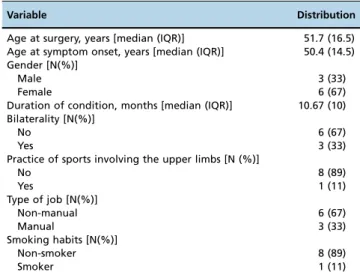
All patients answered a preoperative questionnaire con-cerning gender, age at surgery, age of pain onset, duration of symptoms, bilaterality, suprascapular nerve block, physical activity, type of work and smoking habits (Table 1).
Tissue samples
Tissue samples of approximately 2 mm3were obtained from the anterior-inferior portion of the glenohumeral capsule during the arthroscopic procedure. To reduce sampling variation, only two of the authors (CC and BE) were responsible for collecting the tissue samples. The samples were collected as previously described (30-32). As the synovium is adhered to the capsule, it cannot be separated from the capsule using arthroscopic instruments.
Table 1-Distribution of clinical variables for patients with adhesive capsulitis.
Variable Distribution
Age at surgery, years [median (IQR)] 51.7 (16.5) Age at symptom onset, years [median (IQR)] 50.4 (14.5) Gender [N(%)]
Male 3 (33)
Female 6 (67)
Duration of condition, months [median (IQR)] 10.67 (10) Bilaterality [N(%)]
No 6 (67)
Yes 3 (33)
Practice of sports involving the upper limbs [N (%)]
No 8 (89)
Yes 1 (11)
Type of job [N(%)]
Non-manual 6 (67)
Manual 3 (33)
Smoking habits [N(%)]
Non-smoker 8 (89)
Smoker 1 (11)
To provide immediate stabilization of RNA, all synovium/ capsule specimens were instantly preserved in Allprotect Tissue Reagents(Qiagen, Germany) and then stored at -20o
C.
RNA extraction
An AllPrep DNA/RNA/miRNA Universal Kit (Qiagen, Germany) was used to purify total RNA from the synovium/capsule specimens. Tissue Lyser LT equipment (Qiagen, USA) was used to mechanically lyse the tissue samples. A NanoDrop ND-1000 spectrophotometer (Thermo Scientific, USA) was used to determine RNA concentration and quality. RNA integrity was verified by 1% agarose gel electrophoresis. The RNA samples were stored at -80o
C.
mRNA expression analysis
Reverse transcription-quantitative polymerase chain reac-tion (RT-qPCR) was used to evaluate mRNA expression. First, a High-Capacity cDNA Archive kit (Life Technologies, USA) was used for cDNA synthesis. Then, RT-qPCR was performed as previously described using a ViiA 7 Real-Time PCR System (Life Technologies, USA) (30). To exclude technical variations, target and reference genes (Table 2) were analyzed on the same TaqMan Low-Density Array (TLDA) cards (Life Technologies, USA). All qPCR assays were performed in triplicate. The B2M and HPRT1 genes were used as reference genes to normalize sample input amount. These genes were chosen based on a previous study of suitable internal controls for the evaluation of mRNA expression in shoulder capsule samples (33).
The relative cycle threshold method (Crt method) was used to quantify mRNA expression. In this method, the lower the cycle threshold value (Crt) value, the greater the number of initial target copies in the sample. Thus, low Crt values indicate high gene expression. The expression of the target genes was determined using the following equation:DCrt=target gene Crt–the mean of the reference
genes Crt.
Statistical analysis
All DCrt values are shown as the median with the interquartile range (IQR).
The gender and age distributions between the patients with adhesive capsulitis and the controls were compared using the Chi-square test and the Mann-Whitney test, respectively. The Mann-Whitney test was also applied to compare mRNA levels between the cases and the controls, as well as to investigate the possible associations between mRNA expression and preoperative clinical variables, such as gender, practice of sports involving the upper limbs, type of job (manualversusnon-manual job) and smoking habits. Spearman’s correlation was used to assess possible correla-tions between mRNA levels and duration of symptoms or age at surgery: a value below 0.40 was considered a weak correlation, a value between 0.40 and 0.59 was considered a moderate correlation, a value between 0.6 and 0.79 was considered a strong correlation and a value X0.80 was considered a very strong correlation. For all analyses, a
p-valueo0.05 was considered statistically significant.
Statis-tical analyses were performed using PASW (SPSS) software, version 18 (SPSS Inc., Chicago, USA).
Table 2-Summary of reference gene and target gene assays.
Gene symbol Name Assay*
TGFb1 Transforming growth factor, beta 1 Hs00998133_m1
TGFbR1 Transforming growth factor, beta receptor 1 Hs00610320_m1
LOX Lysyl oxidase Hs00942480_m1
PLOD1 Lysyl hydroxylases 1 Hs00609368_m1
PLOD2 Lysyl hydroxylases 2 Hs01118190_m1
COMP Cartilage oligomeric matrix protein Hs00164359_m1
FN1 Fibronectin 1 Hs00365052_m1
TNC Tenascin C Hs01115665_m1
TNXB Tenascin XB Hs00372889_g1
B2M** Beta-2-microglobulin Hs00984230_m1
HPRT1** Hypoxanthine phosphoribosyl-transferase Hs02800695_m1
* TaqMan probes were purchased as assay-on-demand products for gene expression (Life Technologies, USA). ** Reference genes for target gene expression normalization.
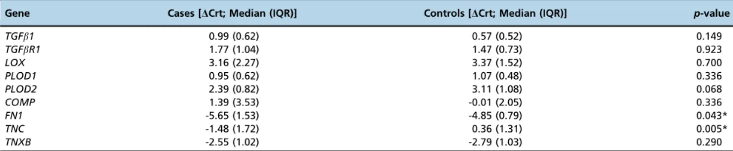
Table 3-Gene expression patterns in the glenohumeral capsules of patients with adhesive capsulitis and in controls.
Gene Cases [DCrt; Median (IQR)] Controls [DCrt; Median (IQR)] p-value
TGFb1 0.99 (0.62) 0.57 (0.52) 0.149
TGFbR1 1.77 (1.04) 1.47 (0.73) 0.923
LOX 3.16 (2.27) 3.37 (1.52) 0.700
PLOD1 0.95 (0.62) 1.07 (0.48) 0.336
PLOD2 2.39 (0.82) 3.11 (1.08) 0.068
COMP 1.39 (3.53) -0.01 (2.05) 0.336
FN1 -5.65 (1.53) -4.85 (0.79) 0.043*
TNC -1.48 (1.72) 0.36 (1.31) 0.005*
TNXB -2.55 (1.02) -2.79 (1.03) 0.290
’ RESULTS
Differences between cases and controls
Table 1 presents the clinical variables of the study participants. In the control group, 1 (12.5%) individual was female and 7 (87.5%) were males. The median age at the time of surgery was 31.44 years (IQR=13.5). The gender distribu-tion did not differ between the study groups (p=0.05). However, the controls were significantly younger than the patients with adhesive capsulitis (p=0.012, Mann-Whitney test).
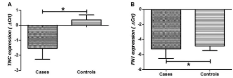
Table 3 shows the median and IQR values for the expression levels of the studied genes in the samples from the patients and the controls. The patients with adhesive capsulitis had higher levels ofTNC(p=0.005; Figure 1A) and
FN1 (p=0.043; Figure 1B) expression compared to the controls. No other significant differences were observed between the patients and the controls (p40.05).
Associations between the clinical characteristics of adhesive capsulitis and mRNA expression
Figure 2 shows the correlations between the duration of adhesive capsulitis symptoms and the expression levels of the studied genes. In the tissue samples, the expression of
TGFbR1 mRNA was significantly and directly correlated with the duration of symptoms (r=-0.731, p=0.025; Figure 2B).
No correlation was found between the age of the patients at the time of surgery and the age at symptom onset (p40.05). Additionally, no association was found between the mRNA levels of the studied genes and any of the clinical features assessed in the patients with adhesive capsulitis (p40.05).
’ DISCUSSION
Although adhesive capsulitis is considered a self-limited disease, some patients show little to no improvement, maintain a limited range of motion and continue to experience shoulder pain. Non-operative treatment is the initial approach used for adhesive capsulitis. However, operative treatment (such as arthroscopic capsular release) may be considered when patients remain in pain and do not regain satisfactory range of motion after prolonged non-operative treatment (34,35).
In this study, we found higher levels of TNC and FN1
expression in glenohumeral synovium/capsule samples collected from patients with adhesive capsulitis compared to those collected from controls. TNC immunoreactivity was previously reported in other shoulder diseases, including in
rotator cuff tendon tears and in the subacromial bursa of patients with impingement syndrome (36,37). In addition, we have previously found that bothTNC andFN1mRNA levels were upregulated in the glenohumeral capsules of patients with traumatic anterior shoulder instability (unpub-lished data). TNC is a large hexameric ECM glycoprotein that has roles in cell adhesion, fibroblast migration and other processes related to tissue remodeling and wound healing (38,39). TNC is specifically expressed following tissue damage, being upregulated within 24 h of injury (38). It is activated after local injury and down-regulated after tissue repair or scarring is completed (40). Persistent expression of TNC is associated with several fibrotic diseases and with chronic non-healing wounds (38).
Therefore, we hypothesize that increased TNC expression may be a marker of capsule injury and the genes involved may participate in inflammatory and fibrotic processes in the glenohumeral capsule.
FN is essential for collagen fibril assembly (41). During the early phase of wound healing, FN is deposited at sites of injury and can induce inflammation; increase ECM deposi-tion, including of FN and collagen; and activate fibroblasts. These pathways can create a vicious cycle that eventually induces keloid formation or fibrosis (42). Additionally, FN has been previously associated with Dupuytren’s contracture (24). In this disease, FN can be found in its oncofetal form (43,44). Additionally, upregulation of FN1 has been asso-ciated with fibrosis in inflammatory orbital diseases (45), hepatic fibrosis (46), idiopathic pulmonary fibrosis (47,48) and liver fibrosis (49). These relationships indicate that this molecule may also be involved in the pathogenesis of other fibrosing diseases. Interestingly, Altrock et al. showed that blocking FN deposition using an FN assembly inhibitor (pUR4) resulted in decreased collagen accumulation and improved liver function during liver fibrogenesis (50). Although only a slight increase in FN1 expression was detected in the glenohumeral capsules of the patients with adhesive capsulitis in the current study, our results suggest that FN1 may play a role in the fibrotic process. Further investigation is still necessary to understand the dynamic transcriptional regulation of FN1 that occurs within the shoulder capsule.
We also observed that the expression ofTGFbR1mRNA in the capsule was directly correlated with symptom duration in the patients with adhesive capsulitis. To the best of our knowledge, only one previous study has evaluated the role of the TGFbreceptor in adhesive capsulitis (5). Rodeo et al. analyzed both TGFb and its receptor in capsule and synovium samples collected from patients with adhesive capsulitis and in those collected from controls (5). They
performed a semi-quantitative analysis by comparing the frequency of positive staining between the groups. The authors described that the synovial and capsular cells of patients with adhesive capsulitis and synovitis showed clear TGFband TGFbR staining, whereas no or minimal staining was observed in the normal tissue specimens. The blood vessels of the affected tissues also presented staining for both proteins. Moreover, there was a higher frequency of positive TGFband TGFbR staining in the synovial cells of the patients with adhesive capsulitis. In addition, there was a greater frequency of positive TGFbstaining in the ECMs of patients with adhesive capsulitis compared to the controls, particu-larly in the capsule tissue.
In the present study, we found no differences in the expression ofTGFb1andTGFbR1mRNA between the cases and the controls. Because the synovium is adhered to the capsule and cannot be separated from the capsule using arthroscopic instruments, our investigation did not discrimi-nate between gene expression in synovial and capsular tissues. However, molecular alterations in both tissues are important in adhesive capsulitis (51). In addition, our study is the first to use a quantitative approach to evaluate the role of TGFbR1 in adhesive capsulitis. Our results suggest that
TGFbR1may have a role in adhesive capsulitis, especially in the long-term disease.
To the best of our knowledge, this study is the first to quantify TGFb1, TGFbR1, LOX, PLOD1, PLOD2, COMP, FN1, TNC and TNXB mRNA expression in the shoulder capsules of patients with adhesive capsulitis. However, this study has some limitations. First, few patients with adhesive capsulitis are surgically treated; as such, there is a limited number of tissue samples available for studies of gene expression (5,35,52-59). Therefore, some of our statistical analyses had reduced power to detect significant differences between the studied groups and false-negative results may have occurred. Second, we included patients who failed in conservative treatment in different phases of frozen shoulder, some in freezing and others in frozen stages. This hetero-geneity may have also contributed to false negatives. Third, molecular alterations may occur in other capsule regions and may have a different etiological role in capsular injury (30-32). We evaluated the AI region because this portion of the capsule presented macroscopic injuries (i.e., a high level of inflammation) during arthroscopic examination of the studied patients. Although we did not detect a correlation between age and gene expression in the tissue samples
collected from the patients with adhesive capsulitis, it is important to highlight that the age distribution between the patients and the controls was different. Thus, we cannot exclude that age might have influenced our findings. Finally, additional analysis of the protein products of the studied genes may be interesting because protein function is also affected by post-transcriptional and post-translational regulation.
Elevated expression ofTNC andFN1 mRNA may be a marker of capsule injury and may be involved in capsule inflammation and fibrosis. Upregulation ofTGFbR1seems to be related to symptom duration in adhesive capsulitis; therefore, TGFb signaling may play a role in this condition.
’ ACKNOWLEDGMENTS
This study was supported by grants and fellowships from the Conselho Nacional de Desenvolvimento Científico e Tecnológico (CNPQ; MC and MACS) and the Fundac¸ão de Amparo à Pesquisa do Estado de São Paulo (FAPESP; MC and MFL). We are grateful to Sintia Iole Belangero, Ph. D, for the scientific project support.
’ AUTHOR CONTRIBUTIONS
Cohen C, Leal MF, Smith MC, Ejnisman B and Faloppa F conceived and designed the experiments. Cohen C, Belangero PS, Figueiredo EA, Andreoli CV, Pochini AC and Ejnisman B were involved in data collection. Cohen C, Belangero PS, Figueiredo EA, Figueiredo EA, Pochini AC and Ejnisman B were responsible for sample collection. Leal MF was involved in the genetic analysis. Cohen C, Leal MF, Belangero PS and Figueiredo EA performed the literature search. Leal MF and Smith MC were involved in data and statistical analyses. Cohen C and Leal MF wrote thefirst draft of the manuscript. All authors listed have contributed to all subsequent drafts and all have approved thefinal manuscript.
’ REFERENCES
1. Manske RC, Prohaska D. Diagnosis and management of adhesive cap-sulitis. Curr Rev Musculoskelet Med. 2008;1(3-4):180-9, http://dx.doi. org/10.1007/s12178-008-9031-6.
2. Tamai K, Akutsu M, Yano Y. Primary frozen shoulder: brief review of pathology and imaging abnormalities. J Orthop Sci. 2014;19(1):1-5, http://dx.doi.org/10.1007/s00776-013-0495-x.
3. Hand GC, Athanasou NA, Matthews T, Carr AJ. The pathology of frozen shoulder. J Bone Joint Surg Br. 2007;89(7):928-32, http://dx.doi.org/ 10.1302/0301-620X.89B7.19097.
4. Bunker TD, Anthony PP. The pathology of frozen shoulder. A Dupuytren-like disease. J Bone Joint Surg Br. 1995;77(5):677-83.
5. Rodeo SA, Hannafin JA, Tom J, Warren RF, Wickiewicz TL. Immunolo-calization of cytokines and their receptors in adhesive capsulitis of the shoulder. J Orthop Res. 1997;15(3):427-36, http://dx.doi.org/10.1002/ jor.1100150316.
6. LeRoy EC, Trojanowska MI, Smith EA. Cytokines and human fibrosis. Eur Cytokine Netw. 1990;1(4):215-9.
7. Clark DA, Coker R. Transforming growth factor-beta (TGF-beta). Int J Biochem Cell Biol. 1998;30(3):293-8, http://dx.doi.org/10.1016/S1357-2725(97)00128-3.
8. Wrana JL, Attisano L, Wieser R, Ventura F, Massague J. Mechanism of activation of the TGF-beta receptor. Nature. 1994;370(6488):341-7, http://dx.doi.org/10.1038/370341a0.
9. Moore-Smith L, Pasche B. TGFBR1 signaling and breast cancer. J Mam-mary Gland Biol Neoplasia. 2011;16(2):89-95, http://dx.doi.org/10.1007/ s10911-011-9216-2.
10. Yoshida M, Fujii K. Differences in cellular properties and responses to growth factors between human ACL and MCL cells. J Orthop Sci. 1999;4 (4):293-8, http://dx.doi.org/10.1007/s007760050106.
11. Knippenberg M, Helder MN, Doulabi BZ, Bank RA, Wuisman PI, Klein-Nulend J. Differential effects of bone morphogenetic protein-2 and transforming growth factor-beta1 on gene expression of collagen-mod-ifying enzymes in human adipose tissue-derived mesenchymal stem cells.
Tissue Eng Part A. 2009;15(8):2213-25, http://dx.doi.org/10.1089/ten. tea.2007.0184.
12. Remst DF, Blaney Davidson EN, Vitters EL, Bank RA, van den Berg WB, van der Kraan PM. TGF-ss induces Lysyl hydroxylase 2b in human synovial osteoarthritic fibroblasts through ALK5 signaling. Cell Tissue Res. 2014;355(1):163-71, http://dx.doi.org/10.1007/s00441-013-1740-5. 13. Witsch TJ, Turowski P, Sakkas E, Niess G, Becker S, Herold S, et al.
Deregulation of the lysyl hydroxylase matrix cross-linking system in experimental and clinical bronchopulmonary dysplasia. Am J Physiol Lung Cell Mol Physiol. 2014;306(3):L246-59, http://dx.doi.org/10.1152/ ajplung.00109.2013.
14. Smith-Mungo LI, Kagan HM. Lysyl oxidase: properties, regulation and multiple functions in biology. Matrix Biol. 1998;16(7):387-98, http://dx.doi.org/10.1016/S0945-053X(98)90012-9.
15. Kagan HM, Li W. Lysyl oxidase: properties, specificity, and biological roles inside and outside of the cell. J Cell Biochem. 2003;88(4):660-72, http://dx.doi.org/10.1002/jcb.10413.
16. Myllyla R, Wang C, Heikkinen J, Juffer A, Lampela O, Risteli M, et al. Expanding the lysyl hydroxylase toolbox: new insights into the localiza-tion and activities of lysyl hydroxylase 3 (LH3). J Cell Physiol. 2007;212 (2):323-9, http://dx.doi.org/10.1002/jcp.21036.
17. Eyre DR, Koob TJ, Van Ness KP. Quantitation of hydroxypyridinium crosslinks in collagen by high-performance liquid chromatography. Anal Biochem. 1984;137(2):380-8, http://dx.doi.org/10.1016/0003-26975. 18. van der Slot AJ, Zuurmond AM, van den Bogaerdt AJ, Ulrich MM,
Middelkoop E, Boers W, et al. Increased formation of pyridinoline cross-links due to higher telopeptide lysyl hydroxylase levels is a general fibrotic phenomenon. Matrix Biol. 2004;23(4):251-7, http://dx.doi.org/ 10.1016/j.matbio.2004.06.001.
19. Rosenberg K, Olsson H, Morgelin M, Heinegard D. Cartilage oligomeric matrix protein shows high affinity zinc-dependent interaction with triple helical collagen. J Biol Chem. 1998;273(32):20397-403, http://dx.doi.org/ 10.1074/jbc.273.32.20397.
20. Haudenschild DR, Hong E, Yik JH, Chromy B, Morgelin M, Snow KD, et al. Enhanced activity of transforming growth factor beta1 (TGF-beta1) bound to cartilage oligomeric matrix protein. J Biol Chem. 2011;286 (50):43250-8, http://dx.doi.org/10.1074/jbc.M111.234716.
21. Farina G, Lemaire R, Korn JH, Widom RL. Cartilage oligomeric matrix protein is overexpressed by scleroderma dermal fibroblasts. Matrix Biol. 2006;25(4):213-22, http://dx.doi.org/10.1016/j.matbio.2006.01.007. 22. Vakonakis I, Campbell ID. Extracellular matrix: from atomic resolution to
ultrastructure. Curr Opin Cell Biol. 2007;19(5):578-83, http://dx.doi.org/ 10.1016/j.ceb.2007.09.005.
23. Dallas SL, Sivakumar P, Jones CJ, Chen Q, Peters DM, Mosher DF, et al. Fibronectin regulates latent transforming growth factor-beta (TGF beta) by controlling matrix assembly of latent TGF beta-binding protein-1. J Biol Chem. 2005;280(19):18871-80, http://dx.doi.org/10.1074/jbc. M410762200.
24. Cordova A, Tripoli M, Corradino B, Napoli P, Moschella F. Dupuytren’s contracture: an update of biomolecular aspects and therapeutic perspec-tives. J Hand Surg Br. 2005;30(6):557-62, http://dx.doi.org/10.1016/ j.jhsb.2005.07.002.
25. Zweers MC, Hakim AJ, Grahame R, Schalkwijk J. Joint hypermobility syndromes: the pathophysiologic role of tenascin-X gene defects. Arthritis Rheum. 2004;50(9):2742-9, http://dx.doi.org/10.1002/art.20488. 26. Carey WA, Taylor GD, Dean WB, Bristow JD. Tenascin-C deficiency
attenuates TGF-ss-mediated fibrosis following murine lung injury. Am J Physiol Lung Cell Mol Physiol. 2010;299(6):L785-93, http://dx.doi.org/ 10.1152/ajplung.00385.2009.
27. Chiquet-Ehrismann R, Tucker RP. Tenascins and the importance of adhesion modulation. Cold Spring Harb Perspect Biol. 2011;3(5), http://dx.doi.org/10.1101/cshperspect.a004960.
28. Mao JR, Taylor G, Dean WB, Wagner DR, Afzal V, Lotz JC, et al. Tenascin-X deficiency mimics Ehlers-Danlos syndrome in mice through alteration of collagen deposition. Nat Genet. 2002;30(4):421-5, http://dx.doi.org/ 10.1038/ng850.
29. Valcourt U, Alcaraz LB, Exposito JY, Lethias C, Bartholin L. Tenascin-X: beyond the architectural function. Cell Adh Migr. 2015;9(1-2):154-65, http://dx.doi.org/10.4161/19336918.2014.994893.
30. Belangero PS, Leal MF, Cohen C, Figueiredo EA, Smith MC, Andreoli CV, et al. Expression analysis of genes involved in collagen cross-linking and its regulation in traumatic anterior shoulder instability. J Orthop Res. 2015, http://dx.doi.org/10.1002/jor.22984.
31. Belangero PS, Leal MF, de Castro Pochini A, Andreoli CV, Ejnisman B, Cohen M. Profile of collagen gene expression in the glenohumeral capsule of patients with traumatic anterior instability of the shoulder. Rev Bras Ortop. 2014;49(6):642-6, http://dx.doi.org/10.1016/j.rbo.2013.10.012. 32. Belangero PS, Leal MF, Figueiredo EA, Cohen C, Pochini Ade C, Smith
MC, et al. Gene expression analysis in patients with traumatic anterior shoulder instability suggests deregulation of collagen genes. J Orthop Res. 2014;32(10):1311-6, http://dx.doi.org/10.1002/jor.22680.
of shoulder instability. PloS One. 2014;9(8):e105002, http://dx.doi.org/ 10.1371/journal.pone.0105002.
34. Holloway GB, Schenk T, Williams GR, Ramsey ML, Iannotti JP. Arthroscopic capsular release for the treatment of refractory postoperative or post-fracture shoulder stiffness. J Bone Joint Surg Am. 2001;83-A(11):1682-7.
35. Hagiwara Y, Ando A, Onoda Y, Takemura T, Minowa T, Hanagata N, et al. Coexistence of fibrotic and chondrogenic process in the capsule of idiopathic frozen shoulders. Osteoarthritis Cartilage. 2012;20(3):241-9, http://dx.doi.org/10.1016/j.joca.2011.12.008.
36. Riley GP, Harrall RL, Cawston TE, Hazleman BL, Mackie EJ. Tenascin-C and human tendon degeneration. Am J Pathol. 1996;149(3):933-43. 37. Hyvonen P, Melkko J, Lehto VP, Jalovaara P. Involvement of the
sub-acromial bursa in impingement syndrome of the shoulder as judged by expression of tenascin-C and histopathology. J Bone Joint Surg Br. 2003;85 (2):299-305, http://dx.doi.org/10.1302/0301-620X.85B2.13124.
38. Trebaul A, Chan EK, Midwood KS. Regulation of fibroblast migration by tenascin-C. Biochem Soc Trans. 2007;35(Pt 4):695-7, http://dx.doi.org/ 10.1042/BST0350695.
39. Snyder JC, Zemke AC, Stripp BR. Reparative capacity of airway epithe-lium impacts deposition and remodeling of extracellular matrix. Am J Respir Cell Mol Biol. 2009;40(6):633-42, http://dx.doi.org/10.1165/ rcmb.2008-0334OC.
40. Midwood KS, Orend G. The role of tenascin-C in tissue injury and tumorigenesis. J Cell Commun Signal. 2009;3(3-4):287-310, http://dx.doi. org/10.1007/s12079-009-0075-1.
41. Kadler KE, Hill A, Canty-Laird EG. Collagen fibrillogenesis: fib-ronectin, integrins, and minor collagens as organizers and nucleators. Curr Opin Cell Biol. 2008;20(5):495-501, http://dx.doi.org/10.1016/j. ceb.2008.06.008.
42. Kelsh RM, McKeown-Longo PJ, Clark RA. EDA Fibronectin in Keloids Create a Vicious Cycle of Fibrotic Tumor Formation. J Invest Dermatol. 2015;135(7):1714-8, http://dx.doi.org/10.1038/jid.2015.155.
43. Ignotz RA, Massague J. Transforming growth factor-beta stimulates the expression of fibronectin and collagen and their incorporation into the extracellular matrix. The Journal of biological chemistry. 1986;261 (9):4337-45.
44. Howard JC, Varallo VM, Ross DC, Faber KJ, Roth JH, Seney S, et al. Wound healing-associated proteins Hsp47 and fibronectin are elevated in Dupuytren’s contracture. J Surg Res. 2004;117(2):232-8, http://dx.doi. org/10.1016/j.jss.2004.01.013.
45. Rosenbaum JT, Choi D, Wilson DJ, Grossniklaus HE, Harrington CA, Dailey RA, et al. Fibrosis, gene expression and orbital inflammatory dis-ease. Br J Ophthalmol. 2015;99(10):1424-9, http://dx.doi.org/10.1136/ bjophthalmol-2015-306614.
46. Modol T, Brice N, Ruiz de Galarreta M, Garcia Garzon A, Iraburu MJ, Martinez-Irujo JJ, et al. Fibronectin peptides as potential regulators of hepatic fibrosis through apoptosis of hepatic stellate cells. Journal of cel-lular physiology. 2015;230(3):546-53, http://dx.doi.org/10.1002/jcp.24714.
47. Singer, II, Kawka DW, Kazazis DM, Clark RA. In vivo co-distribution of fibronectin and actin fibers in granulation tissue: immunofluorescence and electron microscope studies of the fibronexus at the myofibroblast surface. J Cell Biol. 1984;98(6):2091-106, http://dx.doi.org/10.1083/ jcb.98.6.2091.
48. Torr EE, Ngam CR, Bernau K, Tomasini-Johansson B, Acton B, Sandbo N. Myofibroblasts exhibit enhanced fibronectin assembly that is intrinsic to their contractile phenotype. J Biol Chem. 2015 Mar 13;290(11):6951-61, http://dx.doi.org/10.1074/jbc.M114.606186.
49. Hernandez-Gea V, Friedman SL. Pathogenesis of liver fibrosis. Annu Rev Pathol. 2011;6:425-56, http://dx.doi.org/10.1146/annurev-pathol-011110-130246.
50. Altrock E, Sens C, Wuerfel C, Vasel M, Kawelke N, Dooley S, et al. Inhibition of fibronectin deposition improves experimental liver fibrosis. J Hepatol. 2015;62(3):625-33, http://dx.doi.org/10.1016/j.jhep.2014.06.010. 51. Uppal HS, Evans JP, Smith C. Frozen shoulder: A systematic review of therapeutic options. World J Orthop. 2015;6(2):263-8, http://dx.doi.org/ 10.5312/wjo.v6.i2.263.
52. Mullett H, Byrne D, Colville J. Adhesive capsulitis: human fibroblast response to shoulder joint aspirate from patients with stage II disease. J Shoulder Elbow Surg. 2007;16(3):290-4, http://dx.doi.org/10.1016/ j.jse.2006.08.001.
53. Xu Y, Bonar F, Murrell GA. Enhanced expression of neuronal proteins in idiopathic frozen shoulder. J Shoulder Elbow Surg. 2012 ;21(10):1391-7, http://dx.doi.org/10.1016/j.jse.2011.08.046.
54. Ryu JD, Kirpalani PA, Kim JM, Nam KH, Han CW, Han SH. Expression of vascular endothelial growth factor and angiogenesis in the diabetic frozen shoulder. Journal of shoulder and elbow surgery / American Shoulder and Elbow Surgeons [et al. 2006;15(6):679-85.
55. Kabbabe B, Ramkumar S, Richardson M. Cytogenetic analysis of the pathology of frozen shoulder. Int J Shoulder Surg. 2010;4(3):75-8, http://dx.doi.org/10.4103/0973-6042.76966.
56. Kanbe K, Inoue K, Inoue Y, Chen Q. Inducement of mitogen-activated protein kinases in frozen shoulders. J Orthop Sci. 2009;14(1):56-61, http://dx.doi.org/10.1007/s00776-008-1295-6.
57. Nago M, Mitsui Y, Gotoh M, Nakama K, Shirachi I, Higuchi F, et al. Hyaluronan modulates cell proliferation and mRNA expression of adhesion-related procollagens and cytokines in glenohumeral synovial/ capsular fibroblasts in adhesive capsulitis. J Orthop Res. 2010;28(6):726-31, http://dx.doi.org/10.1002/jor.21075.
58. Bunker TD, Reilly J, Baird KS, Hamblen DL. Expression of growth factors, cytokines and matrix metalloproteinases in frozen shoulder. J Bone Joint Surg Br. 2000;82(5):768-73, http://dx.doi.org/10.1302/0301-620X. 82B5.9888.