Contributes to Susceptibility and Prognosis of Lone
Atrial Fibrillation in Chinese Population
Hailong Cao1., Qing Zhou1., Rongfang Lan2., Oluf Dimitri Røe3,4, Xin Chen2, Yijiang Chen3, Dongjin Wang1*
1Department of Thoracic and Cardiovascular Surgery, the Affiliated Drum Tower Hospital of Nanjing University Medical School, Nanjing, China,2Department of Cardiology, the Affiliated Drum Tower Hospital of Nanjing University Medical School, Nanjing, China,3Department of Thoracic and Cardiovascular Surgery, the First Affiliated Hospital of Nanjing Medical University, Nanjing, China,4Department of Cancer Research and Molecular Medicine, Norwegian University of Science and Technology (NTNU), Trondheim, Norway
Abstract
Transforming growth factor-b1 (TGF-b1) is an important mediator of atrial fibrosis and atrial fibrillation (AF). But the involved genetic mechanism is unknown. Herein, theTGF-b1C-509T polymorphism (rs1800469) was genotyped in a case-control study of 840 patients and 845 controls in Chinese population to explore the association between the polymorphism and susceptibility and prognosis of lone AF. As a result, the CT and/or TT genotypes had an increased lone AF risk [adjusted odds ratio (OR) = 1.50 for CT, OR = 3.72 for TT, and OR = 2.15 for CT/TT], compared with the TGF-b1CC genotype. Moreover, patients carrying CT/TT genotypes showed a higher possibility of AF recurrence after catheter ablation, compared with patients carrying CC genotype. In a genotype-phenotype correlation analysis using 24 normal left atrial appendage samples, increasing gradients of atrial TGF-b1 expression levels positively correlated with atrial collagen volume fraction were identified in samples with CC, CT and TT genotypes. Thein vitroluciferase assays also showed a higher luciferase activity of the -509T allele than that of the -509C allele. In conclusion, theTGF-b1C-509T polymorphism is involved in the etiology of lone AF and thus may be a marker for genetic susceptibility to lone AF and predicting prognosis after catheter ablation in Chinese populations. Therefore, we provide new information about treatment strategies and our understanding ofTGF-b1
in AF.
Citation:Cao H, Zhou Q, Lan R, Røe OD, Chen X, et al. (2014) A Functional Polymorphism C-509T inTGFb-1Promoter Contributes to Susceptibility and Prognosis
of Lone Atrial Fibrillation in Chinese Population. PLoS ONE 9(11): e112912. doi:10.1371/journal.pone.0112912
Editor:Kandiah Jeyaseelan, National University of Singapore, Singapore
ReceivedJuly 1, 2014;AcceptedOctober 16, 2014;PublishedNovember 17, 2014
Copyright:ß2014 Cao et al. This is an open-access article distributed under the terms of the Creative Commons Attribution License, which permits unrestricted
use, distribution, and reproduction in any medium, provided the original author and source are credited.
Data Availability:The authors confirm that all data underlying the findings are fully available without restriction. All relevant data are within the paper and its
Supporting Information files.
Funding:This work was supported in part by the National Natural Science Foundation of China [81200133], Jiangsu Top Expert Program in Six Professions
[WSN-032], Medical science project supported by Jiangsu health department [Z201411], Key Project supported by Medical Science and technology development Foundation, Nanjing Department of Health [YKK12056], the Fundamental Research Funds for the Central Universities [021414330063]. The funders had no role in study design, data collection and analysis, decision to publish, or preparation of the manuscript.
Competing Interests:The authors have declared that no competing interests exist.
* Email: [email protected]
.These authors contributed equally to this work.
Introduction
Atrial fibrillation (AF), the most common cardiac arrhythmia, is associated with increased morbidity, mortality and cost [1]. To date, the precise mechanism of AF remains largely unknown. It is particularly true for patients with lone AF (LAF), which is AF in the absence of heart disease or comorbidities predisposing to the arrhythmia. However, increasing evidence showed that the atrial fibrosis is essential for the development and progression of AF [2– 3].
It is accepted that transforming growth factor-b1 (TGF-b1) is the most powerful profibrotic factor, stimulating the secretion of collagen into the extracellular matrix by fibroblasts [4]. Over-expression ofTGF-b1selectively induced atrial fibrosis, leading to increased conduction heterogeneity and AF vulnerability without affecting the cellular electrophysiology [5]. Inhibition ofTGF-b1 expression by Tranilast significantly reduced the atrial fibrosis, as a
result, reduced conduction abnormalities and AF vulnerability were observed [6]. These studies suggest that TGF-b1 plays an essential role in inducing AF.
Materials and Methods
2.1. Ethics statement
This hospital-based case-control study was approved by the Review Boards of the Affiliated Drum Tower Hospital of Nanjing University Medical School and the First Affiliated Hospital of Nanjing Medical University (2007-NJEA-10). All subjects provided written informed consent to be included in the study. The study was conducted according to the Helsinki Declaration and approved by the ethics committees of the respective institutions (2007-NJEA-10). We have complied with the World Medical Association Declaration of Helsinki regarding ethical conduct of research involving human subjects.
2.2. Study Subjects
We consecutively recruited 2786 non-valvular AF patients between October 2007 and June 2013. Each patient donated 5 ml venous blood upon admission to the hospital and was interviewed to collect demographic data and clinical information. Among them, a total of 860 patients were diagnosed as lone AF, which was based on the following criteria [11]: age at first diagnosis of AF,
60 years, no past cardiovascular history, no evidence suggesting ischemic heart disease, no cardiomyopathy, no heart failure, no valvular heart disease, no diabetes, no hypertension within two years of the onset of AF, no hyperthyroidism. Sex- and age-matched 860 AF-free control subjects with written informed consent were genetically unrelated to the cases. They were recruited from healthy subjects without individual history of AF and other chronic diseases. All subjects were genetically unrelated ethnic Han Chinese.
Among all the subjects, genotyping was failed in 20 cases (2.59%) and 15 controls (1.88%) due to DNA quality or quantity.
Therefore, 840 lone AF cases and 845 healthy controls were finally included in the susceptibility analyses. Among the 840 cases, 725 received first-time catheter ablation and were successfully cardioverted to stable SR. During the follow-up, 112 patients were lost and 3 cases died because trauma or cerebrovascular accident. Therefore, a total of 610 patients had complete follow-ups and clinical information.
2.3. Follow-Up
AF-free time was calculated from the date of ablation to the date of recurrence or last follow-up. Atrial arrhythmias that occurred during the first 2 months after ablation, which is considered a blanking period [12], were not counted as recurrences. Antiar-rhythmic medications, including amiodarone, metoprolol or propafenone, were generally continued to the end of third month after ablation unless recurrent arrhythmia indicated the need for continued treatment. All patients with documented arrhythmia and those maintained on antiarrhythmics for control of AF beyond the blanking period were counted as recurrences.
Patients had scheduled clinical visits, 12-lead ECG, and 24-hour Holter monitoring at 3, 6, and 12 months after ablation and then yearly after the first year. Moreover, patients would receive ECG monitoring in local clinics at anytime if they had AF-related symptoms. AF recurrence was identified by symptoms with ECG documentation of an atrial tachyarrhythmia lasting$30 seconds on a 12-lead ECG or Holter monitor recording. AF recurrence during the follow-up was considered censored.
2.4. Genotyping
Genomic DNA was isolated from leukocytes of venous blood by proteinase K digestion and phenol/chloroform extraction. The
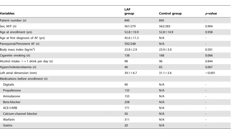
Table 1.Baseline characteristics of subjects with LAF and controls.
Variables
LAF
group Control group p-value
Patient number(n) 840 845
-Sex, M/F (n) 561/279 562/283 0.904
Age at enrollment (yrs) 52.8610.9 52.8614.9 0.938
Age at first diagnosis of AF (yrs) 45.6611.3 N/A
-Paroxysmal/Persistent AF (n) 592/248 N/A
-Body mass index (kg/m2) 23.8
62.9 23.963.0 0.501
Cigarette smoking (n) 138 168 0.066
Alcohol intake.= 1 drink per day (n) 98 96 0.844
Hypercholesterolaemia (n) 46 65 0.067
Left atrial dimension (mm) 39.166.7 31.163.6 ,0.001
Medications before enrollment (n)
Digitalis 90 N/A
-Propafenone 135 N/A
-Amiodarone 155 N/A
-Beta-blocker 258 N/A
-ACE-I/ARB 171 N/A
-Calcium-channel blocker 50 N/A
-Warfarin 311 N/A
-Statins 20 N/A
-Values are presented as mean6SD or number of patients.
TGF-b1C-509T polymorphism was determined using the PCR– restriction fragment length polymorphism method. The PCR primers for the TGF-b1 C-509T polymorphism were 59 -GCTAAGGCATGGCACCGCTT-39 (forward) and 59 -GAAG-GAGGGTCTGTCAACATGGG-39 (reverse). PCR was per-formed in a total volume of 20 mL containing 50 ng genomic DNA, 106Taq buffer, 0.02 mmol?L21of MgCl2, 0.05 mmol?L21 of dNTP mix, 10 pmol?mL21of each primer and 1 U Taq DNA polymerase. After initial denaturation at 95uC for 5 minutes, the reaction was carried out at 95uC denaturation for 30 seconds, annealing for 40 seconds at 62uC and extension for 45 seconds at 72uC for a total of 34 cycles, and a final elongation at 72uC for 10 minutes. The 270-bp PCR products were digested by the restriction enzyme (Eco81I, SauI) (MBI Fermentas, Vilnius, Lithuania) at 37uC overnight. The digested products were then analyzed by electrophoresis in 3% agarose gel stained with 0.5% ethidium bromide and photographed under UV illumination. The C allele was cut into 198-bp and 72-bp fragments, whereas the T allele was not digested. The polymorphism analysis was carried out by two persons independently in a blinded manner. More than 15% of the samples were randomly selected for confirmation, and the results were 100% concordant.
2.5. LAA samples
In order to determine the expression ofTGF-b1and interstitial fibrosis, we collected 24 LAA tissues from healthy heart donors for transplantation. They were trauma victims and were free of cardiovascular pathology and documented AF. LAA specimens were obtained before perfusion. A part of each LAA was fixed in paraformaldehyde for histology, and the others were immediately snap-frozen in liquid nitrogen for laboratory analysis.
2.6. Real-time quantitative RT-PCR
The primers of TGF-b1 (Forward primer [F]: 59 -CTAATGGTGGAAACCCACAACG-39, Reverse primer [R]: 59-TATCGCCAGGAATTGTTGCTG-39, NM_000660) and glyceraldehydes 3-phosphate dehydrogenase (GAPDH) (Forward primer [F]: 59-ATGGGGAAGGTGAAGGTCG-39, Reverse primer [R]: 59-GGGGTCATTGATGGCAACAATA-39, NM_002046) were synthesized by Invitrogen Co., Hong Kong, China. Total RNA was isolated from frozen RAAs by acid-phenol extraction in the presence of chaotropic salts (TRIzol, Invitrogen) and subsequent isopropanol ethanol precipitation. The synthesis of cDNA was according to the manufacturer’s instructions with the reverse transcriptase kit (Promega Co., US). The real-time PCR was performed using the LightCycler 480 system (Roche Diagnostics, Switzerland) with a total volume of 20ml containing 10ml 26Master Mix SYBR Green I (Takara, Japan), 0.25mM forward primers, 0.25mM reverse primers, 2ml cDNA template, and H2O to a final volume of 20ml. The protocol of real-time PCR consisted of 40 cycles, and cycling parameters were as follows: denaturation at 94uC for 10 seconds, annealing at 61uC for 15 seconds and extension at 72uC for 20 seconds. The results were analyzed using Roche LightCycler 480 software. Data of transcripts were calculated relative to GAPDH using the 22DDCt method. The measurements of each sample were performed in triplicate.
2.7. Western blot
Frozen LAAs were used for protein isolation as described previously [13]. Proteins (40mg/lane) were separated by sodium dodecyl sulfate polyacrylamide gel electropheresis and transferred onto Polylinylidene Fluoride membranes using a Bio-Rad semidry transfer system (Bio-Rad). The membranes were blocked with 5%
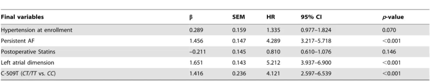
Table 3.Results of Cox multivariate regression analysis on cumulative AF recurrence after catheter ablation.
Final variables b SEM HR 95% CI p-value
Hypertension at enrollment 0.289 0.159 1.335 0.977–1.824 0.070
Persistent AF 1.456 0.147 4.289 3.217–5.718 ,0.001
Postoperative Statins –0.211 0.145 0.810 0.610–1.076 0.146
Left atrial dimension 1.651 0.143 5.212 3.937–6.900 ,0.001
C-509T (CT/TTvs.CC) 1.416 0.236 4.121 2.597–6.539 ,0.001
b, regression coefficient; HR, hazard ratio. doi:10.1371/journal.pone.0112912.t003
Figure 1. Kaplan-Meier survival curves showing freedom from AF recurrence after catheter ablation according toTGF-b1C-509T polymorphism.(A) Survival free from AF recurrence in CC (n = 120), CT (n = 231) and TT (n = 259) groups. (B) Survival free from AF recurrence in CC (n = 120) and CT/TT (n = 490) groups.
non-fat dry milk and then probed with mouse monoclonal
TGF-b1(ab27969, Abcam, USA) and horseradish peroxidase (HRP)-conjugated mouse monoclonal anti-GAPDH (KC-5G5, Kang-Chen Biotech, China). The working dilutions were 1:2000
(TGF-b1) and 1:5000 (GAPDH). The resulting reaction was visualized using HRP-conjugated anti-mouse secondary antibody (Santa-Cruz Biotechnology, the Netherlands), followed by incubation with ECL Western Blot Detection Kit (Amersham, the Nether-lands) for 1 minute. The blots were exposed to Kodak film for 5 minutes and immunoreactive bands developed for quantification using The Discovery Series image analysis software (Bio-Rad) normalized by the corresponding value of GAPDH. Experiments were repeated three times and the mean was scored.
2.8. Masson’s trichrome staining
After fixation with 4% paraformaldehyde in phosphate-buffered saline (PH: 7.4) for 24 h, the tissues were subjected to alcoholic dehydration and embedded in paraffin. 4mm serial sections were sliced and subjected to Masson’s trichrome staining to highlight collagen fibers. Collagen volume fraction (CVF) was determined by the HPISA 100 chromatic color pathological analysis system (Olympus, Japan) using five random images from each slide and five slides per sample, and the mean values of CVF were obtained by one investigator blinded to the groups.
2.9. Promoter functional assay
The isolation and culture of mouse atrial fibroblasts were previously described [14]. The TGF-b1 promoter-luciferase reporter plasmids containing either -509C or -509T sequences were prepared by amplifying the 270-bpTGF-b1promoter region by using primers with restriction sites. The primers were 59
-GCTAAGGCATGGCACCGCTT-39 (forward) and 59 -GAAG-GAGGGTCTGTCAACATGGG-39(reverse), including theKpnI and XhoI restriction sites. The amplified fragments were then sequenced to confirm that there were no errors in matched nucleotides and the plasmid encompassed either -509C or -509T allele. The amplified fragments and luciferase reporter vectors (pGL3)-basic vector (Promega, Madison, WI, USA) were cleaved by using theKpnIand XhoIenzymes (Promega, USA), and the fragments were then cloned into the pGL3-basic vector. After cloning, the vectors were sequenced to confirm the orientation and integrity of the inserts of each construct. For transfections, mouse atrial fibroblasts were seeded onto 24-well plates (100,000 cells per well), and each well was transfected with 1mg of the vector DNA with either -509C or -509T allele, using Lipofectamine 2000 (Invitrogen, Carlsbad, CA, USA). As an internal standard, all plasmids were cotransfected with 8 ng pRL-SV40, which contained the Renilla luciferase gene. The pGL3-basic vector without an insert was used as a negative control. After 48 hours of
Figure 2. Effect of the C-509 T polymorphism in the TGF-b1
promoter activity.(A) Schematic representation of reporter plasmids containing the -509C or -509 T allele, which was inserted upstream of the luciferase reporter gene in the pGL3 basic plasmid. (B) Two constructs were transiently transfected into the mouse cardiac fibroblasts. The luciferase activity of each construct was normalized against the internal control of Renilla luciferase (blank). Values are mean6SD.
doi:10.1371/journal.pone.0112912.g002
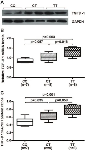
Figure 3. Atrial expression ofTGF-b1 among different geno-types in healthy heart donors. (A) Representative Western immunoblots. (B) Gene expression in left atrial appendages (LAAs). (C) Semi-quantitative protein content in LAAs. Boxes show interquartile ranges, and bars represent the 10th and 90th percentiles.
incubation, cells were collected and analyzed for luciferase activity with the Dual-Luciferase Reporter Assay System (Promega, Madison, WI, USA).
2.10. Statistical analysis
Differences in the distributions of demographic characteristics, clinical variables, and frequencies of genotypes of TGF-b1 C-509T polymorphism between the cases and controls were evaluated using the Student’s t-test (for continuous variables) and
x2 test (for categorical variables). Hardy–Weinberg equilibrium
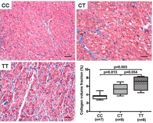
Figure 4. Representative photomicrographs of Masson staining showing the interstitial collagen (stained blue) (6200, bar = 50mm). Collagen volume fraction is used to evaluate the degree of fibrosis.
doi:10.1371/journal.pone.0112912.g004
was tested using a goodness-of-fitx2test. The association between theTGF-b1C-509T polymorphism and AF risk was estimated by computing odds ratios (ORs) and their 95% confidential intervals (CIs) from multivariate logistic model. An allele-specific difference in luciferase activity was also tested using the Student’s t-test. For the comparison of atrial expression ofTGF-b1and the degree of atrial fibrosis, one-way ANOVA test (normally distributed) or Mann-Whitney test (2 groups, non-normally distributed) and Kruskal-Wallis test (n groups, non-normally distributed) were used among the three genotypes. Spearman correlation analysis was applied to assess the association between expression of TGF-b1 and CVF in LAAs. The Kaplan-Meier method, log-rank test, and Cox survival regression model were used to determine factors predictive of AF outcome after ablation.p,0.05 was considered statistically significant, and all statistical tests were two-sided. All the statistical analyses were performed with Statistical Analysis System software (v.9.1.3; SAS Institute, Cary, NC, USA).
Results
3.1. Characteristics of the study population
The characteristics of LAF group and Control group enrolled in this study were shown in Table 1. There were no significant differences in the distribution of the age, sex, body mass index (BMI), cigarette smoking, alcohol intake and hypercholesterolae-mia. Left atrial dimension (LAD) was significantly lager in LAF than control. In LAF group, age at first diagnosis of AF was 45.6611.3 years, and 70.5% was paroxysmal AF. Medications before enrollment in LAF group were Digitalis (n = 90), Propafe-none (n = 135), Amiodarone (n = 155), Beta-blocker (n = 258), angiotensin-converting enzyme inhibitor/angiotensin receptor blocker (n = 171), Calcium-channel blocker (n = 50), Warfarin (n = 311) and Statins (n = 20).
3.2. TGF-b1 C-509T polymorphism and LAF risk
The genotype and allele distributions of theTGF-b1C-509T polymorphism in the cases and controls are shown in Table 2. The observed genotype frequencies for this polymorphism were in Hardy–Weinberg equilibrium in the controls (x2= 0.286, p= 0.593). The frequencies of the CC, CT and TT genotypes were 18.1%, 40.2% and 41.7%, respectively, among the cases, and 32.3%, 47.8% and 19.9%, respectively, among the controls. After adjusting for possible confounders (age, sex, body mass index, smoking status, drinking status, and hypercholesteremia), subjects carrying CT or TT or CT/TT genotypes had an increased risk of LAF (adjusted OR = 1.50(1.17–1.92) for CT, 3.72 (2.83–4.88) for TT and 2.15 (1.71–2.70) for CT/TT; ptrend,0.001), compared with CC homozygote, and the frequency of the T allele being higher than C allele among LAF subjects (p,0.001).
3.3. Factors associated with AF recurrence after ablation Over a median follow-up of 17.5 months (range, 3.5 to n 71 months) after ablation, 207 patients (33.9%) had AF recurrence and 403 patients (66.1%) remained in SR. I multivariate Cox proportional hazards analysis, factors associated with arrhythmia recurrence were found to be persistent AF (versus paroxysmal AF; HR = 4.289 (3.217–5.718); p,0.001), larger left atrial dimension (HR = 5.212 (3.937–6.900);p,0.001), CT/TT genotype (versus CC genotype; HR = 4.121 (2.597–6.539);p,0.001) (Table 3). In addition, Kaplan-Meier survival estimates showed that LAF patients carrying different TGF-b1 C-509T genotypes had different proportion of AF recurrence (p,0.001) (Figure 1A). Moreover, CT/TT genotypes also had a higher proportion of AF
recurrence after ablation, compared with CC genotype (p,0.001) (Figure 1B).
3.4. TGF-b1 C-509T polymorphism and luciferase activity As shown in Figure 2, the vector with the -509T allele had an increase in the relative luciferase activity, compared with that with the -509C allele (p,0.01). These results suggested that the -509T allele may lead to a higher expression levels ofTGF-b1 mRNA than the -509C allele.
3.5. Expression of TGF-b1 and the degree of atrial fibrosis among genotypes
In the 24 LAA specimens, 7 were of the CC genotype, 9 of the CT genotype, and 8 of the TT genotype. As shown in Figure 3B, Real-time quantitative RT-PCR assay showed an increasing gradient of gene expression ofTGF-b1 in the groups carrying CC, CT and TT genotypes, although there was merely a borderline significant difference between CC and CT groups (p= 0.057).
Western blot analysis (Figure 3A, C) showed an increased expression of TGF-b1 in CT and TT groups than CC group, whereas the difference was borderline between CT and TT groups (p= 0.058).
Interstitial collagen (stained blue), revealed by Masson staining and expressed as CVF, was lowest in CC group, followed by CT and TT groups (Figure 4). However, there was merely a borderline significant difference between CT and TT groups (p= 0.054).
The correlation test indicated a strong positive correlation between atrial protein expression ofTGF-b1and CVF in LAAs (r = 0.695, p,0.001; Figure 5).
Discussion
In this case-control study, we investigated the relationships between TGF-b1 C-509T polymorphism and susceptibility and prognosis of LAF in Chinese population. We made several new findings. First, our results revealed that the CT and/or TT genotypes had an increased LAF risk [adjusted OR = 1.50 for CT, OR = 3.72 for TT, and OR = 2.15 for CT/TT], compared with the TGF-b1CC genotype. Second, we demonstrated that LAF patients carrying CT/TT genotypes had a higher possibility of AF recurrence after ablation, compared with patients carrying CC genotype. Furthermore, we validated both in vitro and vivo that T allele had higher expression of TGF-b1 and more aggravated atrial interstitial fibrosis than C allele. However, a recent study determining the same polymorphism in also Chinese Han population found no association between the rs1800469 polymor-phism and the risk of AF under the dominant, recessive and additive genetic models [15]. It could be due to their enrolled patients who had kinds of underlying disease (hypertension, coronary heart disease and so on) and were much older. But herein we only recruited LAF cases who were probably determined by genetics. Moreover, they had much fewer cases. So the inherent selection bias could not be completely excluded. To the best of our knowledge, this is the first study to investigate the potential functional polymorphism C-509T of TGFb-1 promoter in susceptibility and prognosis of AF.
remodeling including myolysis, apoptosis, fibrosis and inflamma-tion, atrial interstitial fibrosis is the most important process in development and progression of LAF [16].TGF-b1is a cytokine that modulates the tissue fibrosis [4]. Recent studies showed that over-expression ofTGF-b1as well as an increase of atrial fibrosis was observed in atrial specimens from patients with AF [17–19]. High-expression of atrial TGF-b1 indicated a higher recurrence rate and a poor recovery of atrial mechanical contraction after surgical maze procedure [20–21]. It was speculated and further confirmed in an transgenic animal model that atrial over-expression ofTGF-b1selectively induced atrial interstitial fibrosis, contributing to AF vulnerability [5,19]. Inhibition of TGF-b1 expression by an antiallergic drug named Tranilast decreased the atrial fibrosis and AF vulnerability [22]. These studies suggest that theTGF-b1attributes to development and recurrence of AF via triggering atrial fibrosis.
The humanTGF-b1gene is located on chromosome 19q13 and can be transcribed and translated to form a 390 amino acid propeptide. The C-509T polymorphism of TGF-b1 gene is located in the promoter region which is relative to the first major transcription start site and was found to be related to transcrip-tional activity and plasma concentration ofTGF-b1[23]. A recent study firstly investigated the polymorphisms ofTGF-b1T+869C at codon 10 and G+915C at codon 25 in susceptibility of AF in essential hypertensive subjects, and found the latter was associated with occurrence of AF and serumTGF-b1level in this population [24]. In this study, our results suggested that the -509T allele leaded to a higher expression level ofTGF-b1bothin vivoand in vitrocorrelated with more severe interstitial fibrosis. Therefore, it is suggested that increasedTGF-b1expression by -509T allele may induce overproduction of extracellular matrix components such as collagen by myofibroblasts, resulting in progressive atrial augmentation, fibrosis, and probably the susceptibility and recurrence of LAF. It is further speculated that TGF-b1 gene polymorphisms could regulate expression ofTGF-b1and play a role in the development and prognosis of LAF.
According to recent guidelines, catheter ablation is considered as initial therapy in selected patients, especially in those with symptomatic paroxysmal AF with no or minimal heart disease and those failed to antiarrhythmic drug therapy [25]. So it is very important to evaluate the prognosis of LAF after catheter ablation. The previous studies demonstrated that bigger left atrial dimen-sion and persistent AF significantly increased the risk of recurrent after ablation [26–28], which was consistent with our findings. Interestingly, we found thatTGF-b1C-509T polymorphism could also be an independent predictor of recurrence after AF ablation in LAF patients. Patients carrying CT/TT genotypes may have higher atrial expression ofTGF-b1 as well as more severe atrial interstitial fibrosis, therefore may be not the suitable candidate for isolated catheter ablation.
4.1. Clinical perspectives
Atrial structural remodeling, particularly interstitial fibrosis, limits the efficacy of existing therapies for AF in clinic. Accordingly, attenuation of structural remodeling, so-called upstream therapy, has increasingly become the focus of attention. It can prevent both the development of AF (primary prevention) and AF recurrence after cardioversion (secondary prevention), therefore becomes a promising approach for AF treatment [29]. Previously, it had been demonstrated thatTGF-b1neutralization via polyclonal antibodies could result in the down-regulation of
extracellular matrix gene expression in rats [30]. Likewise, regulating excessive expression of endogenic TGF-b1 may also help to prevent fibrosis in atria. Taken together, we propose that inhibiting excessive expression of endogenicTGF-b1by targeting the TGF-b1 C-509T polymorphism can be a new promising upstream therapy of AF. Additionally, profile evaluation of
TGF-b1gene polymorphisms has the potential to be used clinically as a routine pre-ablation assessment, and together with other factors including AF type and left atrial diameter, may provide a more integrated picture for physicians to evaluate the clinical status of LAF patients. Paroxysmal AF patients carrying CC genotype of TGF-b1C-509T polymorphism without significantly enlarged left atria may be the optimal candidates for catheter ablation.
4.2. Limitations
Firstly, although 24 LAA specimens were statistically large enough to support our findings, the small sample size indeed limited the statistical power. Secondly, despite regular screening of patients with outpatient visits and ambulatory ECG monitoring, the detection of all episodes of AF recurrence, particularly asymptomatic ones, is very difficult to establish. Therefore, we may have underestimated the true incidence of AF recurrence in our study. Finally, although we applied a rigorous epidemiological design in selecting study subjects and adjusted for confounding factors in further statistical analysis to minimize the potential biases, inherent selection bias cannot be completely excluded.
Conclusion
Our study showed that theTGF-b1C-509T polymorphism was associated with LAF risk and AF recurrence after catheter ablation by affecting the expression level ofTGF-b1as well as the degree of atrial interstitial fibrosis. These findings enhanced our knowledge of the role of TGF-b1 in AF, and suggested that the C-509T polymorphism could be a functional genetic target for developing new treatment strategies and guide the physicians for catheter ablation as a useful marker.
Supporting Information
Data S1 Original data of demographic characteristics for LAF
and SR groups. (XLS)
Data S2 Original data of in vivo and in vitro functional data.
(XLS)
Data S3 Original data of Follow-Up data.
(XLS)
Ethics S4 Ethic certification for this study.
(PDF)
Acknowledgments
The authors thank Dr. Ruyang Zhang (Department of Biostatistics, Nanjing Medical University, Nanjing, China) for his statistical assistance.
Author Contributions
References
1. Nguyen TN, Hilmer SN, Cumming RG (2013) Review of epidemiology and management of atrial fibrillation in developing countries. Int J Cardiol 167: 2412–2420.
2. Burstein B, Nattel S (2008) Atrial fibrosis: mechanisms and clinical relevance in atrial fibrillation. J Am Coll Cardiol 51: 802–809.
3. Goudis CA, Kallergis EM, Vardas PE (2012) Extracellular matrix alterations in the atria: insights into the mechanisms and perpetuation of atrial fibrillation. Europace 14: 623–630.
4. Border WA, Noble NA (1994) Transforming growth factor beta in tissue fibrosis. N Engl J Med 331: 1286–1292.
5. Verheule S, Sato T, Everett T, Engle SK, Otten D, et al (2004) Increased vulnerability to atrial fibrillation in transgenic mice with selective atrial fibrosis caused by overexpression of TGF-beta1. Circ Res 94: 1458–1465.
6. Nakatani Y, Nishida K, Sakabe M, Kataoka N, Sakamoto T, et al (2013) Tranilast prevents atrial remodeling and development of atrial fibrillation in a canine model of atrial tachycardia and left ventricular dysfunction. J Am Coll Cardiol 61: 582–588.
7. Mattey DL, Nixon N, Dawes PT, Kerr J (2005) Association of polymorphism in the transforming growth factor {beta}1 gene with disease outcome and mortality in rheumatoid arthritis. Ann Rheum Dis 64: 1190–1194.
8. Luedecking EK, DeKosky ST, Mehdi H, Ganguli M, Kamboh MI(2000)Ana-)Analysis of genetic polymorphisms in the transforming growth factor-beta1 gene and the risk of Alzheimer’s disease. Hum Genet 106: 565–569.
9. Zhang Y, Liu B, Jin M, Ni Q, Liang X, et al (2009) Genetic polymorphisms of transforming growth factor-beta1 and its receptors and colorectal cancer susceptibility: a population-based case-control study in China. Cancer Lett 275: 102–108.
10. Kikuchi K, Tanaka A, Matsushita M, Kitazawa E, Hosoya N, et al (2007) Genetic polymorphisms of transforming growth factor beta-1 promoter and primary biliary cirrhosis in Japanese patients. Ann N Y Acad Sci 1110: 15–22. 11. Frustaci A, Chimenti C, Bellocci F, Morgante E, Russo MA, et al (1997) Histological substrate of atrial biopsies in patients with lone atrial fibrillation. Circulation 96: 1180–1184.
12. Li Y, Jian Z, Yang ZY, Chen L, Wang XF, et al (2013) Increased expression of connective tissue growth factor and transforming growth factor-beta-1 in atrial myocardium of patients with chronic atrial fibrillation. Cardiology 124: 233– 240.
13. Xiao H, Lei H, Qin S, Ma K, Wang X (2010) TGF-beta1 expression and atrial myocardium fibrosis increase in atrial fibrillation secondary to rheumatic heartdisease. Clin Cardiol 33: 149–156.
14. Rahmutula D, Marcus GM, Wilson EE, Ding CH, Xiao Y, et al (2013) Molecular basis of selective atrial fibrosis due to overexpression of transforming growth factor-b1. Cardiovasc Res 99: 769–779.
15. Zheng W, Yan C, Wang X, Luo Z, Chen F, et al (2013) The TGFB1 functional polymorphism rs1800469 and susceptibility to atrial fibrillation in two Chinese Han populations. PLoS One 8: e83033.
16. Park SJ, On YK, Kim JS, Choi JO, Ju ES, et al (2013) Transforming growth factorb1-mediated atrial fibrotic activity and the recovery of atrial mechanical contraction after surgical maze procedure. Int J Cardiol 164: 232–237.
17. Wang W, Liu L, Li Y, Hu SS, Song YH, et al (2012) Does the expression of transforming growth factor b-1: affect the outcome of the radiofrequency modified mazeprocedure in patients with rheumatic atrial fibrillation? Tex Heart Inst J 39: 17–23.
18. Bunch TJ, Mahapatra S, Bruce GK, Johnson SB, Miller DV, et al (2006) Impact of transforming growth factor-beta1 on atrioventricular node conduction modification by injected autologous fibroblasts in the canine heart. Circulation 113: 2485–2494.
19. Grainger DJ, Heathcote K, Chiano M, Snieder H, Kemp PR, et al (1999) Genetic control of the circulating concentration of transforming growth factor type beta1. Hum Mol Genet 8: 93–97.
20. Wang Y, Hou X, Li Y (2010) Association between transforming growth factor beta1 polymorphisms and atrial fibrillation in essential hypertensive subjects. J Biomed Sci 17: 23.
21. Camm AJ, Kirchhof P, Lip GY, Schotten U, Savelieva I, et al (2010) Guidelines for the management of atrial fibrillation: the Task Force for the Management of Atrial Fibrillation of the European Society of Cardiology (ESC). Europace 12: 1360–1420.
22. den Uijl DW, Delgado V, Bertini M, Tops LF, Trines SA, et al (2011) Impact of left atrial fibrosis and left atrial size on the outcome of catheter ablation for atrial fibrillation. Heart 97: 1847–1851.
23. Tzou WS, Marchlinski FE, Zado ES, Lin D, Dixit S, et al (2010) Long-term outcome after successful catheter ablation of atrial fibrillation. Circ Arrhythm Electrophysiol 3: 237–242.
24. Wokhlu A, Hodge DO, Monahan KH, Asirvatham SJ, Friedman PA, et al (2010) Long-term outcome of atrial fibrillation ablation: impact and predictors of very late recurrence. J Cardiovasc Electrophysiol 21: 1071–1078.
25. Burstein B, Nattel S (2008) Atrial structural remodeling as an antiarrhythmic target. J Cardiovasc Pharmacol 52: 4–10.
26. Tomita H, Egashira K, Ohara Y, Takemoto M, Koyanagi M, et al (1998) Early induction of transforming growth factor-beta via angiotensin II type 1 receptors contributes to cardiacfibrosis induced by long-term blockade of nitric oxide synthesis in rats. Hypertension 32: 273–279.
27. Fuster V, Ryde´n LE, Cannom DS, Crijns HJ, Curtis AB, et al (2006) ACC/ AHA/ESC 2006 guidelines for the management of patients with atrial fibrillation-executive summary: a report of the American College of Cardiolo-gy/American Heart Association Task Force on Practice Guidelines and the European Society of Cardiology Committee for Practice Guidelines (Writing Committee to Revise the 2001 Guidelines for the Management of Patients with Atrial Fibrillation). Eur Heart J 27: 1979–2030.
28. Hussein AA, Saliba WI, Martin DO, Shadman M, Kanj M, et al (2011) Plasma B-type natriuretic peptide levels and recurrent arrhythmia after successful ablation of lone atrialfibrillation. Circulation 123: 2077–2082.
29. Adam O, Theobald K, Lavall D, Grube M, Kroemer HK, et al (2011) Increased lysyl oxidase expression and collagen cross-linking during atrial fibrillation. J Mol Cell Cardiol 50: 678–685.