Histopathological Findings in Extended Prostate Biopsy with
PSA ≤ 4 ng/mL
Katia R. Leite, Miguel Srougi, Marcos F. Dall’Oglio, Adriana Sanudo, Luiz H. Camara-Lopes
Laboratory of Medical Investigation - LIM55 (KRL, MS, MFD, AS), Division of Urology, University
of Sao Paulo, USP, and Laboratory of Surgical and Molecular Pathology (KRL, LHCL), Sirio
Lebanese Hospital, Sao Paulo, SP, Brazil
ABSTRACT
Objective: Cancer detection has been reported in up to 27% of patients when lowering the PSA cutoff to 2.5 ng/mL. Although this practice could increase the number of biopsies performed, it also could lead to more frequent detection of
signiicant prostate cancers at an organ-conined stage and/or a less aggressive state. This study describes the incidence of malignancy and tumor characteristics in extended prostate biopsies with PSA ≤ 4 ng/mL.
Materials and Methods: Prostate biopsies from 1081 patients where examined, 275 (25.4%) patients had PSA level ≤ 4 ng/mL.
Results: Cancer was diagnosed in 32.0% and 35.7% of patients with PSA ≤ 4 ng/mL and > 4 ng/mL, respectively (p =
0.906). The median Gleason score was 7 independent of PSA > or ≤ 4 ng/mL (p = 0.078). The median number of cores positive for tumor was 4 and 3, respectively, for PSA > 4 ng/mL and PSA ≤ 4 ng/mL (p = 0.627). There was a difference in the total percent of tumors involving all cores, 11% and 7% for PSA > or ≤ 4 ng/mL (p = 0.042). Fifty-six patients underwent radical prostatectomy, 12 had PSA ≤ 4 ng/mL. In both groups, a diagnosis of cancer was accurate with no dif-ferences in Gleason score, tumor volume or staging for both groups.
Conclusion: When PSA is below 4 ng/mL, cancer is detected in a proportion equal to the proportion diagnosed with a
PSA > 4 ng/mL, and tumor characteristics are similar between the two groups. Only clinically signiicant tumors were diagnosed following radical prostatectomy.
Key words: PSA; prostate cancer; biopsy; diagnosis; Gleason score; tumor volume
Int Braz J Urol. 2008; 34: 283-92
INTRODUCTION
Numerous investigators have demonstrated the detection of an increasing proportion of early-stage prostate cancer (CaP) and improvement in biochemical outcome after treatment in the Prostate-speciic antigen (PSA) era (1-4). It is also believed to be at least partially responsible for the recent decline in prostate cancer mortality rates in the US and in some European countries (5,6).
Traditionally, a PSA cutoff of 4.0 ng/mL has been used to recommend prostate biopsy (7). How-ever, one third of men with PSA level between 4 and
10 ng/mL and more than one half with PSA greater
than 10 ng/mL are found to have cancer that has ex-tended to the surgical margins or to the extraprostatic tissue (8).
When the PSA cutoff level is lowered to 2.5
than 4.0 ng/mL may increase the number of biopsies performed, studies have shown that it also leads to more frequent detection of signiicant CaP at an or-gan-conined stage and/or a less aggressive state with no excessive increase in the detection of clinically insigniicant cancers (9-12).
Another matter of debate is the contemporary strategy of extended prostate biopsy, which increases
the number of needle cores from 8 to 13, which is a practice that could lead to a greater detection of clini
-cally insigniicant cancers. Conversely studies have
shown that this practice is responsible for an increase of more than 30% in cancer detection not related to
clinically insigniicant cancer (13).
Histopathological indings and tumor charac-teristics have not been well characterized when the PSA cut-off is below 4 ng/mL in the extended prostate biopsy era. To our knowledge complete data including tumor volume have not been previously reported. The aim of this study was to compare the histopathologi-cal indings of extended prostate biopsy and radihistopathologi-cal prostatectomy in men with PSA levels lower or higher than 4 ng/mL.
MATERIALS AND METHODS
From January 1st 2005 to October 31st 2006, 1587 biopsies were examined in our laboratory. All information was available for 1081 patients. The mean age was 61.7 years, median 61 (range 31-93). The mean PSA was 7.43 ng/mL, median 5.5 ng/mL (range 0.3-146.0 ng/mL). The mean size of the prostate was
57.6 cm3, median 48 cm3 ranging from 15 to 275 cm3, and the mean number of cores taken in each biopsy section was 15.5, median 14, ranging from 6 to 47.
Of the 1081 patients, 275 (25.4%) had PSA levels ≤ 4 ng/mL. The median age was 59 years (range 31-78), the mean size of the prostate was 40.6 cm3 (SD 21.5) and the median number of cores taken in each biopsy section was 14, ranging from 6 to 27. The characteristics of the patients according the PSA levels are in Table-1.
The reason for the biopsy in the men with PSA under 4 ng/mL was available for only 71 (25.8%) patients. Abnormalities in the digital rectal examina-tion was the primary cause, described in 33 (46.5%)
patients, followed by suspicious or pre-malignant (prostate intraepithelial neoplasia (PIN) and atypical small acinar proliferation (ASAP)) lesion in previous biopsies in 27 (38.0%), persistent elevation of PSA in 5 (7.1%), family history of prostate cancer in 4 (5.6%) and cancer previously diagnosed in transurethral re-section in 2 (2.8%).
Transrectal ultrasound guided prostate biop-sies were routinely processed and examined by only one pathologist (KRL). Diagnosis was classiied as: 1) benign; 2) suspicious but not conclusive for cancer, also known as ASAP; 3) PIN, and 4) cancer. When the diagnosis was adenocarcinoma, the Gleason score
was used for histological differentiation and the tumor
extension was shown by the number of cores posi-tive for tumor and total percent of tumor in all cores
seen.
A subset of 56 patients, from the 376 who
were found to have cancer, underwent radical pros-tatectomy at our institution. The pathologic analyses of the prostatectomy specimens were completely sampled as described previously in detail (14). Organ-conined disease was deined as tumor that did not extend through the capsule, invade seminal vesicles, or metastasize to lymph nodes. Gleason score was used for grading. The tumor volume was determined as a percentage of the prostate gland involved by
carcinoma, as estimated using the grid as described
by Humphrey and Vollmer (15) and extrapolated to
cm3 for analysis. Staging followed the TNM 2002 recommendations (16).
The differences between the pathologic
features were compared between patients whose
cancers were detected at a PSA level between 0 and 4.0 ng/mL and those whose cancers were detected after the PSA level rose to greater than 4.0 ng/mL. Standard statistics, chi-square or Fisher’s exact test, and Mann-Whitney test analysis were used to compare
the data.
RESULTS
1.1 to 2 ng/mL in 34 (12.4%), 2.1 to 3 ng/mL in 82 (29.8%) and 3.1 to 4 ng/mL in 138 (50.2%) patients. The remaining 806 (74.6%) had a PSA higher than 4.0 ng/mL, with a mean of 8.99 ng/mL (SD 9.5 ng/mL), and a median of 6.6 ng/mL, ranging from 4.01 to 146.0
ng/mL.
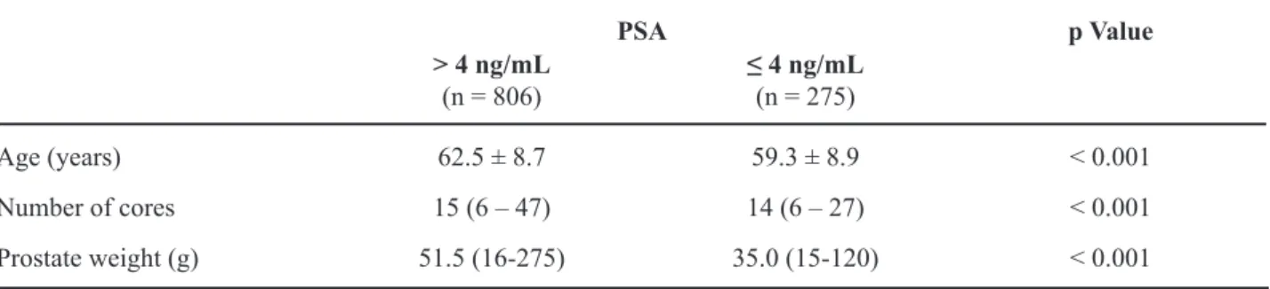
Patients with PSA ≤ 4 ng/mL were
signiicant-ly younger, with a mean age of 59.3 years (p < 0.001),
and had lighter prostate glands 35.0g compared with
51.5g when PSA > 4 ng/mL (p < 0.001) (Table-1). Considering the diagnosis, except for PIN, that was more frequently diagnosed in men with PSA ≤ 4 ng/mL, there was no statistical difference between
the diagnosis of benign, ASAP and adenocarcinoma
(p = 0.906) (Table-2).
Stratifying PSA levels for men with PSA ≤ 4.0 ng/mL, cancer was diagnosed in 1/21 (4.8%)
patients with PSA level ≤ 1.0 ng/mL, 10/34 (29.4%) with PSA 1.1 to 2 ng/mL, 23/82 (28.0%) with PSA 2.1 to 3 ng/mL and 54/138 (39.1%) with PSA 3.1 to 4 ng/mL.
The cancer characteristics were similar for both groups (Table-3). The median Gleason score was 7 for both (p = 0.078), the median of number of cores positive for tumor was 4 and 3, respectively, for PSA > 4 ng/mL and PSA ≤ 4 ng/mL (p = 0.627). Considering the total percent of tumor involving all cores, patients with PSA > 4 ng/mL had a median of 11% versus 7% for patients with PSA ≤ 4 ng/mL (p = 0.042).
Considering the 71 patients who had infor
-mation about the reason of the biopsy, we studied
the characteristics of those 33 who were clinical
staged T2 comparing with the 38 where digital rectal
Table 1 – Characteristics of 1081 patients submitted to prostate biopsy between January 2005 and October 2006 consid-ering the PSA level.
PSA p Value
> 4 ng/mL
(n = 806) ≤ 4 ng/mL(n = 275)
Age (years) 62.5 ± 8.7 59.3 ± 8.9 < 0.001
Number of cores 15 (6 – 47) 14 (6 – 27) < 0.001
Prostate weight (g) 51.5 (16-275) 35.0 (15-120) < 0.001
Table 2 – Diagnosis of prostate biopsy in 1081 patients with PSA ≤ 4 ng/mL and PSA > 4 ng/mL.
PSA
Diagnosis > 4 ng/mL ≤ 4 ng/mL Total
Benign 357 (44.3%) 112 (40.7%) 469 (43.4%)
PIN 120 (14.9%) 64 (23.3%) 184 (17.0%)
ASAP 41 (5.1%) 11 (4.0%) 52 (4.8%)
Adenocarcinoma 288 (35.7%) 88 (32.0%) 376 (34.8%)
Total 806 (74.6%) 275 (25.4%) 1081 (100.0%)
examination was normal. Among patients that had abnormalities in the digital rectal examination, cancer was diagnosed in 13 (39.4%), comparing with only 8 (21.1%) in 38 without abnormalities in the digital rectal examination (p < 0.0001). PSA levels were simi-lar for both groups, 2.54 ng/mL for T2 patients and 2.73 ng/mL for T1, as was the Gleason score, mean 6.7 for the T2 and 6.1 for T1. Tumors were larger for T2 lesions, with mean number of cores positive for tumor 3.9 and mean total percentage 13.0%, versus 2.3 cores and 2.7% for T1 lesions.
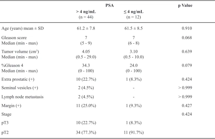
Fifty-six patients underwent radical pros-tatectomy and the indings are shown in Table-4. Twelve had PSA ≤ 4 ng/mL. There was no statistical difference between Gleason score and tumor volume for both groups of patients. The median Gleason score was 7 for both groups (p = 0.068), and tumor volume was 10% or 3.1 cm3 and 11% or 4.05 cm3 for ≤ 4 ng/mL and PSA > 4 ng/mL, respectively (p = 0.689 for percentage and p = 0.639 for cm3). There
were no differences between the groups regarding
extra-prostatic extension (p = 0.424), seminal vesicles iniltration (p > 0.999), lymph node metastasis (p > 0.999) and positive surgical margins (p = 0.427). One (8.3%) patient was stage pT3 with PSA ≤ 4 ng/mL and 10 were staged at this level (22.7%) with PSA > 4 ng/mL (p = 0.424).
In the group of patients with PSA ≤ 4 ng/mL there was no insigniicant cancer as deined by Ep-stein et al. (17) as a tumor volume of less than 0.5
cm3, Gleason score less than 7, and organ-conined. Additionally, one patient was stage pT3a, showing ex-tra-prostatic extension and positive surgical margin.
COMMENTS
In order to minimize economic impact in the health system and maximize the effectiveness of detecting and treating CaP, various studies have aimed to ind the best levels of PSA and its variations, especially PSA density and PSA kinetics (18). CaP screening programs have shown that using 4.0 ng/mL as a cutoff results in only clinically signiicant tumors
being detected, and one third of the men treated for
radical prostatectomy disease that had progressed beyond the prostate (8). Lowering PSA levels to 2.5 ng/mL seems to better detect organ-conined tumors, enhancing chances of disease-free and overall sur-vival, particularly in younger men (19,20). In asso-ciation with lowering PSA levels, the current practice of more intensive biopsy regimens could lead to the detection of non-signiicant tumors. It was the aim of our study to describe histopathological indings in extended prostate biopsy in patients with PSA levels lower than 4 ng/mL.
We have shown that cancer was diagnosed
in 32% of patients, which is the same proportion of
patients diagnosed with cancer with a PSA > 4 ng/mL, which is the traditional cutoff for prostate biopsy. We also observed that cancer was diagnosed even in pa-tients with a very low level of PSA, below 1 ng/mL, and there was signiicantly worse disease as PSA lev-els rose. Malignancy was observed in 29.4%, 28.0% and 39.1% of patients with PSA levels from 1.1 to 2 ng/mL, 2.1 to 3 ng/mL, and 3.1 to 4 ng/mL, respec-tively. Our numbers were even higher than those re-ported by the Prostate Cancer Prevention Trial, which
Table 3 – Tumor characteristics in prostate biopsies of 376 patients with PSA ≤ 4 ng/mL and PSA> 4 ng/mL.
PSA p Value
> 4 ng/mL
(n = 288) ≤ 4 ng/mL(n = 88)
Gleason score 7 (4 - 10) 7 (5 - 9) 0.078
Number of cores positive for tumor 4 (1 - 18) 3 (1 - 14) 0.627
indicated that the overall cancer detection was 15.2%. They found cancer in 6.6% of patients when PSA was
less than 0.5 ng/mL, 10.1% when it was between 0.6 to 1.0 ng/mL, 17.0% from 1.1 to 2 ng/mL, 23.9%, from 2.1 to 3.0 ng/mL, and 26.9% when PSA was 3.1
to 4 ng/mL (21). The median PSA value for men in their 40s and 50s is approximately 0.7 ng/mL and 0.9 ng/mL, respectively, and a baseline PSA level greater
than the median for each age group was related to a 12
to 22-fold greater risk of having CaP (22). Although the American Cancer Society Guidelines recommend screening for CaP before age 50 only in men with risk factors for CaP, including African-American descent or a strong family history of CaP, authors have recom-mended the measurement of baseline PSA at age 40, which could allow the determination of PSA kinetics, and is a sensitive marker for prostate cancer diagnosis
and prognostic prediction (22). This knowledge may be changing the standard practice of urology since in this present study patients with PSA ≤ 4 ng/mL were signiicantly younger. Bill-Axelson et al. (23) have claimed that initiating screening before age 50 and detecting cancer earlier should prevent death, especially because patients undergoing radical pros-tatectomy younger than 65 years-old have reduced CaP-speciic mortality. Sun et al. (18) have previously shown that in patients younger than 50, PSA levels of 2.5 ng/mL have speciicity of 94% for detecting cancer, and strongly recommend measuring PSA in younger men. Together with the number of patients we have just found, biopsy should be recommended when PSA is higher than the median for that speciic
age, since almost one third of men will be diagnosed CaP.
Table 4 – Patient age and tumor characteristics in radical prostatectomy specimens when PSA was ≤ 4 ng/mL or > 4 ng/mL.
PSA p Value
> 4 ng/mL
(n = 44) ≤ 4 ng/mL(n = 12)
Age (years) mean ± SD 61.2 ± 7.8 61.5 ± 8.5 0.910
Gleason score
Median (min - max) (5 - 9)7 (6 - 8)7 0.068
Tumor volume (cm3)
Median (min - max) (0.5 - 29.0)4.05 (0.5 - 10.0)3.10 0.639 %Gleason 4
Median (min - max) (0 - 100)34.3 (0 - 100)24.0 0.079
Extra prostatic (+) 10 (22.7%) 1 (8.3%) 0.424
Seminal vesicles (+) 2 (4.5%) - > 0.999
Lymph node metastasis 2 (4.5%) - > 0.999
Margin (+) 11 (25.0%) 1 (9.3%) 0.427
Stage 0.424
pT3 10 (22.7%) 1 (8.3%)
Gleason score is the most important isolated prognostic factor, and we observed no difference in the Gleason score between the groups with PSA lower or higher than 4 ng/mL, which both had a median Gleason score of 7. Furthermore, tumor volume in prostate biopsy has been addressed as a very important predictor of cancer extension and outcome. Multiple measurements have been used, including number of positive cores, total millimeters of cancer amongst all cores, percentage of each core occupied by cancer and total percent of cancer in the entire specimen. The best method for determining tumor burden is not yet clear, but estimating a percentage is easy and has been dem-onstrated to be a useful predictor of tumor extension and cancer-free survival rate (24,25). In the current study we showed no difference considering the num-ber of cores compromised by tumor, but tumors were
smaller when PSA ≤ 4 ng/mL, with a total percentage
of 7% against 11% when PSA > 4 ng/mL. Smaller tumors are also more likely to be organ-conined. This was not conirmed for patients undergoing radical prostatectomy where tumor characteristics, including volume were very similar to those with PSA higher than 4 ng/mL. One explanation for this fact is a bias considering the choice of treatment. Urologists could have preferred surgery for those patients with other associated adverse characteristics leading to similar results, mostly taking into account tumor volume. This data needs to be clariied in further series.
The detection of organ-conined cancer when PSA is lower than 4 ng/mL could cause some appre-hension in treating “harmless” or insigniicant cancer. Insigniicant cancer is deined as tumor with Gleason pattern less than 4 or 5, organ-conined and volume
less than 0.5 cm3 (17). Reports of fewer than 10% of insigniicant cancers have been published, and our
series is in agreement with the literature since we did
not ind any clinically insigniicant cancer. In addition
to the low number of patients who underwent radical
prostatectomy with PSA ≤ 4 ng/mL, our data show tumors that can not be considered insigniicant, with mean Gleason score 6.6, median 7, ranging from 6 to 8. In addition it is known that presence of tertiary Gleason 4 or 5, and the percent of a higher Gleason pattern impact the prognosis of prostate cancer. The mean percent of Gleason pattern 4 for this group of
patients was 32%, which means a 30% reduction in
disease free survival in 10 years (26). Considering tumor volume, McNeal (27) had found good prognosis for tumors with volume less than 4 cm3. The mean tumor volume of our surgical specimens was 3.9 cm3,
but 33% were higher than 5 cm3, with one 10 cm3,
which could be considered a huge tumor, with a 33%
probability of recurrence in 10 years (26).
One limitation of our study was the lack of data of PSA velocity (PSAV). PSAV measurement has been shown to be very helpful, as clinically signiicant prostate cancer is more likely to be found in men with a rapidly rising PSA. Studies suggest that for men with a total PSA higher than 4 ng/mL, a PSA velocity of 0.75 ng/mL/year is an indication for biopsy. However, in men whose total PSA level is lower than 4 ng/mL, an ideal cutoff has not yet been determined and should range from 0.1 to 0.5 ng/mL/year (28-30). It has been demonstrated that for each 0.1 ng/mL per year increase in PSA, the likelihood of death from prostate cancer in-creases 15%. For men with a consistent increase in PSA of 0.35 ng/mL per year or higher, the relative risk of dying of prostate cancer is 5 times higher in the next 2 to 3 decades than for men with lower PSA increases(31). Nevertheless, this weak point may be overcome by the indings recently published by Yu X et al. (32). These authors have shown a correlation between total PSA and PSAV, describing a PSAV of more than 2 ng/mL per year in only 1% and 14% of patients whose PSA total levels were lower than 2.5 ng/mL or between 2.5 ng/mL and 4 ng/mL, respectively, indicating a less aggressive and more curable disease.
In conclusion, our indings show that in the extended biopsy era cancer will be detected in 32% of patients when biopsy is performed with PSA below 4 ng/mL. Gleason score and number of cores positive for cancer are similar to those with PSA > 4 ng/mL.
Although cancer characteristics in radical prostatec
CONFLICT OF INTEREST
None declared.
REFERENCES
1. Amling CL, Blute ML, Lerner SE, Bergstralh EJ, Bostwick DG, Zincke H: Inluence of prostate-spe-ciic antigen testing on the spectrum of patients with prostate cancer undergoing radical prostatectomy at a large referral practice. Mayo Clin Proc. 1998; 73: 401-6.
2. D’Amico AV, Whittington R, Malkowicz SB, Fon-durulia J, Chen MH, Tomaszewski JE, et al.: The combination of preoperative prostate speciic antigen and postoperative pathological indings to predict prostate speciic antigen outcome in clinically local-ized prostate cancer. J Urol. 1998; 160: 2096-101. 3. Stephenson RA: Population-based prostate cancer
trends in the PSA era data from the Surveillance, Epi-demiology, and End Results (SEER) Program. Monogr Urol. 1998; 19: 3-19.
4. Newcomer LM, Stanford JL, Blumenstein BA, Brawer MK: Temporal trends in rates of prostate cancer: de-clining incidence of advanced stage disease, 1974 to 1994. J Urol. 1997; 158: 1427-30.
5. Catalona WJ, Loeb S: The PSA era is not over for prostate cancer. Eur Urol. 2005; 48: 541-5.
6. Han M, Partin AW, Piantadosi S, Epstein JI, Walsh PC: Era speciic biochemical recurrence-free survival following radical prostatectomy for clinically localized prostate cancer. J Urol. 2001; 166: 416-9.
7. Catalona WJ, Smith DS, Ratliff TL, Dodds KM, Coplen DE, Yuan JJ, et al.: Measurement of prostate-speciic antigen in serum as a screening test for prostate cancer. N Engl J Med. 1991; 324: 1156-61. Erratum in: N Engl J Med 1991; 325: 1324.
8. Catalona WJ, Smith DS, Ratliff TL, Basler JW: De-tection of organ-conined prostate cancer is increased through prostate-speciic antigen-based screening. JAMA. 1993; 270: 948-54.
9. Catalona WJ, Ramos CG, Carvalhal GF, Yan Y:
Low-ering PSA cutoffs to enhance detection of curable
prostate cancer. Urology. 2000; 55: 791-5.
10. Krumholtz JS, Carvalhal GF, Ramos CG, Smith DS, Thorson P, Yan Y, et al.: Prostate-speciic antigen
cutoff of 2.6 ng/mL for prostate cancer screening is
associated with favorable pathologic tumor features. Urology. 2002; 60: 469-73; discussion 473-4.
11. Schröder FH, van der Cruijsen-Koeter I, de Koning HJ, Vis AN, Hoedemaeker RF, Kranse R: Prostate cancer detection at low prostate speciic antigen. J Urol. 2000; 163: 806-12.
12. Zhu H, Roehl KA, Antenor JA, Catalona WJ: Biopsy of men with PSA level of 2.6 to 4.0 ng/mL associated with favorable pathologic features and PSA progres-sion rate: a preliminary analysis. Urology. 2005; 66: 547-51.
13. Siu W, Dunn RL, Shah RB, Wei JT: Use of extended pattern technique for initial prostate biopsy. J Urol. 2005; 174: 505-9.
14. Epstein JI, Amin M, Boccon-Gibod L, Egevad L, Humphrey PA, Mikuz G, et al.: Prognostic factors and
reporting of prostate carcinoma in radical prostatec
-tomy and pelvic lymphadenec-tomy specimens. Scand J Urol Nephrol Suppl. 2005; 216: 34-63.
15. Humphrey PA, Vollmer RT: Intraglandular tumor ex-tent and prognosis in prostatic carcinoma: application of a grid method to prostatectomy specimens. Hum Pathol. 1990; 21: 799-804.
16. Greene FL, Page DL, Fleming ID: AJCC Cancer Stag-ing Manual. 6th edition. New York, SprStag-inger; 2002. 17. Epstein JI, Walsh PC, Carmichael M, Brendler CB:
Pathologic and clinical indings to predict tumor extent of nonpalpable (stage T1c) prostate cancer. JAMA. 1994; 271: 368-74.
18. Sun L, Moul JW, Hotaling JM, Rampersaud E, Dahm P, Robertson C, et al.: Prostate-speciic antigen (PSA) and PSA velocity for prostate cancer detection in men aged <50 years. BJU Int. 2007; 99: 753-7.
19. Dall’Oglio MF, Crippa A, Passerotti CC, Nesrallah LJ, Leite KR, Srougi M: Serum PSA and cure perspective
for prostate cancer in males with nonpalpable tumor.
Int Braz J Urol. 2005; 31: 437-44.
20. Catalona WJ, Ramos CG, Carvalhal GF, Yan Y:
Low-ering PSA cutoffs to enhance detection of curable
prostate cancer. Urology. 2000; 55: 791-5.
21. Thompson IM, Pauler DK, Goodman PJ, Tangen CM, Lucia MS, Parnes HL, et al.: Prevalence of prostate cancer among men with a prostate-speciic antigen level < or =4.0 ng per milliliter. N Engl J Med. 2004; 350: 2239-46. Erratum in: N Engl J Med. 2004; 351: 1470.
22. Loeb S, Roehl KA, Antenor JA, Catalona WJ, Suarez BK, Nadler RB: Baseline prostate-speciic antigen compared with median prostate-speciic antigen for age group as predictor of prostate cancer risk in men younger than 60 years old. Urology. 2006; 67: 316-20.
EDITORIAL COMMENT
This is a well conducted study concluding that when PSA is below 4 ng/mL, cancer is detected
in a proportion equal to the proportion diagnosed
with a PSA > 4 ng/mL, and tumor characteristics are similar between the two groups. These find-ings are supported by other studies. Krumholtz et al. (1) evaluated the pathologic characteristics of clinical stage T1c prostate cancers detected in the 2.6 to 4.0 ng/mL PSA range and compared them with cancers concurrently detected in the 4.1 to 10.0 ng/mL. The authors found that men detected
at the 2.6 to 4.0 ng/mL PSA range had significantly smaller cancer volumes however, no difference was found in the proportion of tumors that met previ-ously published criteria of “clinically insignificant” (organ confined, less than 0.2 cm3 tumor volume, and Gleason sum 6 or less) or “clinically unimport-ant” (organ confined, less than 0.5 cm3 tumor vol-ume, and Gleason sum 6 or less) tumors. Using the
lower PSA cutoff point resulted in the detection of
a significantly higher percentage of organ-confined tumors. The authors conclude that the use of a 2.6
versus watchful waiting in early prostate cancer. N Engl J Med. 2005; 352: 1977-84.
24. Rubin MA, Bassily N, Sanda M, Montie J, Strawder-man MS, Wojno K: Relationship and signiicance of greatest percentage of tumor and perineural invasion on needle biopsy in prostatic adenocarcinoma. Am J Surg Pathol. 2000; 24: 183-9.
25. Freedland SJ, Csathy GS, Dorey F, Aronson WJ: Clini-cal utility of percent prostate needle biopsy tissue with cancer cutpoints to risk stratify patients before radical prostatectomy. Urology. 2002; 60: 84-8.
26. Stamey TA, McNeal JE, Yemoto CM, Sigal BM, John-stone IM: Biological determinants of cancer progres-sion in men with prostate cancer. JAMA. 1999; 281: 1395-400.
27. McNeal JE: Prostate cancer volume. Am J Surg Pathol. 1997; 21: 1392-3.
28. Fang J, Metter EJ, Landis P, Carter HB: PSA velocity for assessing prostate cancer risk in men with PSA
levels between 2.0 and 4.0 ng/ml. Urology. 2002; 59: 889-93; discussion 893-4.
29. Berger AP, Diebl M, Steiner H, Bektic J, Pelzer A,
Leon-hartsberger N et al., Longitudinal PSA changes in men
with and without prostate cancer: assessment of prostate cancer risk. J Urol. 2005; 173 (Suppl. 4): 402.
30. Catalona WJ, Loeb S: The PSA era is not over for prostate cancer. Eur Urol. 2005; 48: 541-5.
31. Carter HB, Ferrucci L, Kettermann A, Landis P, Wright EJ, Epstein JI, et al.: Detection of life-threatening prostate cancer with prostate-speciic antigen veloc-ity during a window of curabilveloc-ity. J Natl Cancer Inst. 2006; 98: 1521-7.
32. Yu X, Loeb S, Roehl KA, Han M, Catalona WJ: The association between total prostate speciic antigen concentration and prostate speciic antigen velocity. J Urol. 2007; 177: 1298-302; discussion 1301-2.
Accepted after revision: February 12, 2008
Correspondence address: Dr. Katia R. M. Leite Rua Adma Jafet, 91
Sao Paulo, SP, 01308-050, Brazil Fax: +55 11 3231-2249
ng/mL PSA threshold for screening resulted in the
more frequent detection of small, organ-confined tumors without over detecting possibly clinically insignificant ones. Obviously, additional studies in larger populations with longer follow-up are
needed to confirm these findings.
REFERENCE
1. Krumholtz JS, Carvalhal GF, Ramos CG, Smith DS, Thorson P, Yan Y, et al.: Prostate-speciic antigen
cutoff of 2.6 ng/mL for prostate cancer screening is
associated with favorable pathologic tumor features. Urology. 2002; 60: 469-73; discussion 473-4.
Dr. Athanase Billis
Full-Professor of Pathology State University of Campinas, Unicamp Campinas, Sao Paulo, Brazil E-mail: [email protected]
EDITORIAL COMMENT
Early in the PSA era patients with a serum prostate-speciic antigen (PSA) level > 4.0 ng/mL and a normal digital rectal examination (DRE) were recommended to undergo prostate biopsy because of a 20-30% risk of prostate cancer at a pre-speciied sensitivity of 95%. The majority of such patients have clinically important cancers and the rate of indolent disease, deined as specimen Gleason score 2-6, no extra-prostatic extension, and no Gleason pattern 4/5 is generally < 20%. Many have argued that a PSA threshold for biopsy of 4.0 is more frequently associated with under- rather than over-diagnosis as rates of non-organ-conined cancer (25-35%) are 2 to 4 times higher than indolent cancers, whereas cancers detected in the 2.6-4.0 PSA range are more likely to be organ-conined without substantial dif-ferences in the rate of low-grade or indolent cancer. In a longitudinal screening study, a decreased risk of PSA-deined biochemical recurrence was observed for patients treated by radical prostatectomy after lowering the PSA threshold for biopsy from 4.0 to 2.5 (1). As such, a lowering of the PSA level for biopsy to 2.5 has been advocated to increase the detection of clinically signiicant cancers at a more curable stage,
and this had been adopted in the guidelines of some
professional societies (2).
The Prostate Cancer Prevention Trial (PCPT) has challenged the validity of any PSA threshold for biopsy as no speciic PSA value had suficient sen-sitivity and speciicity for the detection of prostate cancer to be clinically useful (3). Based on the results of patients who had an end-of-study biopsy without
usual clinical implications, there was a continuum of
cancer risk at all PSA values. Among patients with a PSA < 1.0, 1.1-2.0, and 2.1-3.0, the cancer detection rate was 9%, 17%, and 24%, respectively and the corresponding proportion of cancers graded as Glea-son 7-10 was 11%, 12% and 19% (4). These results indicate that there is no PSA below which the risk of having cancer is zero.
In the current study, Leite et al. report on the biopsy and pathological characteristics of a cohort of patients biopsied with a PSA < 4. Reasons for biopsy included abnormal DRE, prior biopsies showing
atyp-ical small acinar proliferation or prostatic intraepithe
-lial neoplasia, persistently elevated PSA and negative prior biopsy, or a positive family history, so that the population studied is not fully representative of the general population usually subjected to opportunistic screening. Nonetheless the indings are illuminating,
demonstrating similar rates of prostate cancer in those
in tumor grade (median score 7 in both groups), and slightly fewer positive cores in the PSA < 4 group. In the small subset of patients who underwent radi-cal prostatectomy, there was no difference in tumor volume, grade, or pathological stage. Surprisingly, and unlike our own experience with similar patients
where the incidence of indolent cancers is higher in
men with PSA < 4, the authors found no indolent can-cers as deined by Epstein’s criteria of organ-conined tumors of volume < 0.5 cc and grade < 7. This likely relects the indications for biopsy in this population
and less widespread and repeated screening than in
the United States.
What then is the optimal PSA cutoff for rec
-ommending biopsy in 2008? The theoretical answer is that the optimal threshold is one that maximizes detection of biologically signiicant but curable can-cers, reduces prostate-cancer-speciic mortality, and minimizes over-diagnosis of indolent disease. The practical answer is one that recognizes that PSA represents a continuum of risk that is also impacted by many other factors, and that the best way decides whom to biopsy includes a consideration of all of the relevant factors. At the Cleveland Clinic, we have
stopped reporting a “normal” cutoff for PSA on our
lab reports and substituted the following: “Published data from the Prostate Cancer Prevention Trial dem-onstrated that there is no PSA level below which the risk of having prostate cancer is zero. For an indi-vidual patient, the signiicance of a PSA level should be interpreted in a broad clinical context, including age, race, family history, digital rectal exam, prostate size, results of prior prostate biopsy, and use of 5 alpha
reductase inhibitors. Considering the high incidence
of asymptomatic cancer in the general population that may not pose an ultimate risk to the patient, the deci-sion to recommend urological evaluation or prostate biopsy should be individualized after considering all of these factors.” We have encouraged the use of the PCPT risk calculator (available at www.compass. fhcrc.org/edrnnci/bin/calculator/main.asp) as one tool (validated published nomograms for this purpose also exist) to achieve the goal of deining individual risk prior to recommending biopsy. Using this calculator, a 55 year old Caucasian male with a negative DRE, a PSA of 1.5, and no family history of prostate cancer has a 19% risk of having prostate cancer but only a
2% risk of having high grade (Gleason 7 or greater) disease, information that can give the patient a more precise estimate of the risks and beneits of undergo-ing biopsy before decidundergo-ing whether to have it done. For a 55 year old African American man with a nor-mal DRE, a positive family history, and PSA of 2.4, the calculator estimates a risk of any cancer at 31% and of high grade cancer at 8%; here the risk: beneit ratio probably justiies biopsy even though his PSA is generally considered below the current threshold. Adoption of this approach outside of the U.S. requires construction and validation of similar models on local populations; ultimately, proof of the utility of PSA
screening at all awaits the reporting of large screen
-ing trials (the ERSPC and PLCO) currently near-ing
completion.
REFERENCES
1. Jang TL, Han M, Roil KA, Hawkins SA, Catalona WJ: More favorable tumor features and progression-free survival rates in a longitudinal prostate cancer screen-ing study: PSA era and threshold-speciic effects. Urology. 2006; 67: 343-8.
2. Smith RA, Cokkinides V, Eyre HJ: American Cancer Society guidelines for the early detection of cancer, 2006. CA Cancer J Clin. 2006; 56: 11-25; quiz
49-50.
3. Thompson IM, Ankerst DP, Chi C, Lucia MS, Good-man PJ, Crowley JJ, et al.: Operating characteristics of prostate-speciic antigen in men with an initial PSA level of 3.0 ng/ml or lower. JAMA. 2005; 294: 66-70.
4. Thompson IM, Pauler DK, Goodman PJ, Tangen CM, Lucia MS, Parnes HL, et al.: Prevalence of prostate cancer among men with a prostate-speciic antigen level < or =4.0 ng per milliliter. N Engl J Med. 2004; 350: 2239-46. Erratum in: N Engl J Med. 2004; 351: 1470.