QUANTIFICATION OF TUMOR EXTENSION IN PROSTATE BIOPSIES –
IMPORTANCE IN THE IDENTIFICATION OF CONFINED TUMORS
KÁTIA R.M. LEITE, MIGUEL SROUGI, RUY G. BEVILACQUA, MARCOS DALL’OGLIO,
CÁSSIO ANDREONI, JOSÉ R. KAUFMANN, LUCIANO NESRALLAH, ADRIANO
NESRALLAH, LUIZ H. CAMARA-LOPES
Laboratory of Surgical and Molecular Pathology, Syrian Lebanese Hospital, Sao Paulo, Discipline of Urology, Paulista School of Medicine, Federal University of Sao Paulo (UNIFESP), and Department of
Surgery, University of Sao Paulo School of Medicine (USP), SP, Brazil
ABSTRACT
Objective: To assess the importance of quantifying the adenocarcinoma in prostate biopsies when determining the tumor’s final stage in patients who undergo radical prostatectomy. To identify the best methodology for obtaining such data.
Patients and Methods: Prostate biopsies from 132 patients were examined, with determina-tion of Gleason histological grade and tumor volume in number of involved fragments, tumor extent of the fragment mostly affected by the tumor and the total percentage of tumor in the specimen. Theses parameters were statistically correlated with the neoplasia’s final stage following the evalua-tion of radical prostatectomy specimens.
Results: An average of 12 and a median of 14 biopsy fragments were evaluated per patient. In the univariate analysis the Gleason histological grade, the largest tumor extent in one fragment and the total percentage of tumor in the specimen were correlated with tumor stage of the surgical speci-men. In the multivariate analysis, the Gleason histological grade and the total percentage of tumor were strongly correlated with the neoplasia’s final stage. The risk of the tumor not being confined was 3 for Gleason 7 tumors and 10.6 for Gleason 8 tumors or above. In cases where the tumor involved more than 60% of the specimen, the risk of non-confined disease was 4.4 times. Among 19 patients with unfavorable histological parameters, Gleason > 7 and extension greater than 60% the tumor final stage was pT3 in 95%.
Conclusion: When associated to the Gleason histological grade, tumor quantification in pros-tate biopsies is an important factor for determining organ-confined disease, and among the methods, total percentage of tumor is the most informative one. Such data should be included in the pathologi-cal report and must be incorporated in future nomograms.
Key words: prostate neoplasms; biopsy, needle; pathology; quantitative evaluation; neoplasms staging Int Braz J Urol. 2003; 29: 497-501
INTRODUCTION
The selection of the best treatment for the prostate cancer patient depends basically on the sta-tus of the primary tumor. Curative therapies are indi-cated exclusively for confined tumors, with
histologi-cal grade. Currently the literature has discussed the importance of quantifying the tumor in the prostate biopsy (2-4). Such assessment can be made in sev-eral ways, such as the measurement of the neoplasia in millimeters, the analysis of percentage of tumor in every fragment, the percentage of the most involved fragment and the number of fragments that are infil-trated by the neoplasia.
The objective of this study is to assess the importance of quantifying the carcinoma in prostate biopsies, when determining the tumor final stage.
PATIENTS AND METHODS
The study comprises the retrospective analy-sis of prostate biopsies from 132 patients with mean age of 63 years, who underwent radical prostatectomy between January 1999 and March 2001. The mean number of analyzed fragments was 12, the median 14, ranging from 5 to 14. The mean and the median values for fragment length was 13 mm, ranging from 7 to 18 mm. Biopsies were fixed in formalin 10%, embedded in paraffin, stained by hematoxylin-eosin and analyzed by one only pathologist (KRML). The Gleason histological grade was used for evaluating the histological differentiation and for statistical analysis, and was divided in 3 groups, ≤ 6, 7, and > 7. For quantifying the tumor, the following were ana-lyzed: 1) Relationship between the number of posi-tive fragments over the total of biopsied fragments, 2) Percentage of a fragment that is more involved by the neoplasia, 3) Total percentage of tumor in the frag-ments, that is, the arithmetic mean between the per-centages of each isolated fragment.
The specimens of radical prostatectomy were fixed in buffered formalin 10% for a period of 4 to 16 h. Each gland was submitted to histological study in accordance to the previously described recommen-dations (5). After weighting and measuring the gland, thin transversal sections were performed in the surgi-cal margins related to the bladder neck and the pros-tate apex. The seminal vesicles were sectioned in the base and longitudinal sections were submitted to his-tological examination. The entire gland was included for study after having their margins painted with In-dia ink. The right and left lobes were separated, with
sequential transversal sections being performed ev-ery 3 mm, designed from the proximal region towards the distal one. Between 10 and 12 sections from each lobe were included for histological study. The lymph nodes from the fat related to the resection of the iliac chain were dissected and sections representative of each nodular structure were included for study. The specimens of radical prostatectomy underwent the usual processing with inclusion in paraffin. Sections of 4 to 6 µm were stained by hematoxylin-eosin, and analyzed by one only pathologist as well (KRML).
The Gleason histological grade was used for evaluating the histological differentiation. The assess-ment of tumor extent was performed with the aid of the grid card, as described by Humphrey & Vollmer (6). The invasion of adipose tissue and the peripros-tatic neurovascular plexus was considered as involve-ment of extra-prostatic tissue and, therefore, non or-gan-confined disease. The staging system was TNM 2002 (7).
Non-parametric analyses (Mann-Whitney) were performed for assessing the significance between the biopsy variables and the neoplasia’s final stage. The qui-square test was used for evaluating the Gleason score and the status of the surgical speci-men. The multivariate logistic regression determined the relative risk of non organ-confined disease for the multiple variables. The tests were performed in the software SPSS version 11 (SPSS Inc. Chicago, IL).
RESULTS
Table 1 – Pre-operative clinical and pathological charac-teristics of the 132 patients under study.
Patients (N)
Mean Age ± SD
Gleason Score (%)
5 –6 7 8 – 9 Mean ± SD Median
% Positive Fragments
Mean ± SD Median
Total % of Tumor
Mean ± SD Median
% of Tumor in 1 Fragment
Mean ± SD Median
132
63 + 8.4
67 (50.8) 26 (19.7) 39 (29.5) 6.7 + 1.2 6
29 + 19.4 25
35 + 29.2 25
57 + 28.8 60
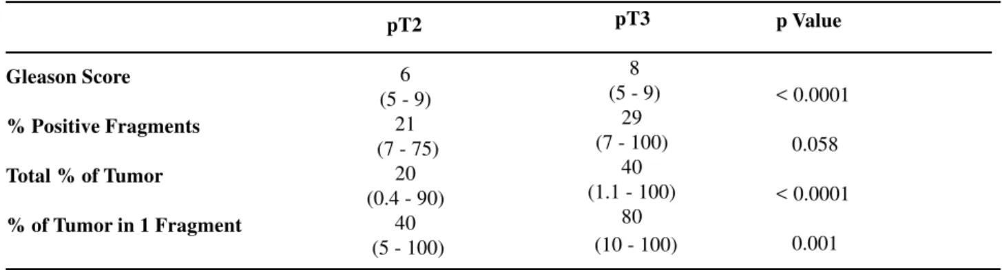
Table 2 – Univariate relationbetween pre-operative factors and final pathological stage *. Median and range.
Gleason Score
% Positive Fragments
Total % of Tumor
% of Tumor in 1 Fragment
pT2
6 (5 - 9)
21 (7 - 75)
20 (0.4 - 90)
40 (5 - 100)
pT3
8 (5 - 9)
29 (7 - 100)
40 (1.1 - 100)
80 (10 - 100)
p Value
< 0.0001
0.058
< 0.0001
0.001
* The qui-square test was used to calculate the significance of the Gleason score. For the other variables, the Mann-Whitney non-parametric test was used.
The univariate analysis demonstrated a sta-tistically significant difference for confined and non-confined tumors concerning larger tumor extension in one single biopsy fragment, total percentage of tumor and Gleason histological grade. Results are shown in Table-2.
Results from the multivariate analysis showed that there was statistical significance only for total percentage of tumor and Gleason histological grade concerning the tumor’s final stage. The risk of non-confined disease was 3 times higher for Gleason 7 tumors and 10.6 times for adenocarcinomas with Gleason > 7 (p < 0.0001). The risk of involvement of extra-prostatic tissue was 4.4 times higher for those tumors that occupied more than 60% of the specimen (p = 0.002).
Nineteen cases were considered unfavorable, since they presented Gleason > 7 and total percent-age above 60%. Ninety five percent of these tumors were classified as pT3.
DISCUSSION
Our results show the power of tumor quanti-fication for determining the final stage of prostate adenocarcinomas. The current nomograms of Partin et al. (1) and the recently validated nomogram of Graefen et al. (8) include in the equation one single biopsy information (Gleason histological grade), with-out considering the tumor volume.
num-ber is 4 or above the percentage of non-confined tu-mor ranges from 47 to 100% (9-11).
Rubin et al. had already demonstrated the relation between different methods for quantifying the prostate carcinoma in biopsies and adverse patho-logical aspects of the surgical specimen (12). In univariate analysis, they showed that the probability of a tumor being no longer confined was 77% for tumors that involve more than 80% of a single bi-opsy fragment. Subsequently, Gao et al. confirmed the importance of such determination for low risk patients. While studying 62 patients, they showed that 38% of the tumors were no longer confined when there was an involvement of 25% or more in the extent of a single biopsy fragment (13). In our casuistry, the higher percentage of tumor in one single biopsy frag-ment was significant for determining the final stage only in univariate analysis. The median of the percent-age of tumor in one fragment was 40% in confined tumors and 80% in non-confined tumors (p = 0.001). In multivariate analysis, we demonstrated the value of total percentage of tumor in biopsies, together with Gleason histological grade for predicting of the tumor’s final status. The median of the total percent-age of tumor in biopsies was 20% for confined tu-mors and 40% for non-confined tutu-mors (p < 0.0001). More interestingly, the logistic regression analysis demonstrated a risk 4.4 times higher of non organ-confined disease for tumors involving 60% of biop-sies or more. Freedland et al. (14,15), were able to stratify patients with intermediate risk (Gleason 7 and/ or PSA of 10 to 20 ng/ml) and high risk (Gleason higher than 7 and/or PSA above 20 ng/ml) in sub-groups when they considered tumor extent in the bi-opsies. For patients with intermediate risk, the indexes of biochemical recurrence following radical prostate-ctomy were significantly higher in patients with in-volvement of more than 20% of the biopsy specimen. For high-risk patients, those with involvement of more than 55% of the specimen had higher indexes of bio-chemical recurrence following surgery.
Our data confirm those from the literature with an important differential that is the number of analyzed fragments. The mean and median of biop-sies analyzed per patient were respectively 12 and 14, twice as those analyzed by Freeland et al. (14).
Currently, the sextant biopsies with representation of only 6 fragments is considered insufficient for diag-nosing prostate tumors (16), and are being replaced by wider representations of the gland, thus our data are important for directing new analyses.
In addition to tumor quantification, we con-firmed the importance of the Gleason histological grade for identifying the final status of the tumor. We demonstrated a risk of non-confined disease that is 3 times higher for Gleason 7 tumors and 10.6 times for tumors with Gleason grade equal or higher than 8.
An interesting event was the identification of a group that we called unfavorable, whose histologi-cal grade was higher than 7 and total percentage of tumor was higher than 60%. Nineteen patients had tumors with such characteristics, and 95% of them were classified as pT3 after radical prostatectomy.
We concluded that tumor quantification in prostate biopsy is important for identifying non-con-fined tumors, and among the studied parameters, the total percentage of tumor was the most informative one, along with the Gleason histological grade. We suggest that this data is incorporated to the patho-logical report and that it is considered in the design of new nomograms.
REFERENCES
1. Partin A, Kattan MW, Subong EN, Walsh PC, Wojno KJ, Oesterling JE, et al.: Combination of prostate spe-cific antigen, clinical stage, and Gleason score to pre-dict pathological stage of localized prostate cancer: a multi-institutional update. JAMA. 1997; 277: 1445-51.
2. Graefen M, Haese S, Pichlmeier U, Hammerer PG, Noldus J, Butz K, et al.: A validated strategy for side specific prediction of organ confined prostate cancer: A tool to select for nerve sparing radical prostatec-tomy. J Urol. 2001; 165: 857-63.
3. Epstein JI, Potter SR: The pathological interpretation and significance of prostate needle biopsy findings: Implications and current controversies. J Urol. 2001; 166: 402-10.
5. Bostwick DG, Foster CS: Examination if radical pros-tatectomy specimens: Therapeutic and prognostic sig-nificance. In: Foster W, Bostwick D (eds.), ogy of Prostate, Series Major Problems in Pathol-ogy. Philadelphia, WB Saunders Co. 1998; vol. 34, p. 172.
6. Humphrey PA, Vollmer RT: Intraglandular tumor ex-tent and prognosis in prostatic carcinoma: aplication of a grid method to prostatectomy specimens. Hum Pathol. 1990; 21: 799-804.
7. AJCC CANCER Staging Manual, Chicago, Lippincott Raven. 2002, 6th. ed.
8. Graefen M, Karakiewicz PI, Cagiannos I, Quinn DI, Henchall SM, Grygiel JJ, et al.: International valida-tion of a preoperative nomogram for prostate cancer recurrence after radical prostatectomy. J Clin Oncol. 2002; 20: 3206-12.
9. Peller PA, Young DC, Marmaduke DP, Marsh WL, Badalament RA: Sextant prostate biopsies: a histo-pathologic correlation with radical prostatectomy specimens. Cancer. 1995; 75: 530-8.
10. Wills ML, Sauvageot J, Partin AW, Gurganus R, Epstein JI: Ability of sextant biopsies to predict radi-cal prostatectomy stage. Urology. 1998; 51: 759-64. 11. Sebo TJ, Bock BJ, Cheville JC, Lohse C, Wollan P,
Zincke H: The percent of cores positive for cancer in prostate needle biopsy specimens is strongly preditive
of tumor stage and volume at radical prostatectomy. J Urol. 2000; 163: 174-8.
12. Rubin MA, Bassily N, Sanda M, Montie J, Strawderman MS, Wojno K: Relationship and signifi-cance of greatest percentage of tumor and perineural invasion on needle biopsy in prostatic adenocarcinoma. Am J Surg Pathol. 2000; 24: 183-9.
13. Gao X, Mohideen N, Flanigan RC, Waters B, Mojcik EM, Leman CR: The extent of biopsy involvement as an independent predictor of extraprostatic extension and surgical margin status in low risk prostate cancer: implications for treatment selection. J Urol. 2000; 164: 1982-6.
14. Freedland SJ, Csathy GS, Dorey F, Aronson WJ: Clini-cal utility of percent prostate needle biopsy tissue with cancer cutpoints to risk stratify patients before radical prostatectomy. Urology. 2002; 60: 84-8.
15. Freedland SJ, Aronson EJ, Csathy GS, Kane CJ, Amling CL, Presti JC, et al.: Comparison of percent-age of total prostate needle biopsy tissue with cancer to percentage of cores with cancer for predicting PSA recurrence after radical prostatectomy: results from the search database. Urology. 2003; 61: 742-7.
16. Chab TY, Chan DY, Stutzman KLRE, Epstein JI: Does increases needle biopsy sampling of prostate detect a higher number of potentially insignificant tumors? J Urol. 2001; 166: 2181-4.
Correspondence address:
Dr. Katia Ramos Moreira Leite Rua Dona Adma Jafet, 91 São Paulo, SP, 01308-050, Brazil Fax: + 55 11 3231-2249
E-mail: [email protected]