395
On routine physical examination thepa-tient’s physician noted microscopic hematuria The inding was reconirmed by 2 Dipsticks over an interval of 4 months. The patient was a known diabetic, controlled by diet. Otherwise the patient was asymptomatic, without signiicant past medi -cal history at the time of this work-up, the 47 year old Caucasian male appeared to be in good general health. Laboratory data showed Hb of 15.1gm/dL, HCT 45%, RBC 4.8 million/uL, WBC 6200,Neu 62%, BUN 18 mg/dL, Creatinine 1.1 mg/dl, GRF 94 mL/min, A/G ratio 1.4, Glu 128 mg/dl, K 4.2 mmoL/L, Na 145 MMOL/L Cl 108 mmoL/L Urine analysis, spec grav 1018, 3-5 RBC/hpf, no WBC or bacteria on hpf, no casts, urine culture negative x 2. A KUB (Flat plate of abdomen) showed no opaque calculi nor other abnormalities. Cystos-copy and blue light cystosCystos-copy revealed no abnor-malities.
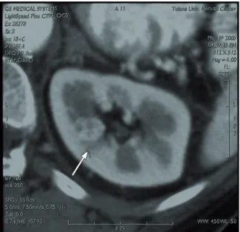
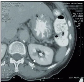
An enhanced 4 phase MDCT was per-formed The pre-enhancement phase was entirely unremarkable; no parenchymal lesions were de-tected. Following administration of 100 ml non-ionic contrast medium at a low rate of 5 mL/sec, the 12 second delayed arterial phase Ct demon-strated a relatively poorly enhancing 1.6 cm mass at the cortico-medullary junction (Figure-1), the lesion is much better shown on reversal image. Both the parenchymal phase CT( 50 second delay) and the excretory phase CT (4 minutes delay Fig-ure-2) demonstrate a non-enhancing 16 mm mass at the cortico-medullary junction (Figure-3).
In the light of a clinical history of diabe-tes and microscopic hematuria, the non-enhancing hypovascular mass seen on parenchymal and
ex-cretory phase CTs in the medulla might have been written off as Medullary Necrosis (With character-istic CT indings of a negative pre-enhancement phase CT, but a non-enhancing lesion shown on parenchymal and excretory phase; an early avas-cular necrosis) (1). However, the reversal image of the arterial phase CT clearly shows an enhancing lesion, though somewhat hypovascular for a RCC. The hypo-density on parenchymal and excretory phase CT relects the characteristic “wash-out” phenomenon of RCCs in these phases. The tumor having no tubules is less dense then adjacent nor-mal parenchyma The correct diagnosis was made, and a laparoscopic resection carried out.
Radiology Page
International Braz J Urol
The Elusive Renal Cell Carcinoma: Reversal Imaging of Arterial
Phase to Improve Acuity
Erich K. Lang, Karl Zhang, Quan Nguyen, Daniel Thorner, Ernest Rudman
Department Radiology and Urology, SUNY Downstate Medical School, Brooklyn, New York, NY, USA
Vol 37 (3): 395-396, May - June, 2011
396
Radiology PageREFERENCES
1. Lang EK, Macchia RJ, Thomas R, Davis R, Ruiz-Deya G, Watson RA, et al.: Multiphasic helical CT diagnosis of early medullary and papillary necrosis.
J Endourol. 2004; 18: 49-56.
2. Israel GM, Bosniak MA: How I do it: evaluating renal masses. Radiology. 2005; 236: 441-50.
_____________________ Correspondence address: Dr. Erich K. Lang
Departments of Urology and Radiology SUNY, Downstate Health Science Center 455 Lenox Road
Brooklyn, NY, 11203, USA E-mail: [email protected]
Figure 2 - lateral extension of the gas-dissection in the