Comprehensive Analysis of Etiology on the Prognosis of Urethral
Strictures
Rajkumar Mathur, Gaurav Aggarwal, Bhaskar Satsangi, Fareed Khan, Sudarshan Odiya
ABSTRACT
Department of Surgery, M.G.M Medical College & M.Y.H group of Hospitals, Indore, Madhya
Pradesh, India
Introduction: Urethral strictures remain a reconstructive dilemma, due to high incidence of recurrence and less than satisfactory outcomes. Even experienced surgeons following strict surgical principles have not achieved optimal results, leading us to think whether
the etiology of strictures dictate the outcome . We evaluated this “cause-effect” relationship highlighting the signiicance of the etiology on the overall prognosis of urethral strictures.
Materials and Methods: A total of 302 males with urethral strictures were assessed (both retrospectively and prospective-ly) over a period of ten years. The preoperative evaluation was performed by retrograde urethrogram, urethrosonogram, and urolowmetry and categorized, based on etiology: a) as post traumatic, b) post infective, c) iatrogenic or d) unknown. Traumatic strictures were subjected to pelvic X-ray and sub-categorized into grades A, B and C, following the TILE clas -siication. Patients were operated; with tunica albuginea urethroplasty for anterior strictures and U shape prostato-bulbar anastomosis for posterior strictures.
Results: Traumatic strictures accounted for 54% of cases. 127 of the 302 patients were treated using Tunica Albuginea Urethroplasty, while U shaped Prostatobulbar Anastomosis was performed on others. Post traumatic strictures had best outcome whereas post infective strictures had the worse outcome. Among strictures following pelvic fractures, TILE grades A and B had a better post operative course as compared to TILE C. Overall complication rate was 13.24%. Conclusion: Our study demonstrated that etiology of urethral strictures may play a vital role for the overall prognosis of urethral strictures.
Key words: Prognosis; urethral stricture; treatment outcome; etiology. Int Braz J Urol. 2011; 37: 362-370
INTRODUCTION
Urethral Stricture Disease encompasses a spectrum of divergent ailments that cause obliteration of the urethral lumen and slowing or blocking urinary low, secondary to ibrosis and healing of the urethral mucosa and surrounding tissues.
Urethral strictures have been a reconstructive dilemma for many years due to the high incidence of recurrence and less than satisfactory outcomes. A
thorough preoperative evaluation, appropriate surgi-cal planning, and adherence to basic surgisurgi-cal princi-ples, even in the hands of the most experienced sur-geon, have failed to achieve the desired results.
This has shifted the entire onus on the impact of a yet unexplored entity- the etiology.
Strictures are a common iatrogenic sequel to urethral instrumentation and catheterization. In -fective strictures have taken a back seat when com-pared with the rate of pelvic trauma and urethral manipulation, as well as with the advent of newer antibiotics and increasing public awareness as re-gards to sexually transmitted diseases (1).
Despite advancements in the ield of recon -structive urology, there is no consensus as the best mode of treating this complex entity, in order to op-timize the results.
We evaluated the “cause- effect” relation-ship between etiology of urethral stricture and out-come post-urethroplasty.
MATERIALS AND METHODS
This study included 302 male patients pre-senting with signs and symptoms of urethral stric-ture, for the irst time, over a period of ten years, both retrospectively as well as prospectively, from 1999 to 2009 [RKM, GA, BS, FK, SO]. Congenital strictures, as well as, patients managed without sur-gical reconstruction were excluded from the study.
A detailed preoperative assessment includ-ed careful history-taking and physical examination; followed by stricture evaluation via retrograde ure-throgram, urethrosonogram, and urolowmetry. Pa -tients were categorized, based on their stricture eti -ologies, as post traumatic, post infective, iatrogenic
(post catheterization and post instrumentation), or unknown.
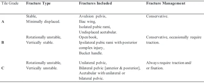
Patients with traumatic urethral strictures were then subjected to pelvic X-rays in both antero-posterior and lateral views, to identify the presence, as well as, type of pelvic fracture, and further sub-categorize them into grades A, B and C, of the TILE classiication (Table-1).
Irrespective of the etiology, if any patient had chronic retention of urine or complete outlow obstruction not amenable to urethral drainage, a urinary diversion in the form of suprapubic cystos-tomy was done.
A minimum wait of 6 weeks was allowed before urethroplasty to patients that required supra-pubic cystostomy. Tunica albuginea urethroplasty (TAU) was the procedure of choice for anterior ure-thral strictures and U shaped prostato-bulbar anas-tomosis (USPBA) for posterior urethral strictures, in order to minimize the inluence of the surgical procedure on the outcome (2,3). No modiications were performed as regards to the surgical tech-niques, over the course of the study.
Post urethroplasty, a silicon catheter was kept for three weeks in case of simple strictures and for six weeks in complex strictures. Complex cases included the combined strictures of both the ante-rior and posteante-rior urethra and the posteante-rior urethral strictures with false passages resulting from multiple attempts of urethral instrumentation before the pa-tients were referred to our center.
Table 1 - TILE Classiication of Pelvic Fractures.
TILE Classification of Pelvic Fractures.
Tile Grade Fracture Type Fractures Included Fracture Management
A
Stable,
M inimally displaced.
Avulsion pelvis, Iliac wing,
Isolated pubic rami, Undisplaced acetabular.
Conservative.
B
Rotationally unstable, Vertically stable.
Open book,
Ipsilateral pubic rami with posterior complex injury,
Bucket handle.
Conservative, occasionally require traction.
C
Rotationally unstable, Vertically unstable.
Unilateral pelvic,
Bilateral pelvic [anterior & posterior], Acetabular with unilateral or bilateral pelvic.
Patients were scheduled for an initial postop -erative assessment at the end of 3 months, with con-trast urethrogram, urethrosonogram, and urolometry. Post operative results were assessed by comparing pre and postoperative investigations and patient satisfac-tion (Table-2) and similarly at subsequent follow ups, at 6, 12 and 24 months to evaluate the long term re-sults and complications.
RESULTS
The results are exempliied both descriptively and statistically. Most of our patients were young with a mean age of 35.5 years old (range: 15-65 years).
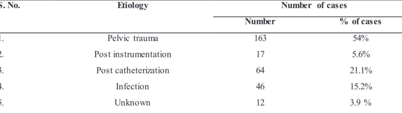
Traumatic strictures were the most common etiology found, accounting for 54% of all cases (Ta-ble-3). Although the most common stricture loca-tion was the membranous urethra (35%), the bulbar (30%) and penile (27.5%) urethra were equally af-fected and 7.5% patients had multiple urethral stric-tures.
Preoperative maximum low rate (Qmax) analysis showed a range of 3.0 to 12.5 mL/s, the mean low rate was 7.6 mL/s, which improved to 32 mL/s post operatively, ranging from 18 mL/s to 46 mL/s.
Average stricture length measured intra-operatively was 3.2 cm, ranging from 2.5 to 12.5 cm. A total of 127 patients (42%) were treated us-ing Tunica Albuginea Urethroplasty, while U shape Table 2 - Postoperative Result Assessment Criteria.
Postoperative Result
Criteria
GOOD FAIR POOR
Retrograde Urethrogram Good caliber. Partial narrowing
at stricture site.
Persistent stricture.
Urethrosonogram Patent and distensible
lumen.
Patent lumen with
decreased distensibility
Stricture present.
Uroflowmetry Qmax > 20 mL/sec Qmax: 15- 20 mL/sec Qmax < 15 mL/sec
Patient Satisfaction Satisfactory voiding, No instrumentation
needed.
Satisfactory voiding
but required
dilatations.
Not satisfied, not voiding or
voiding with thin stream,
need multiple dilatations or
repeat surgery.
Prostatobulbar Anastomosis was performed in 175 patients (48%).
Results were quantiied at regular intervals on the basis of the criteria enlisted in Table-1, evalu-ating patient’s satisfaction.
Loss of follow-up is a well recognized chal -lenge in any study but we collected data in 82% of our patients for two years.
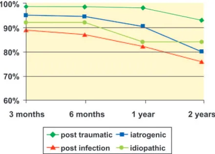
A comparison of the etiology regarding to success rates depicted that post traumatic strictures had the best overall outcome. Whereas, post infec-tive variety had the poorest results (Figure-1).
Among the strictures following pelvic frac-tures, those in TILE grades A and B had a better
post operative course as compared to TILE C vari -ety (Figure-2).
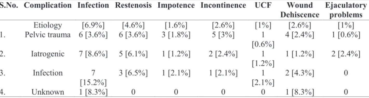
Overall complication rate in our study was 13.24% (40 patients). Re-stenosis occurred in 4.6% patients second only to post operative infection (6.9%). Analyses of complications based on the pri-mary cause are shown in Table-4.
DISCUSSION
Various studies have been done to identify the major etiologies for stricture formation, notwith-standing their actual impact on the long term out-come post surgical intervention.
Table 3 - Various etiologies encountered.
was published, with urethritis being the most com-mon cause at that time, in 40% of the cases (4). Other major series in the developed world, also report a 32% to 79% rate of iatrogenic causes (5,6). Jordan and Schlossberg, in 2002, suggested that most stric-tures in the recent era are a result of external trauma (7). Our study also observed such transition to the etiology of strictures, with 54% incidence of urethral strictures occurring post trauma.
Traumatic strictures most frequently involve the posterior urethra. Pelvic fracture causing disrup -tion at the bulbo-membranous junc-tion. Studies have reported an incidence of such strictures up to 31%, in countries with poor road conditions, as well as, inad-equate trafic regulations (8). Casselman and Schil -linger hypothesized that the mechanism of injury to the urethra in major pelvic fracture cases involves compression of the pelvic ring laterally, resulting in an increase in the antero-posterior diameter, leading to superior bladder displacement and consequent urethral stretching and avulsion (9). In our study, 54% of strictures encountered were of the traumatic etiology, with 84% of these patients having radio -logical evidence of pelvic fractures. Various types of pelvic fractures were seen, majority of injuries clas-siied as grades A and B (83.2%) of the TILE classi-ication system (10), with involvement of the inferior pubic rami being the most common type.
The TILE classiication of pelvic fractures is based on progressive instability of the pelvis (10). Type A fractures are stable with respect to rotation and vertical displacement, whereas type B fractures are rotationally unstable and vertically stable. Type C fractures are both vertically and rotationally unstable.
Classifying by level of instability is valuable in pre-dicting morbidity and mortality, as the pattern of ure-thral injury can be predicted on the basis of the type of pelvic fracture. The highest risk of urethral injury is found in a straddle fracture combined with diastasis of the sacroiliac joint (11). For every 1 mm increase of the pubic symphyseal diastasis, or displacement of the infero-medial pubic bone fracture fragments, the risk of urethral injury increases by 10% (12).
Post traumatic strictures tend to be short, oc -curring exclusively in the bulbar (5) and membranous urethra 87.73% of traumatic strictures in our study were seen to involve these sites, and 85% of these were less than 4 cm in length.
Management of post traumatic strictures usually involves preliminary diversion by suprapu-bic cystostomy, followed by deinitive repair several weeks later. The delayed urethroplasty after 3 months after initial trauma reduces complications of impo-tence and incontinence. In addition, the major advan -tage of delayed urethral reconstruction is that it can be done under controlled conditions when the patient has recovered from major associated injuries (13).
In our study, 6 weeks post suprapubic cystos -tomy, these patients were taken up for surgery; with tunica albuginea urethroplasty (TAU) being our pro-cedure of choice for anterior urethral strictures and U shaped prostato-bulbar anastomosis (USPBA) for posterior urethral strictures (2,3).
Postoperatively, periodic evaluation revealed a decline in the success rate over a period of time as depicted in our previous studies (2,3). A similar trend was evident in post traumatic strictures in the present study. Furthermore, the results were much better in
S. No. Etiology Number of cases
Number % of cases
1. Pelvic trauma 163 54%
2. Post instrumentation 17 5.6%
3. Post catheterization 64 21.1%
4. Infection 46 15.2%
patients falling in TILE category A, with 92% success rate at 2 years follow-up, when compared with 91.5% for TILE grade B and 77% in cases of TILE C, at the same time.
Numerous studies support the occurrence of complications after urethral injury following a pelvic fracture, due to the injury itself rather than the surgi-cal technique (14-16). We encountered complications in 26 patients with post traumatic strictures: infection,
re-stenosis, impotence, urinary incontinence, urethra-cutaneous istulae, wound dehiscence and ejaculatory disturbances, with rates acceptable in comparison to other studies (17,18).
Iatrogenic urethral injuries producing urethral strictures result from urethral instrumentation, ure-thral catheterization, and rarely following abdomino-perineal resection for carcinoma of the rectum (19). Prolonged catheterization and transurethral surgery Figure 1 - Etiology and success rates.
Iatrogenic strictures frequently require elabo -rate reconstructive procedures (25). Repeated ure-thral dilatation exacerbates short strictures, making them longer (26). The reconstructive urologist must consider the etiology, as well as, the prior treatments when planning whether to excise or graft the stenotic urethral segment.
Post operative short term follow-up results were encouraging for post traumatic strictures but the long term results were not so good.
Infection and re-stenosis were the most fre -quent complications encountered s, more so in the case of post catheterization strictures, whereas im -potence, incontinence, urethra-cutaneous istulae, wound dehiscence and ejaculatory disturbances were also seen.
Nevertheless, adequate and strict indications for urinary catheterization, skilled urethral catheter insertion and consideration of suprapubic catheter placement whenever prolonged catheterization is an -ticipated, should decrease the incidence of these iatro-genic strictures, which often tend to be multifocal or panurethral (27), and thus more dificult to treat.
Inlammatory strictures may be seen with gonorrheal infection, nonspeciic infections by chla -mydia and ureaplasma urealyticum, as well as, lichen sclerosus, usually beginning with inlammation of glans and inevitably leading to meatal stenosis. Pro -gression of stricture may involve the anterior and pos-terior urethra (28).
We encountered 46 patients (15.2%) with post infective strictures, with 66% of these located in the penile and bulbar urethra. Escherichia Coli was the most common isolate from the urine culture in these patients, suggesting that urethral obstruction are the main reasons for the ischemic urethral insult.
A rate of 32% to 79% of iatrogenic causes has been re-ported in different studies (5,6). We recorded a 26.8% incidence of iatrogenic urethral strictures.
Urethral stricture is a major late complication of TUR (2.2% to 9.8% of cases) (20, 21), as well as, radical (8.4%) (22) and simple (1.9%) (23) prostatec -tomy. Numerous causes of stricture formation post-TUR have been suggested, such as improper traumat-ic insertion of the resectoscope with perforation of the bulbous urethra and instrument friction at the peno-scrotal angle, eventually exacerbated by the narrow urethral caliber as well as monopolar current “leak” due to insuficient resectoscope isolation (20). More -over, patients undergoing such urological procedures are also catheterized in the post operative period. The exact cause, however, remains undetermined to date.
Prolonged catheterization, as in multiple trauma or burn patients, leads to urethral inlamma -tion and ischemia, and ultimately stricture forma-tion (5). Moreover, improper urethral catheter insertion is a preventable source of injury, with a recent report suggesting that 3.2 urethral injuries per 1000 inpa-tients occurred due to improper urethral catheteriza -tion (24).
Post instrumentation strictures are usually short and well deined, commonly located in the bul -bomembranous urethra, whereas those occurring post catheterization tend to be long and irregular, and more common at the penoscrotal junction (19), correlating well with our indings. A total of 60% of post instru -mentation strictures encountered by us were at the membranous and bulbar urethra, while 48% of post catheterization strictures were located at the penoscro -tal junction.
Complication rates based on stricture etiologies.
S.No. Complication Infection Restenosis Impotence Incontinence UCF Wound Dehiscence
Ejaculatory problems
Etiology [6.9%] [4.6%] [1.6%] [2.6%] [1%] [2.6%] [1%]
1. Pelvic trauma 6 [3.6%] 6 [3.6%] 3 [1.8%] 5 [3%] 1
[0.6%] 4 [2.4%] 1 [0.6%] 2. Iatrogenic 7 [8.6%] 5 [6.1%] 1 [1.2%] 2 [2.4%] 1
[1.2%]
1 [1.2%] 2 [2.4%]
3. Infection 7
[15.2%] 3 [6.5%] 1 [2.1%] 1 [2.1%] [2.1%]1 2 [4.3%] 0
4. Unknown 1 [8.3%] 0 0 0 0 1 [8.3%] 0
may induce organisms which are cleared rapidly from the normal urinary tract, to cause bacteruria, bactere-mia and even pyelonephritis (29).
Post urethroplasty evaluation at 6 months showed 86.95% success rate, which reduced to 75.67% at 2 years. A high rate of infection (15.2%) and re-stenosis (6.5%) was seen in the follow-up of inlammatory strictures, correlating with other simi-lar studies. None of the patients, however, faced any ejaculatory problems. Post infective strictures was the worst in terms of overall prognosis.
Idiopathic strictures or strictures without any apparent reason have been observed in different stud-ies (5,7,29). 3.9% of our patients fell into this catego-ry.
Although several theories have formulated, its mechanism remains unknown. But it may occur as a delayed manifestation of unrecognized child-hood trauma, even presenting as long as 18 years after perineal trauma (1). Many idiopathic strictures may develop as a consequence of inadequate canalization, when the part of the urethra derived from the uro-genital sinus joins the part derived from the urouro-genital folds, becoming symptomatic with growth (30). In elderly men, another explanation postulated is that id-iopathic strictures are mainly ischemic in origin (29).
These strictures are signiicantly more prev-alent in the bulbar area and more common in the younger age group (31). A total of 59% of idiopathic strictures in our study involved the bulbar urethra.
Our study demonstrates that post traumatic urethral strictures, secondary to pelvic fractures of the TILE category A, are shorter; mainly involve the bul-bar and membranous urethra, post surgical interven-tion have the best long term outcome, as well as, the lowest complication rate, and the best prognosis.
CONCLUSIONS
Post urethroplasty success has been attrib-uted to sevral factors,i.e.; location and length of the stricture, degree of spongioibrosis, the choice of sur-gical procedure and sursur-gical expertise. However, the impact of etiology regarding to outcome has remained unexplored. Our study demonstrated that the etiology may playsa vital role to the overall prognosis of ure-thral strictures treatment.
CONFLICT OF INTEREST
None declared.
REFERENCES
1. Baskin LS, McAninch JW: Childhood urethral inju -ries: perspectives on outcome and treatment. Br J Urol. 1993; 72: 241-6.
2. Mathur RK, Himanshu A, Sudarshan O: Technique of anterior urethra urethroplasty using tunica albuginea of corpora cavernosa. Int J Urol. 2007; 14: 209-13. 3. Mathur RK, Aggarwal H, Odiya S, Lubana PS:
U-shaped prostatobulbar anastomosis for urethral injury after pelvic trauma. ANZ J Surg. 2008; 78: 605-9. 4. De Sy WA, Oosterlinck W, Verbaeys A: Le traitement
du retrecissement de l uretre masculine. Acta Urol Belg. 1981; 49: 101-2.
5. Fenton AS, Morey AF, Aviles R, Garcia CR: Anterior urethral strictures: etiology and characteristics. Urol -ogy. 2005; 65: 1055-8.
6. Barbagli G, Guazzoni G, Lazzeri M: One-stage bulbar urethroplasty: retrospective analysis of the results in 375 patients. Eur Urol. 2008; 53: 828-33.
7. Jordan GH, Scholssberg SM: Surgery of the penis and urethra, (In) Walsh PC, Retik AB, Vaughan ED Jr, et al. (ed.), Campbell’s Urology, 8th ed. WB Saunders, Philadelphia, 2002; pp. 3886-952.
8. Tazi K, Nouri M, Med Moudouni S, Koutani A, Bena -tyaa A, Hachimi M, et al.: Traitment des stenoses in -lammatoires de l uretre par uretrotomie endoscopique. Ann Urol 2000; 34: 184-8.
9. Casselman RC, Schillinger JF: Fractured pelvis with avulsion of the female urethra. J Urol. 1977; 117: 385-6.
10. Tile M: Pelvic ring fractures: should they be ixed? J Bone Joint Surg Br. 1988; 70: 1-12.
11. Mouraviev VB, Santucci RA: Cadaveric anatomy of pelvic fracture urethral distraction injury: most inju -ries are distal to the external urinary sphincter. J Urol. 2005; 173: 869-72.
12. Basta AM, Blackmore CC, Wessells H: Predicting ure -thral injury from pelvic fracture patterns in male pa-tients with blunt trauma. J Urol. 2007; 177: 571-5. 13. Koraitim MM: Pelvic fracture urethral injuries: the un
-resolved controversy. J Urol. 1999; 161: 1433-41. 14. Kotkin L, Koch MO: Impotence and incontinence after
-anitis xerotica obliterans. BJU Int. 2000; 86: 459-65. 30. Andrich DE, Mundy AR: What is the best technique
for urethroplasty? Eur Urol. 2008; 54: 1031-41. 31. Lumen N, Hoebeke P, Willemsen P, De Troyer B, Pi
-eters R, Oosterlinck W: Etiology of urethral stricture disease in the 21st century. J Urol. 2009; 182: 983-7.
____________________ Accepted after revision:
November 16, 2010
______________________ Correspondence address: Dr. Rajkumar Mathur
Head, Department of Surgery
M.Y. Hospital and M.G.M Medical College Indore, Madhya Pradesh, 452001, India Fax: + 967 098 2604-0088
E mail: [email protected]
EDITORIAL COMMENT
Mathur and co-workers present a very exten-sive series on urethral stricture disease and its man-agement, as well as, prognostic factors. In their anal -ysis they found that more than 54% of the strictures were posttraumatic. Moreover they are able to even correlate pelvic fracture patterns with individual out-come. Overall complication rate with less than 15% is remarkably low in such a complex cohort of patients. In addition to the well-known technique of prostato-bulbar anastomosis, the authors employed their own technique of for anterior reconstruction. The so-called tunica albuginea urethroplasty offers the advantage of locally available substitute tissue even in case of oral disease precluding the use of buccal mucosa (1). As such we do require alternative graft sources in patients with oral disease allowing for comparable complica-tion rates (2). Genital skin remains such an alterna-tive donor site being important for the reconstrucalterna-tive armamentarium. The authors clearly demonstrate that post-inlammatory strictures yielded a poor outcome as compared to the posttraumatic ones. In fact this ob-servation is most likely due to underestimating the de-sult of injury or management? J Urol. 1996; 155:
1600-3.
15. Shenfeld OZ, Kiselgorf D, Gofrit ON, Verstandig AG, Landau EH, Pode D, et al.: The incidence and causes of erectile dysfunction after pelvic fractures associated with posterior urethral disruption. J Urol. 2003; 169: 2173-6.
16. Koraitim MM: On the art of anastomotic posterior ure -throplasty: a 27-year experience. J Urol. 2005; 173: 135-9.
17. Santucci RA, Mario LA, McAninch JW: Anastomotic urethroplasty for bulbar urethral stricture: analysis of 168 patients. J Urol. 2002; 167: 1715-9.
18. Elliott SP, Metro MJ, McAninch JW: Long-term fol -lowup of the ventrally placed buccal mucosa onlay graft in bulbar urethral reconstruction. J Urol. 2003; 169: 1754-7.
19. McCallum RW, Rogers JM, Alexander MW: The ra -diologic assessment of iatrogenic urethral injury. J Can Assoc Radiol. 1985; 36: 122-6.
20. Rassweiler J, Teber D, Kuntz R, Hofmann R: Com -plications of transurethral resection of the prostate (TURP)--incidence, management, and prevention. Eur Urol. 2006; 50: 969-79; discussion 980.
21. Lumen N, Oosterlinck W: Challenging non-traumatic posterior urethral strictures treated with urethroplasty: a preliminary report. Int Braz J Urol. 2009; 35: 442-9. 22. Elliott SP, Meng MV, Elkin EP, McAninch JW, Duch
-ane J, Carroll PR, et al.: Incidence of urethral stricture after primary treatment for prostate cancer: data From CaPSURE. J Urol. 2007; 178: 529-34; discussion 534. 23. Varkarakis I, Kyriakakis Z, Delis A, Protogerou V,
Deliveliotis C: Long-term results of open transvesical prostatectomy from a contemporary series of patients. Urology. 2004; 64: 306-10.
24. Kashei C, Messer K, Barden R, Sexton C, Parsons JK: Incidence and prevention of iatrogenic urethral inju -ries. J Urol. 2008; 179: 2254-7; discussion 2257-8. 25. Wessells H, Morey AF, McAninch JW: Single stage
reconstruction of complex anterior urethral strictures: combined tissue transfer techniques. J Urol. 1997; 157: 1271-4.
26. Park S, McAninch JW: Straddle injuries to the bulbar urethra: management and outcomes in 78 patients. J Urol. 2004; 171: 722-5.
27. Ruutu M, Alfthan O, Talja M, Andersson LC: Cytotox -icity of latex urinary catheters. Br J Urol. 1985; 57: 82-7.
28. Venn SN, Mundy AR: Urethroplasty for balanitis xe -rotica obliterans. Br J Urol. 1998; 81: 735-7.
-gree and extension of spongioibrosis. Ultrasound has been shown to be a valuable tool for this task before urethroplasty (3).
Nevertheless, Mathur et al remain to be con-gratulated for the comprehensive analysis of stricture etiology, pelvic fracture pattern and individual surgical outcome. Being aware of the relation between these features and surgical success allows us for a more ac-curate preoperative counseling in our patients.
REFERENCES
1. Mathur RK, Himanshu A, Sudarshan O: Technique of anterior urethra urethroplasty using tunica albuginea of corpora cavernosa. Int J Urol. 2007; 14: 209-13. 2. Schwentner C, Seibold J, Colleselli D, Alloussi SH,
Schilling D, Sievert KD, et al.: Dorsal onlay skin graft urethroplasty in patients older than 65 years. Urology. 2010; 76: 465-70.
3. Mitterberger M, Christian G, Pinggera GM, Bartsch G, Strasser H, Pallwein L, et al.: Gray scale and color Doppler sonography with extended ield of view tech -nique for the diagnostic evaluation of anterior urethral strictures. J Urol. 2007; 177: 992-6; discussion 997.
Dr. C. Schwentner
Department of Urology
Eberhard-Karls-University Tuebingen
Tuebingen, Germany E-mail: [email protected]
EDITORIAL COMMENT
This paper evaluated 302 patients with ure-thral strictures discussing its causes, the importance on prognosis and surgical outcomes according to treatment options. The lack of prospective studies in this segment of reconstructive urology represents a barrier on deining the best treatment modality for a very common subject among us urologists.
A recent publication from this group has shown the importance of urethral stricture related to pelvic fractures after a representative study on
patients presenting pelvic trauma (1). Focus on the detailed diagnosis of the fractures and their co-mor-bidity was given, being possibly the reason for a research extension directed to etiology and surgical outcomes. This relation was also seen in the works of Bjurlin (2) and Pavelka (3) reinforcing the rel -evance of these indings.
Tunica albuginea urethroplasty and U shaped prostato-bulbar anastomosis for urethral strictures are extremely challenging surgical exer-cises even in the best of hands. Excellent long-term results can be expected especially when adequate excision of ibrotic tissue as well as wide-caliber, tension-free, bulboprostatic anastomosis is done. The outcomes reported in this study do corroborate with this afirmation, evidencing the experience of the authors.
Although a descriptive etiology related study is proposed, potential limitations of the paper are seen in the study design and number of surgeons who took part in this 10 year follow-up, as well as, incomplete post urethroplasty data. These results are extreme valuable and we encourage the authors to deepen their interest in this direction.
REFERENCES
1. Mathur R, Aggarwal G, Satsangi B, Khan F, Odiya S: Prognosis of urethral strictures following pelvic frac -ture urethral distraction defects-A single centre study. Int J Surg. 2010; 29. [Epub ahead of print]
2. Bjurlin MA, Fantus RJ, Mellett MM, Goble SM: Geni -tourinary injuries in pelvic fracture morbidity and mor-tality using the National Trauma Data Bank. J Trauma. 2009; 67: 1033-9.
3. Pavelka T, Houcek P, Hora M, Hlavácová J, Linhart M: Urogenital trauma associated with pelvic ring fractures. Acta Chir Orthop Traumatol Cech. 2010; 77: 18-23
Dr. R. Rossi Neto Urologische Universitätsklinik Essen
Hufelandstrasse 55 45122, Essen, Germany