online | memorias.ioc.fiocruz.br
Oxamniquine, praziquantel and lovastatin association in the
experimental Schistosomiasis mansoni
Neusa Araújo/+, Ana Carolina Alves de Mattos, Ana Karine Sarvel/1, Paulo Marcos Zech Coelho/1,
Naftale Katz/1
Laboratório de Esquistossomose, Instituto René Rachou-Fiocruz, Av. Augusto de Lima 1715, 30190-002 Belo Horizonte, MG, Brasil 1Santa Casa de Belo Horizonte, Belo Horizonte, Brasil
The activity of lovastatin associated with oxamniquine or praziquantel against schistosomiasis mansoni was evaluated in mice infected with Schistosoma mansoni. Forty days after infection, mice were treated with lovastatin, 400 mg/kg for five consecutive days by oral route, and on the last day of this sequence with 50 mg/kg oxamniquine or with 200 mg/kg praziquantel, both by oral route, single dose. Fifteen days later, the animals were perfused in paral-lel with an untreated control group. Studies were carried out in vitro, using lovastatin in culture medium containing
S. mansoni worms proceeding from experimentally infected mice. In the in vivo trials, the association of lovastatin with oxamniquine or praziquantel did not show any additive action, but there were oogram changes when lovastatin was associated with oxamniquine. In vitro lovastatin was able to interrupt the maturation of S. mansoni eggs, which remained at the 1st or 2nd stages, depending on the dose used. The total number of morphologically dead eggs found in culture of worms exposed to 2 µg/ml or 4 µg/ml concentrations of lovastatin was significantly higher than the number of viable eggs. Using the probe Hoescht 33258 it was observed that 70% of the eggs considered morphologi-cally viable in the treated groups (against 16% in the control group) were labeled, indicating that the majority of the viable eggs had membrane permeability increased due to lovastatin action.
Key words: Schistosoma mansoni - lovastatin - drug association
In practice, chemotherapy is the main measure uti-lized for the control of schistosomiasis in endemic coun-tries. The schistosomicidal drugs used, oxamniquine and praziquantel, present few side effects and a high schis-tosomicidal activity, thus contributing for the treatment of infection, as well as for the control of morbidity and transmission of the disease. In the 1970s, oxamniquine was very used for individual and mass treatment of schistosomiasis, presenting satisfactory results regard-ing efficacy and tolerance (Katz 1980). At the end of that decade, praziquantel became available for the treatment of schistosomiasis, showing good therapeutic activity and also few side effects as well. Praziquantel is now the drug of choice for the treatment of the disease caused by the three main species of the parasite that infect humans (Doenhoff et al. 2002). Taking into account the possible rise of resistance to treatment, the tolerance level of the worm to the drug, and the therapeutic failures, it is very important to search for new therapeutic alternatives for schistosomiasis. This search is now imperative, especial-ly considering the possible interruption of oxamniquine production as a result of its higher cost, when compared to praziquantel.
The association of different drugs for use in the thera-peutics of various infectious diseases can serve as a mech-anism to avoid, or to delay, the rise of drug resistance.
Financial support: FIOCRUZ and FAPEMIG +Corresponding author: [email protected] Received 27 January 2008
Accepted 4 July 2008
In the present study, in vivo and in vitro trials were car-ried out on the efficacyof the treatment with lovastatin against the Schistosoma mansoni adult worms, when administered in association with oxamniquine or prazi-quantel in the mouse model.
MATERIALS AND METHODS
Experimental chemotherapy - Swiss mice (weighting 20 g on average), infected with 100 ± 10 S. mansoni
cercariae (LE strain), by subcutaneous route, were used. The guidelines of the Ethical Committee for the use of experimental animals of the Fiocruz were followed. Forty days post infection (pi), groups of animals were treated with 400 mg/kg/day, oral route lovastatin (Mevacor ®, Merck Sharp & Dohme), for five consecutive days, and on the last day with a single oral dose of 50 mg/kg oxam-niquine or asingle oral dose of 200 mg/kg praziquantel. The animals were sacrificed by cervical fracture, and perfused 15 days after the end of treatment. The same procedures were used for the control group, which was comprised of infected and untreated mice (Pellegrino & Siqueira 1956). The female worms were observed with an optical microscope to observe the presence of eggs. The eggs in various stages of maturity were studied using the oogram pattern (Pellegrino et al. 1962).
In vitro trials - Mice infected with S. mansoni cer-cariae were sacrificed using sodium pentobarbital 3%
(300 μl/mice), and perfused according to the technique
100 µg/ml streptomycin. The worms were exposed to lovastatin, diluted in culture medium RPMI-1640, at the concentrations of 0.5 µg/ml, 1.2 µg/ml and 4µg/ml for 24h, and kept in an incubator at 37oC and 5% CO
2. Af-terwards, the worms were washed with culture medium, and maintained under the same conditions, without addi-tion of the drug. Simultaneously, a control group was or-ganized with worms that had been maintained under the same conditions, except for the presence of drug. Using an inverted microscope, observations were performed, and daily photos were taken from 24h up to seven days after the beginning of culture. The culture medium was changed on alternate days.
Analysis of egg viability - The eggs obtained in cul-ture were stained with the probe Hoescht 33258, at the concentration of 1 mg/ml. For each milliliter of culture medium with eggs, 10 µl of the probe was added. Slides were then prepared and observed under fluorescent mi-croscope (K-Zeiss), using magnification of 10 and 40 times, for an 8-day-culture period (Sarvel et al. 2006).
In vitro/in vivo trials - The worms obtained from mice treated with lovastatin (400 mg/kg/day for 5 days) and sac-rificed three days after the end of treatment were cultured in vitro (in vivo/in vitro trials) in parallel with a group of worms exposed to 4 µg/ml lovastatin in vitro, as well as with a control group (worms not exposed to drug).
Analysis of data -The Student´s t test, the chi-square test or the analysis of variance, with the significance
level of p ≥ 0.05 were applied. RESULTS
The results obtained in S. mansoni-infected mice treated with 400 mg/kg lovastatin, for five consecutive days, starting on the 40th day pi, associated with 50 mg/ kg oxamniquine or 200 mg/kg praziquantel, single oral dose administered on the last day of treatment can be seen in Table. The percentage of worms in the group of animals treated with lovastatin was distributed as fol-lows: 86.4% in the mesentery, and 13.6% in the liver. In the group treated with oxamniquine, these values, were 49.1% in the mesentery and 50.9% in the liver. In the group treated with lovastatin and oxamniquine in asso-ciation, 30.6% and 69.4% of the worms were recovered from the mesentery and liver, respectively. When the animals were treated with oxamniquine or with oxam-niquine/lovastatin in association, the mortality rates of the worms were 43.6% and 60.3%, respectively. No sig-nificant difference has been observed in these results. As far as the oogram change is concerned, the value in-creased from 75.0% (oxamniquine administered alone) to 92.3% (when oxamniquine and lovastatin were ad-ministered in association) (p > 0.05). When praziquantel was administered, the mortality of parasites was 45.1% with oogram changes giving a value of 54.5% in com-parison when it was administered associated with lovas-tatin (36.7% worm mortality; 57.1% oogram changes). The percentage of worms in the group of animals treated with praziquantel was distributed as follows: 49.5% from the mesentery, and 50.5% from the liver. When
T A B L E R e su lt s obt a in e d in ex p e ri m e n ta ll y in fe ct e d m ic e w it h 10 0 ± 10 S . m a n so n i ce rc a ri a e (L E st ra in ), tr e at e d w it h 4 0 0 m g /k g /d ay fo r fi v e d ay s lo v a st a in (L O V ) o ra ll y, 4 0 d ay s af te r in fe ct io n , a n d w it h 2 0 0 m g /k g /d ay p ra z iq u a n te l (P Z Q ), si n g le d o se , o r 5 0 m g /k g o x a m n iq u in e (O X A ). P Z Q a n d O X A w e re a d m in is te re d al o n e o r w it h L O V o n th e la st d ay (5 th ) o f tr e at m e n t w it h L O V . M ic e w e re s a c ri fi ce d 1 5 d ay s a ft e r t h e e n d o f t re at m e n t T re at m e n t N u m b e r o f a n im al s M e a n o f D is tr ib u ti o n o f w o rm s ( % ) D e a d w o rm s i n O o g ra m W o rm s w it h W o rm s w it h o u t D ru g m g /k
g x d
ay s T re at e d E x a m in e d w o rm s M e se n te ri u m L iv e r th e l iv e r ( % ) ch a n g e s ( % ) in tr a-u te ri n e e g
g n (
% ) in tr a-u te ri n e e g
g n (
% ) P Z Q 2 0
0 x 1
14 11 8. 3 4 9. 5 5 0 .5 4 5 .1 5 4 .5 1 2 ( 6 3 .2 ) 7 ( 3 6 .8 ) O X A 5
0 x 1
14 8 6 .9 4 9. 1 5 0 .9 4 3 .6 7 5 .0 2 ( 11 .1 ) 16 ( 8 8. 9 ) b L O V 4 0
0 x 5
14 9 1 3 .1 8 6 .4 1 3 .6 0 .0 4 4 .4 5 ( 8. 3) 5 5 ( 91 .7 ) b L O
V + P
Z
Q
4
0
0 x 5 + 2
0
0 x 1
14 7 11 .3 5 4 .4 4 5 .6 3 6 .7 5 7. 1 1 3 ( 5 0 .0 ) 1 3 ( 5 0 .0 ) L O
V + O
X
A
4
0
0 x 5 + 5
0 x 1
14 1 3 8. 5 3 0 .6 6 9. 4 6 0 .3 9 2 .3 a 1 ( 3 .7 ) 2 6 ( 9 6 .3 ) b C o n tr o l - - 14 11 .9 91 .0 9 .0 0 .0 0 .0 5 4 ( 6 9. 2 ) 2 4 ( 3 0 .8 ) a
: p < 0
.0 5 w h e n c o m p a re d t o t h e g ro u p t re at e d w it h o x a m n iq u in e a lo n e; b
: p < 0
the animals were treated with the association of prazi-quantel and lovastatin the percentages were 54.4% dead worms in the mesentery, and 45.6% in the liver. The dif-ferences were not statistically significant. The group of animals treated with lovastatin alone presented 44.4% of oogram changes (a statistically significant result, when compared with the control group), with dead worms in the liver, and with a worm distribution in the mesentery and liver similar to that of the control group.
The number and respective percentage of female worms presenting intra-uterine egg in the different groups are in Table. When the three drugs were admin-istered alone, lovastatin and oxamniquine presented a great activity regarding the cessation of oviposition (absence of intra-uterine egg). In fact, 91.7% and 88.9% of S. mansoni females treated with lovastatin or oxam-niquine did not show any intra-uterine eggs, a signifi-cant result when compared to 30.8% of the control group (p < 0.05). As regard praziquantel, no difference could be detected when compared to the control group. When lovastatin was used in association with oxamniquine or praziquantel, it was observed that the number of females without intra-uterine eggs varied from 36.8% to 50% (praziquantel association), and from 88.7% to 96.3% (oxamniquine association), but the differences were not significant, i.e., there was no additive or synergistic ac-tion as a result of the associaac-tion of these drugs.
In vitro trials - The worms exposed to lovastatin, at any drug concentration, did not show any morphological change under observation using an inverted microscope, using 100X magnification. The tegument was found to be apparently complete, and the worm motion similar to that of the control group. Eggs could be seen in all the groups. However, the eggs laid by worms exposed to lovastatin did not develop throughout the experiment,
i.e., eggs at the lst and 2nd stages, or rarely at the 3rd stage, were found dead at the end of the experiment, in the group exposed to 0.05µg/ml lovastatin. On the other hand, in the control group, eggs at all the stages, including mature eggs, and hatched miracidia could be seen (Fig. 1). In the groups submitted to lovastatin at the
concentrations of 2 and 4 μg/ml, the 1st stage-eggs were
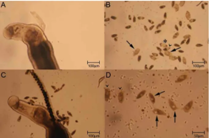
not able to develop. After an 8-day-period of observa-tion, the total number of morphologically dead eggs in the groups exposed to 2 µg/ml and 4 µg/ml lovastatin in vitro was 44 (73.3%) and 348 (94.8%), respectively; in the control group it was 144 (27.5%). Using the probe Hoechst 33258, a fluorescent marker that label dead eggs (Sarvel et al 2006) it was observed that in the group ex-posed to 2 µg/ml lovastatin, 69% of the morphologically viable eggs were labeled by the probe, whereas in the group exposed to 4 µg/ml lovastatin, 74% of the eggs were labeled. In the control group, 16% of the morpho-logically viable eggs were labeled by the probe (p < 0.05). The labeled eggs are shown in Fig. 2.
Fig. 1: evaluation of the in vitroactivity of lovastatin on Schistosoma mansoni worms after a 7-day-culture period. A, B: control. A: adult paired worms without apparent morphological change; B: eggs at all stages; mature eggs (arrow), 3rd stage (head of the arrow), 4th stage (*); C: paired worms exposed to 2 µg/ml lovastatin and without ap-parent morphological change, and eggs at the 1st and 2nd stages; D: eggs laid by worms exposed to 4 µg/ml lovastatin; eggs at the 1st stage (arrow) and dead (head of the arrow).
The worms obtained from mice treated with lovas-tatin (400 mg/kg/day for 5 days) and sacrificed three days after the end of treatment were cultured in vitro (in vivo/in vitro trials) in parallel with a group of worms exposed to 4 µg/ml lovastatin in vitro, as well as with a control group (worms not exposed to drug). Morphologi-cal changes could not be observed throughout the 8-day-culture, regarding the motion and egg-laying among the worms treated in vivo/ in vitro, as well as among the worms of the control group. In both cases, the eggs reached maturation, and hatching of miracidia could also be seen. The worms exposed to lovastatin in vitro presented results similar to those of the former experi-ment, as above mentioned.
DISCUSSION
Vandewaa et al. (1989) working with mevinolinate, farnesol and mevinolin, in vivo and in vitro, suggest that egg-production in schistosomiasis is associated with the activity of the enzyme 3-hydroxy 3-methylglutaryl coen-zyme A (HMG-CoA) reductase, and that non-sterol lip-ids produced in biochemical metabolism, modulated by this enzyme, stimulate egg-production. Chen et al. (1990) observed that egg-laying by the parasite females was dis-continued in mice infected with S. mansoni and treated with low doses of mevinolin. The administration of 0.2% mevinolin in the diet of infected mice (14 days) resulted in elimination of 96% of the parasites. The same dose admin-istered two days before and 15 days pi resulted in 93-95% reduction in the number of adult parasites. When mevino-lin was administered with 0.5% mevinomevino-linate, there was inhibition of the anti-schistosomicidal activity of the for-mer, suggesting that mevinolinate and/or metabolites are of the utmost importance for inhibiting S. mansoni egg-production, as well as promoting the parasite´s survival.
Lovastatin, a lactone, inactive form of the corre-sponding open hydroxylic acid, is a potent inhibitor of the endogenous synthesis of cholesterol. After gastroin-testinal absorption, lovastatin is rapidly hydrolisated to open hydroxyacid, a competitive inhibitor of HMG-CoA reductase, an enzyme that catalyzes a regulatory step in the biosynthesis of cholesterol. In clinical studies (Araújo et al. 2002), lovastatin reduced the total plasmatic choles-terol concentrations and lipoprotein of low density, as well as lipoprotein of very low density bound to cholesterol.
Araújo et al. (2002) investigated the activity of lovas-tatin on the oviposition of the adult worm in experimen-tally infected mice with S. mansoni. These authors dem-onstrated that, despite the relatively low mortality rate (~30%) of the worms, when the beginning of treatment was on the 30th day pi, with an oral dose of 400 mg/kg, for five consecutive days, there was a reduction in the oviposition of S. mansoni females, presenting oogram changes in up to 80% of mice, and an average number of eggs in the jejunum and liver significantly lower in the treated animals, when compared to the control group. The morphological analysis of the worms revealed de-generative changes, mainly in the reproductive appara-tus of the worms, with reduction and alteration in the vitelline follicles and in the ovary of females, as well as changes in the male testis.
The role of cholesterol in schistosomiasis mansoni was experimentally evaluated by Neves (2006), using four groups of animals: infected, fed on a diet rich in cholesterol (29% of lipids); infected, fed on a standard diet (12% of lipids); uninfected, fed on a diet rich in cho-lesterol; uninfected, fed on a standard diet. It was ob-served that infected mice that received a diet rich in cho-lesterol presented a higher number of viable S. mansoni
eggs, a greater maturation and peak numbers of eggs in feces, when compared to the group of infected mice fed on a standard diet. It was also observed that the adult worms recovered from the group of infected mice fed on a diet rich in cholesterol (29% lipids) presented a higher number of morphological changes in the reproductive apparatus of male worms, mainly in relation to supra-numerary lobe, seminal vesicle and spermatheca. In this group, the ovary of females presented a higher number of oocytes being discharged than in females belonging to the group with 12% lipids in the diet. It seems that a site rich in cholesterol is beneficial for reproduction and discharge of S. mansoni eggs (Neves 2006).
Soliman & Ibrahim (2005) studied the activity of atorvastatin (cholesterol lowering agent) alone, and currently with medoxyprogesterone (intramuscular con-traceptive) on S. haematobium adult worms, in experi-mentally infected hamsters. Atorvastatin administered by oral route, at the dose of 0.9 mg/kg starting from day 35 pi, for 49 consecutive days, and with medoxyproges-terone acetate in association, at the dose of 0.1 ml/kgon days seven and 35 pi, intramuscularly, produced dam-ages in the tegument of male and female worms. Loss or rupture of tubercles, collapseof tissues, and erosion of the tegument and of the oral suckers, degenerations or collapses of sensorial organelles, were some of the observed changes. Administration of atorvastatin alone, according to the same schedule previously described, produced less severe damages in the tegument of male worms, and only a few females presented tegumental changes. Both treatment schedules (atorvastatin con-currently with medoxyporogesterone acetate or alone) contributed to a significant reduction in the total num-ber of recovered worms, 46.2% and 51.3%, respectively. The two drugs in association produced oogram changes, with a high number of dead eggs.
viabil-ity of S. mansoni eggs showed a significant difference when a fluorescent probe was used. In fact, the average of morphologically viable eggs, labeled by the probe, was 70% approximately in the groups exposed to 2 µg/ ml or 4 µg/ml lovastatin. In the control group, it was observed that only 16% of the morphologically viable eggs were labeled by the probe. Since egg labeling by the probe indicates that the egg is dead, the difference in the classification of viable eggs is evident when the probe is used in comparison when only the morphology of the egg is observed under an optic microscope. When applied in assays without drug utilization, the methodol-ogy recommended by Sarvel et al. (2006) with the aim to confirm the traditional morphological classification, 83% of corroboration between the two methods was de-tected. Nevertheless, in the trials related to drug activity was much lower (30%), i.e., only 10 out of 35 morpho-logically viable eggs present in the groups exposed to 2 µg/ml or 4 µg/ml lovastatin were not labeled by the probe. The use of the probe Hoechst 33258 enhances the sensitivity of the trials aiming to evaluate drug action on viable S. mansoni eggs.
The data obtained corroborate the action of lovasta-tin in S. mansoni egg-laying, in both in vivo and in vitro experiments. Nevertheless, the synergistic or additive schistosomicidal action of this drug concurrently with other known antischistosomal drugs (oxamniquine or praziquantel) could not be observed.
ACKNOWLEDGEMENT
To Dr. John R. Kusel for his suggestions and review of the manuscript.
REFERENCES
Araujo N, Kohn A, Oliveira AA, Katz N 2002. Schistosoma mansoni: ação da lovastatina no modelo murino. Rev Soc Bras Med Trop 35: 35-38.
Chen GZ, Foster L, Bennett JL 1990. Antischistosomal action of mevinolin evidence that 3-hydroxy-methylglutaryl-coenzime a reductase activity Schistosoma mansoni is vital for parasite sur-vival. Naunyn Schmiedebergs Arch Pharmacolol 342: 477-482.
Doenhoff MJ, Kussel JR, Coles GC, Cioli D 2002. Resistance of
Schistosoma mansoni to praziquantel: is there a problem? Trans R Soc Trop Med Hyg96: 465-469.
Katz N 1980. Experiência com quimioterapia em grande escala no controle da esquistossomose no Brasil. Rev Inst Med TropSao Paulo22: 40-51.
Neves RH 2006. Avaliação do papel da dieta rica em colesterol na es-quistossomose mansônica experimental de Mus musculus Swiss Webster, PhD Thesis, Instituto Oswaldo Cruz, Rio de Janeiro, 161 pp.
Pellegrino J, Oliveira CA, Faria J, Cunha AS 1962. New approach to screening of drugs in experimental Schistosomiasis mansoni in mice. Am J Trop Med Hyg 11: 201-215.
Pellegrino J, Siqueira AF 1956. Técnica de perfusão para colheita de
Schistosoma mansoni em cobaias experimentalmente infestadas.
Rev Bras Malariol Doenças Trop 8: 589-597.
Sarvel AK, Kusel JR, Araujo N, Coelho PMZ, Katz N 2006. Com-parison between morphological and staining characteristics of live and dead eggs of Schistosoma mansoni. Mem Inst Oswaldo Cruz 101 (Suppl. I): 289-292.
Smithers SR, Terry RJ 1965. The infection of laboratory hosts with cercariae of Schistosoma mansoni and the recovery of adults worms. Parasitology55: 695-700.
Soliman MFM, Ibrahim MM 2005. Antischistosomal action of ator-vastatin alone and concurrently with medroxyprogesterone ace-tate on Schistosoma haematobium harboured in hamster: surface ultraestructure and parasitological study. Acta Trop 93: 1-9.
Vandewaa EA, Mills G, Foster LA, Bennett JL 1989. Physiological role of HGM-CoA reductase in regulating egg production by