J. bras. pneumol. vol.34 número5 en v34n5a05
Texto
Imagem
Documentos relacionados
In vitro/in vivo trials - The worms obtained from mice treated with lovastatin (400 mg/kg/day for 5 days) and sac- rificed three days after the end of treatment were cultured
Residual levels of Sb in tissues after a 21-day course of treatment with MA - Tissue concentrations of Sb were determined in rats killed 24 h after the last dose of MA and in a
After 30 days, the percentage of explants forming calluses and their colour were evaluated for each treatment; after 60 days the percentage of explants that regenerated buds, the
Cleome dendroides in vitro germination took place after 7-8 days of culture, by the rupture of the tegument in the hilum and root protrusion.. After 8 days, hypocotyl
Sheep 7 and 8, from group 3, challenged 15 days after the end of the resistance induction period, showed early clinical signs two days after the ingestion of a single dose of
It was observed signifi cant increased diameter of the ophthalmic artery after 7 and 30 days of treatment and decreased mean arterial pressure after 7, 15 and 30 days of
Based on the results of ANOVA on collagen 1 after 7 and 30 days, there were significant differences in the expression of collagen 1 between the control group and the
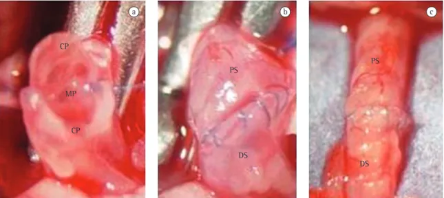
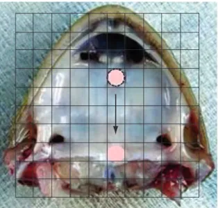
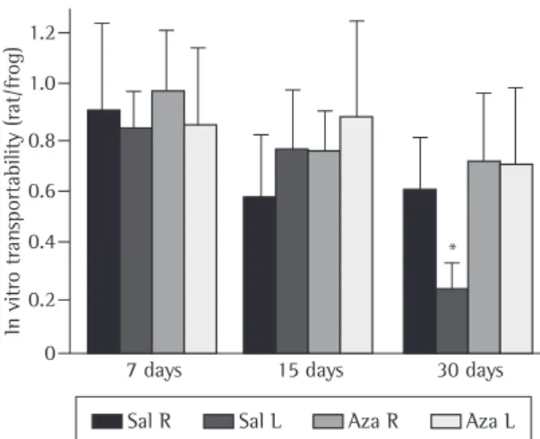
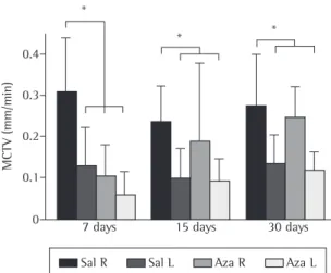
After 7, 15 and 30 days of treatment, six animals from each group were killed, after which in situ mucociliary transport velocity, in vitro mucus transportability, and contact angle
