J Vasc Bras. 2012;11(3):305-309.
Fabio Henrique Rossi1, Camila Baumann Beteli2, Mabel Barros Zamorano3, Lilian Mary da Silva4, Patrik Bastos Metzger5, Cybelle Bossolani Onofre6, Edir Branzoni Leal7, Akash Kuzhiparambil Prakasan8, João Italo Dias França9,
Nilo Mitsuru Izukawa10, Amanda Rego Souza11
Abstract
Objective: To determine the importance of the variables: Energy Intensity (I), Power (P), and Time of Application (T) in the histological changes which occurred in varicose veins of the lower limbs that underwent endovascular electrocauterization. Method: A prospective trial conducted in patients undergoing intravenous electrocauterization of a proximal saphenous vein fragment, according to a randomization table – GI: I = 0J, P = 0W, T = 15 s; GII: I = 300 J, P = 60 W, T = 5 s; GIII: I = 600 J, P = 60 W, T = 10 s; GIV: I = 900 J, P = 60 W, T = 15 s, GV: I = 450 J, P = 90 W, T = 5 s; GVI: I = 900 J, P = 90 W, T = 10 s; GVII: I = 1350 J, P = 90 W, T = 15 s; GVIII: I = 600 J, P = 120 W, T = 5 s; GIX: I = 1200 J, P = 120 W, T = 10 s; GX: I = 1800 J, P = 120 W, T = 15 s. he fragments were submitted to histopathology in order to analyze the depth of tissue necrosis, classiied as follows: Group A – endothelium and media, Group B – endothelium, media, and adventitia. Results: he depht of histological necrosis – Groups A and B – which occurred in the fragments were proportional to the Energy Intensity of electrocauterization (p = 0.0001). his linear association could also be checked for the variables Power (p = 0.017) and Time of Application (p = 0.0001). Spearman’s correlation coeicient was higher for the variable Time of Application: 0.42269 (p = 0.002) when compared with the variable Power of Energy (P): 0.3542 (p = 0.005). Conclusion: he Time of Application of Energy is a stronger predictor than the Power of electrocauterization, in determining the depth of the histological efects observed in the walls of lower limb varicose veins, for the same electrocauterization Energy Intensity applied.
Keywords: varicose veins; endovascular procedures; and catheter ablation.
Resumo
Objetivo: Determinar a importância das variáveis: Intensidade de Energia (I), Potência (P) e Tempo de Aplicação (T) nas alterações histológicas ocorridas em varizes de membros inferiores submetidas à eletrocauterização endovascular. Método: Estudo prospectivo experimental realizado em pacientes submetidos à eletrocauterização endovenosa de fragmento proximal da veia safena magna, de acordo com uma tabela de aleatorização – GI: I = 0J, P = 0 W, T = 15 s; GII:I = 300 J, P = 60 W, T = 5 s;GIII:I = 600 J, P = 60 W, T = 10 s; GIV: I = 900 J, P = 60 W, T = 15 s; GV: I = 450 J, P = 90 W, T = 5 s;GVI: I = 900 J, P = 90 W, T = 10 s; GVII: I = 1350 J, P = 90 W, T = 15 s;GVIII: I = 600 J, P = 120 W, T = 5 s; GIX:I = 1200 J, P = 120W, T = 10s;GX: I = 1800J, P = 120W, T = 15 s. Os fragmentos foram submetidos a estudo anatomopatológico com o objetivo de analisar a profundidade das alterações tissulares, assim classiicadas: Grupo A – endotélio e média,Grupo B – endotélio, média e adventícia. Resultados: A intensidade das alterações histológicas – Grupo A e B – ocorridas nos fragmentos foram proporcionais à Intensidade de Energia de eletrocauterização (p = 0,0001). Essa associação linear também pode ser veriicada para as variáveis Potência (p = 0,017) e Tempo de Aplicação (p = 0,0001). O índice de correlação de Spearman foi maior para variável Tempo de Aplicação: 0,42269 (p = 0,002) quando comparada com a variável Potência de Energia: 0,3542 (p = 0,005). Conclusão: O Tempo de Aplicação de Energia é mais importante do que a Potência de Energia utilizada para uma mesma energia de eletrocauterização, na determinação da profundidade dos efeitos histológicos observados na parede das varizes de membros inferiores.
Palavras-chave: varizes; procedimentos endovasculares; ablação por cateter.
Immediate efects of endovascular electrocautery in lower
limb varicose veins
Efeitos imediatos do eletrocautério endovascular em varizes de membros inferiores
Study carried out at the Instituto Dante Pazzanese de Cardiologia – São Paulo (SP), Brazil.
1 Doutor em Medicina pela Universidade de São Paulo (USP); Médico assistente da seção médica de Cirurgia Vascular e membro do Centro de Intervenções Endovasculares (CIEV) do Instituto
Dante Pazzanese de Cardiologia – São Paulo (SP), Brazil.
2 Médica residente da seção médica de Cirurgia Vascular do Instituto Dante Pazzanese de Cardiologia – São Paulo (SP), Brazil. 3 Chefe da seção médica de Anatomia Patológica do Instituto Dante Pazzanese de Cardiologia – São Paulo (SP), Brazil.
4 Médica colaboradora da seção médica de Anatomia Patológica do Instituto Dante Pazzanese de Cardiologia – São Paulo (SP), Brazil. 5 Médico aprimorando do CIEV do Instituto Dante Pazzanese de Cardiologia – São Paulo (SP), Brazil.
6 Tecnóloga em saúde do setor de Bioengenharia do Instituto Dante Pazzanese de Cardiologia – São Paulo (SP), Brazil. 7 Engenheiro do setor de Bioengenharia do Instituto Dante Pazzanese de Cardiologia – São Paulo (SP), Brazil.
8 Médico assistente da seção médica de Cirurgia Vascular; Membro do CIEV do Instituto Dante Pazzanese de Cardiologia – São Paulo (SP), Brazil. 9 Estatístico do Laboratório de Epidemiologia e Estatística (LEE) do Instituto Dante Pazzanese de Cardiologia – São Paulo (SP), Brazil.
10 Doutor em Medicina pela Universidade de São Paulo (USP), Médico chefe da seção médica de Cirurgia Vascular e membro do CIEV do Instituto Dante Pazzanese de Cardiologia –
São Paulo (SP), Brazil.
11 Médica, Livre Docente pela USP; Diretora Técnica do Instituto Dante Pazzanese de Cardiologia – São Paulo (SP), Brazil.
Introduction
Chronic venous insuiciency of the lower limbs afects 20% of the Western adult population and its main cause
are the primary varicose veins.1 Currently, endovascular
treatment (lasers2,3 and radiofrequency4-6), thermal energy
is released in the lumen of the vessel afected, causing destruction of its wall and interruption of blood low inside. Endovascular electrocauterization can cause selective destruction of the layers of a vein.7 his efect is proportional
to the energy used which, in turn, is dependent on the Power and Time of Application.
he aim of this study was to determine the importance of the variables: Energy Intensity (I), Power (P), and Time of Application (T) in the immediate histological changes observed in patients afected by lower limb varicose veins who underwent endovascular electrocauterization.
Method
his prospective trial was carried out by Vascular Surgery, Pathology, and Bioengineering sectors of Instituto Dante Pazzanese de Cardiologia - Sao Paulo, study protocol approved by the Ethics in Research Committee and sponsored by FAPESP.
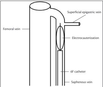
Forty-two patients with lower limb varicose veins and great saphenous vein insuiciency, documented by venous Duplex Scanning preoperatively, underwent conventional surgical varicose veins treatment. Ater surgical exposure of the great saphenous vein hiatus and pre-malleolar segments, a 6F diagnostic catheter was placed proximally, just below the supericial epigastric tributary vein. he electrocautery was then introduced through the catheter until its inal position, and then set back, enough to expose its distal “head”, composed of four stainless steel rods 2.0 cm long by 1.5 cm in diameter (Figure 1). Ater exposure, and prior to leboextraction, endovenous electrocauterization was performed on this proximal fragment of the saphenous vein, according to a randomization table presented below (Table 1).
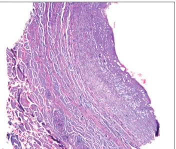
hese venous fragments were extracted and submitted to histopathological examination in order to analyze the depth of tissue necrosis which occurred within the layers, classiied as follows: Group A - endothelium and media; Group B - endothelium, media, and adventitia (Figure 2).
he parameters used to verify the presence of the efects of electrocauterization were: presence of necrosis (nuclear rarefaction, cytoplasmic shrinkage), presence of thrombus, vacuolization, coagulation, tissue loss, and perforation.
All the patients underwent postoperative Duplex Scanning, with the aim of identifying possible presence
Figure 1. Electrocauterization of the proximal segment of the saphe-nous vein.
Table 1. Randomization table of electrocauterization.
Randomization table
Group Intensity (J) Power (W) Time (s)
I 0 0 15
II 300 60 5
III 600 60 10
IV 900 60 15
V 450 90 5
VI 900 90 10
VII 1350 90 15
VIII 600 120 5
IX 1200 120 10
X 1800 120 15
of femoral vein thrombosis in the region submitted to electrocauterization, and had monthly follow-ups in outpatient visits.
To evaluate the existence of linear association between the variables Energy Intensity, Power and Time of Application of electrocauterization with the histological efects, we performed the chi-square test for linear tendency.
To analyze the relevance of the variables: Power and Time of Application, we used the dose-response model and Spearman correlation index, using as the dependent variable the destruction of the vessel layers studied by electrocauterization. We considered statistically signiicant when p < 0.05.
Results
A total of 60 proximal fragments, obtained in 42 patients who underwent saphenous electrocauterization, were analyzed. Mean age was 46 years, and 65% of patients were female. he average temperature outside the region submitted to electrocauterization was 47.3 °C (37-62). Patients had follow-ups for an average period of 4.5 months, and serious complications related to the procedure were not identiied.
Histological evaluation showed necrosis of the intima in all the patients (Group A) (Figure 3), involvement of all layers in 53.3% (Group B) (Figure 4), and rupture in 1.6% of the cases.
We observed that the intensity of histological changes – Group A and B – that occurred in the fragments were proportional to the Energy Intensity of the electrocauterization (p = 0.0001) applied (Chart 1).
Figure 3. Necrosis present in intima and media of the proximal frag-ment of the saphenous vein (HE staining; magniication 60×).
Figure 4. Necrosis present in intima, media, and adventitia in the proxi-mal fragment of the saphenous vein (HE staining, magniication 40×).
his linear association can also be checked for variables Power (p = 0.017) (Chart 2) and Time of Application (Chart 3) (p = 0.0001) when studied in isolation.
he Spearman correlation coeicient was more signiicant for the variable Time of Application (T): 0.42269 (p = 0.002) when compared with the variable Power Energy of electrocauterization (P): 0.3542 (p = 0.005).
Discussion
Endovascular treatment of varicose veins of the lower limbs is a relatively new method and presents some
by improving the equipment and application techniques of endovascular Power Energy.9,10
In a recent study, we demonstrated that electrocauterization can cause selective destruction of
a blood vessel.7 Some studies have demonstrated the
possibility of applying electrical energy to treat varicose veins of the lower limbs in the past, but inconclusive results and the lack of skills in catheterization of blood vessels by past vascular surgeons have discouraged clinical application.11-16
Today, with the paradigm shit in forms of treatment and a better understanding of the advantages and disadvantages of endovascular treatment of varicose veins of the lower limbs, electrocauterization can become an alternative method, and perhaps an advantageous therapeutic modality.
When electrical current is conducted through a tissue, it can induce cell death by increasing temperatures (thermomechanical phenomena) – dissipation of energy in the form of heat – and also by the simple passage of this current, when the interaction of electrons with ions and molecules of biological tissues (electromechanical phenomenon) occur. his phenomenon is quite similar to what occurs when detergent substances are applied in lipid membranes and endothelium appears to be especially sensitive to it.17-19
In our study we surprisingly observed that the temperatures reached in the venous fragments subjected to electrocauterization were well below those achieved by endovascular treatment which is currently carried out (laser, radiofrequency). his brings us to hypothesize that the electromechanical phenomena may play an important role in the mechanism of necrosis induced by electrocauterization in the fragments studied. hus, apparently, electrocauterization can cause destruction of the inner layers of a vessel with lower temperatures and, possibly, with lower complication rates.
he electrocauterization Intensity of Energy in Joules depends on the Power in Watts and the Time of its application in seconds. We know that the degree of destruction of the walls of a vessel is proportional to the Energy Intensity,7 but
did not know if this was true for the Power and Time of Application, and which of these two variables had greater power to determine the degree of destruction of the wall in a vessel.
In this study, we found that the Intensity of Energy of electrocauterization presents a positive correlation with the depth of destruction of the layers of a vessel (p = 0.0001) (Chart 1). he higher this variable, the greater the number of cases in which lesion of the adventitial layer (Group B) advantages compared to conventional surgery. he two
main methods – laser and radiofrequency – use thermal ablation of the inner layers of the vessel to cause occlusion. he degree of destruction of these layers, as well as the therapeutic success in short, medium, and long term, is proportional to the temperature reached.8
However, high temperatures can cause damage to structures and organs adjacent to the treated vessel, maybe causing complications such as pain, skin burns, nerve damage, thrombosis in vessels of the deep venous system, and perforation, leading to the formation of hematomas. A series of current work is trying to reduce these complications
Chart 2. Power of electrocauterization and depth of immediate histo-logical changes in varicose veins of the lower limbs.
8. Gloviczki P, Comerota AJ, Dalsing MC, et al. he care of patients with varicose veins and associated chronic venous diseases: Clinical practice guidelines of the Society for Vascular Surgery and the American Venous Forum. J Vasc Surg. 2011;53:2S-48S. PMid:21536172.
9. Lohr, J, Kulwicki A. Radiofrequency ablation: evolution of a treatment. Semin Vasc Surg. 2010;23:90-100. http://dx.doi. org/10.1053/j.semvascsurg.2010.01.004
10. Ash JL, Moore CJ. Laser treatment of varicose veins: order out of chaos. Semin Vasc Surg. 2010;23:101-6. http://dx.doi.org/10.1053/j. semvascsurg.2010.01.005
11. Araújo M, Velasco FCG. Métodos físicos utilizados para oclusão de varizes dos membros inferiores. J Vasc Bras. 2006;5:139-46. http:// dx.doi.org/10.1590/S1677-54492006000200010
12. Hejhal L, Firt P, Livora D. Endovascular electrocoagulation of supericial varices of leg. Rozhl Chir. 1959;38:418-25. PMid:14400800.
13. Musaev SM. Intravascular electrocoagulation of dilated subcutaneous varicose veins of the lower extremities. Eksp Khir Anesteziol. 1963;27:36-7. PMid:14068803.
14. Politowski M, Zelazny T. Complications and diiculties in electrocoagulation of varices of the lower extremities. Surgery. 1966;59:932-4.
15. Watts GT. Endovenous diathermy destruction of internal saphenous. Br Med J. 1972;4:53. http://dx.doi.org/10.1136/ bmj.4.5831.53
16. O’Reilly K. Letter: endovenous diathermy sclerosis as a unit of the armamentarium for the attack on varicose veins. Med J Aust. 1974;1:900.
17. Lee RC. Injury by electrical forces: pathophysiology, manifestations and therapy. Curr Probl Surg. 1977;34:679-758. PMid:9365421.
18. Akinlaja J, Sachs F. he breakdown of cell membranes by electrical and mechanical stress. Biophys J. 1998:75:247-54.
19. Lee RC, Kolodney MS. Electric injury mechanisms: Electrical breakdown of celular membranes. Plast Reconst Surg. 1987;80:862-7. PMid:3671558.
Correspondence
Fabio Henrique Rossi Av. Dr. Dante Pazzanese, 500 – Ibirapuera CEP: 04012-909 – São Paulo (SP), Brazil E-mail: [email protected]
Authors’ contributions
Conception and design: FHR Analysis and interpretation: FHR Data collection: FHR, CBB, MBZ, LMS, PBM Writing the article: FHR Critical revision of the article: CBO, EBL, AKP, JIDF, NMI, ARS Final approval of the article*: FHR, CBB, MBZ, LMS, PBM, CBO, EBL, AKP, JIDF,
NMI, ARS Statistical analysis: JIDF Overall responsibility: FHR *All authors have read and approved the inal version submitted to J Vasc Bras.
was found. his could also be observed when the variables Power (p = 0.017) (Chart 2) and Time of Application (p = 0.0001) (Chart 3) were studied separately.
To evaluate the importance of each of these variables in the destruction of the inner layers of the vessel studied, we found that the time of application of energy (Spearman: 0.42269, p = 0.002) have greater inluence than the energy output (Spearman: 0.3542, p = 0.005). As we know that in endovascular treatment of lower limb varicose veins we ideally need to destroy intima and media layers of the vessel, and not the adventitia. his knowledge possibly brings us an important practical application: to minimize the possibility of injury to the adventitia layer, and possibly rupture and injury to adjacent structures, we must use a power application of electrocauterization that will provide the shortest possible time of application.
hus, we conclude that the time of application of energy by electrocauterization is more important than the power of energy used for the same intensity of energy applied to determine the degree of histological efects observed on the wall of varicose veins of the lower limbs.
References
1. Meissner MH, Eklof B, Lohr JM, Lurie F, Kistner R, Wakeield TW. Preface: acute and chronic venous disease. Current status and future directions. J Vasc Surg. 2007;Suppl:1S-3S. PMid:18068559.
2. Navarro L, Min RJ, Boné C. Endovenous laser: a new minimally invasive method of treatment for varicose veins-preliminary observations using an 810 nm diode laser. Dermatol Surg. 2001;27:117-22. PMid:11207682.
3. Proebstle TM, Krummenauer F, Gül D, Knop J. Nonocclusion and early reopening of the great saphenous vein after endovenous laser treatment is luence dependent. Dermatol Surg. 2004;30:174-8. PMid:14756646.
4. Manfrini S, Gasbarro V, Danielsson G, et al. Endovenous management of saphenous vein relux. Endovenous Relux Management Study Group. J Vasc Surg. 2000;32:330-42. PMid:10917994.
5. Goldman MP. Closure of the greater saphenous vein with endoluminal radiofrequency thermal heating of the vein wall in combination with ambulatory phlebectomy: preliminary 6-month follow-up. Dermatol Surg. 2000;26:452-6. PMid:10816234.
6. Rautio T, Ohinmaa A, Perälä J, et al. Endovenous obliteration versus conventional stripping operation in the treatment of primary varicose veins: a randomized controlled trial with comparison of the costs. J Vasc Surg. 2002;35:958-65. PMid:12021712.