Transposition of Great Arteries with Intramural
Coronary Artery: Experience with a Modified
Surgical Technique
Amit Mishra
1, M.Ch, F.P.C.S; Anil Jain
1, M.Ch; Manish Hinduja
1, M.Ch; Vivek Wadhawa
1, M.Ch; Ramesh Patel
2, MD; Nikunj
Vaidhya
1, MS; Dayesh Rodricks
3, DPT; Hardik Patel
4, M.B.B.S
Abstract
Objective: Transposition of the great arteries is a common congenital heart disease. Arterial switch is the gold standard operation for this complex heart disease. Arterial switch operation in the presence of intramural coronary artery is surgically the most demanding even for the most experienced hands. We are presenting our experience with a modified technique for intramural coronary arteries in arterial switch operation.
Methods: This prospective study involves 450 patients undergoing arterial switch operation at our institute from April 2006 to December 2013 (7.6 years). Eighteen patients underwent arterial switch operation with intramural coronary artery. The coronary patterns and technique used are detailed in the text.
Results: The overall mortality found in the subgroup of 18 patients having intramural coronary artery was 16% (n=3). Our first patient had an accidental injury to the left coronary artery
and died in the operating room. A seven-day old newborn died from intractable ventricular arrhythmia fifteen hours after surgery. Another patient who had multiple ventricular septal defects with type B arch interruption died from residual apical ventricular septal defect and sepsis on the eleventh postoperative day. The remainder of the patients are doing well, showing a median follow-up duration of 1235.34±815.26 days (range 369 - 2730).
Conclusion: Transposition of the great arteries with intramural coronary artery is demanding in a subset of patients undergoing arterial switch operation. We believe our technique of coronary button dissection in the presence of intramural coronary arteries using coronary shunt is simple and can be a good addition to the surgeons’ armamentarium.
Keywords: Heart Defects, Congenital, Surgery. Transposition of Great Vessels. Coronary Artery Disease. Ventricular Function.
DOI 10.5935/1678-9741.20160003
1Department of Cardiovascular and Thoracic Surgery, U.N. Mehta Institute of Cardiol-ogy and Research Center (Affiliated to B. J. Medical College), Civil Hospital Campus, Asarwa, India.
2Department of Cardiac Anesthesia, U.N. Mehta Institute of Cardiology and Research Center (Affiliated to B. J. Medical College), Civil Hospital Campus, Asarwa, India. 3Department of Perfusion, U.N. Mehta Institute of Cardiology and Research Center (Affiliated to B. J. Medical College), Civil Hospital Campus, Asarwa, India.
4Medical Officer, U.N. Mehta Institute of Cardiology and Research Center (Affiliated to B. J. Medical College), Civil Hospital Campus, Asarwa, India.
This study was carried out at U.N. Mehta Institute of Cardiology and Research Center (Affiliated to B. J. Medical College), Civil Hospital Campus, Asarwa, India.
Financial support: This work was supported by the U.N.Mehta Institute of Cardiology and Research Center itself and received no specific grant from any funding agency, commercial or not-for-profit sectors.
No conflict of interest.
Correspondence Address: Amit Mishra
U. N. Mehta Institute of Cardiology and Research Center (Affiliated to B. J. Medical Col-lege), New Civil Hospital Campus – Asarwa – Ahmedabad-380016 – Gujarat – India E-mail: [email protected]
Article received on October 28th, 2015 Article accepted on January 4th, 2016
Abbreviations, acronyms & symbols
ASO CHDs ECG IVS TGA VSD
= Arterial switch operation = Congenital heart defects = Electrocardiogram = Intact ventricular septum = Transposition of the great arteries = Ventricular septal defect
INTRODUCTION
Transposition of the great arteries (TGA) is a congenital cardiac anomaly in which the aorta arises entirely or largely from the right ventricle and the pulmonary trunk arises entirely or largely from the left ventricle, also known as discordant
ventriculoarterial connection[1]. It is the most frequent cyanotic
The term “intramural coronary artery” refers to the coronary patterns in which there is an intimate relationship between aortic and coronary arterial walls; histologically, the aortic and coronary
medial walls are attached without interposed adventitia[2].
Intramural coronary arteries are rare in patients with normal ventriculoarterial connections, but they are proportionally more common in TGA, with a reported incidence of 3% to 5%. The intramural artery usually originates from the wrong sinus and runs between the great arteries. Sometimes the intramural artery originates from its normal sinus, but above the sinotubular junction. These abnormal coronary patterns might complicate
coronary transfer during ASO[2]. We present our modified surgical
technique with a prospective analysis of the data.
METHODS
This prospective study was conducted at U. N. Mehta Institute of Cardiology and Research Centre for 7.6 years. After receiving approval from the institutional ethics committee, a total of 450 patients who underwent ASO from April 2006 to December 2013 were included in the study (Table 1). All of the patients were diagnosed with two-dimensional echocardiography. Two hundred and seventy six patients had TGA with intact ventricular septum (IVS), 141 patients had TGA with ventricular septal defect (VSD) (six of them had multiple VSD), and 33 patients had Taussig-Bing anomaly (one had multiple VSD). Three patients in the TGA/
VSD group and two with Taussig-Bing anomaly had type-B arch interruption. Eighteen patients (Table 2) had intramural coronary artery (Figure 1), eleven of which were from the TGA/VSD group and seven from the TGA/IVS group (one was on prostaglandin infusion and two underwent balloon atrial septostomy on the
Table 1.Details of the patients who underwent arterial switch operations.
Total: 450 patients
Diagnosis Number of
Patients (%)
Transposition of the Great Arteries with
Intact Ventricular Septum 276 (61.33%)
Transposition of the Great Arteries with
Ventricular Septal Defect 141 (31.33%)
Taussig-Bing anomaly 33 (7.3%)
Total: 18 (4%) Patients with Intramural Coronary Artery
Transposition of the Great Arteries with
Ventricular Septal Defect 11 (61.11%)
Transposition of the Great Arteries with
Intact Ventricular Septum 7 (38.88%)
Fig. 1 - Intramural coronary artery pattern observed in eighteen patients.
A- Intramural left coronary artery with normal origin of right coronary artery (9 patients). B- Intramural left and right coronary artery with separate openings (1 patient).
C- Intramural left and right coronary arteries with single ostia (7 patients).
Table 2. Demographic details of patients with intramural coronary artery.
No Age (days) Sex Weight Diagnosis Coronary
Pattern
Great Artery
Relationship Outcome
1 45 M 3.1 D-TGA VSD 2 LRCX Aorta Anterior
to PA Doing well on regular follow-up
2 38 M 3.0 D-TGA IVS 2 LRCX Aorta Anterior
to PA
Died due to accidental injury to left intramural coronary artery
3 42 F 3.0 D-TGA VSD 2 LRCX Aorta Anterior
to PA Doing well on regular follow-up
4 62 M 3.4 D-TGA VSD 2 LRCX Aorta Anterior
to PA Doing well on regular follow-up
5 25 F 3.1 D-TGA IVS 2 LRCX Aorta Anterior
to PA
Died from intractable ventricular arrhythmias fifteen hours after
surgery
6 36 F 2.6 D-TGA VSD 2 LRCX Aorta Anterior
to PA Doing well on regular follow-up
7 42 F 3.2
D-TGA, Multiple VSD & type B arch interuption
2 LRCX Aorta Anterior
to PA
Died secondary to sepsis and residual VSD
8 90 F 3.9 D-TGA VSD 2 LRCX Aorta Anterior
to PA Doing well on regular follow-up
9 55 M 3.0 D-TGA VSD 2 LRCX Aorta Anterior
to PA Doing well on regular follow-up
10 48 F 2.9 D-TGA VSD 2 LRCX Aorta Right &
Anterior to PA Doing well on regular follow-up
11 42 F 3.2 D-TGA VSD 2 LRCX Aorta Anterior
to PA Doing well on regular follow-up
12 38 M 3.2 D-TGA VSD 2 LRCX Aorta Anterior
to PA Doing well on regular follow-up
13 27 M 3.1 D-TGA IVS 2 LRCX Aorta Anterior
to PA Doing well on regular follow-up
14 19 F 2.7 D-TGA IVS 2 LRCX Aorta Anterior
to PA Doing well on regular follow-up
15 33 F 2.4 D-TGA IVS 2 LRCX Aorta Anterior
to PA Doing well on regular follow-up
16 27 F 2.2 D-TGA IVS 2 LRCX Aorta Anterior
to PA Doing well on regular follow-up
17 18 M 2.4 D-TGA IVS 2 LRCX Aorta Anterior
to PA Doing well on regular follow-up
18 44 F 3 D-TGA VSD 2 LRCX Aorta Right &
Anterior to PA Doing well on regular follow-up
D-TGA=dextro transposition of the great arteries; IVS=intact ventricular septum; VSD=ventricular septum defect; LRCX=left-right circumflex
4th and 6th day of life). None of the patients from the Taussig-Bing
anomaly group had intramural coronary artery. Similarly, none of the eighteen patients had preoperative diagnosis/suspicion of intramural coronary pattern. Eight patients with intramural coronary arteries had both left and right coronary arteries having
Table 3. Demographic details and surgical outcomes of
patients with intramural coronary artery.
Variables Mean±SD
Age (days) 40.61±16.94
Weight (kg) 2.96±0.40
Height (cm) 49.66±3.08
Sex M=7
F=11
Mortality 3 (16.66%)
Survival duration (days) Mean: 1245.94±836.7
Median: 1788.5 (369 – 2920)
Morphologic and demographic characteristics of the patients with intramural coronary artery are given in Figure 1 and Table 3.
Statistical analysis
The statistical calculations were performed using SPSS software v.20.0 (Chicago, IL, USA) Quantitative data were expressed as mean±SD whereas qualitative data were expressed as percentage. Survival statistics were performed using Kaplan-Meier survival curve.
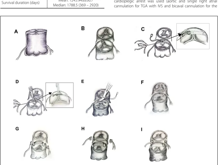
Technique (Figure 2)
The standard technique of cardiopulmonary bypass and cardioplegic arrest was used (aortic and single right atrial cannulation for TGA with IVS and bicaval cannulation for the
Fig. 2 – Anatomical patterns of the coronary arteries. A- External appearance of the intramural coronary artery. B- Single ostial opening in sinus 2.
C- Coronary shunt showing the direction of the left intramural coronary artery with a stay stitch on ostial opening. D- Unroofing of the intramural left coronary artery.
E- Unroofing of the intramural right coronary artery.
F- Unroofed left and right coronary arteries with their ostia in their respective sinuses. G- Excision of coronary buttons.
TGA/VSD group; multiple dose ST Thomas cardioplegia was used in the first nine patients and Del nido cardioplegia in the other nine). The aorta was transected at the level of the pulmonary artery bifurcation and it was inspected from above (Figures 2A and 2B) in both sinuses for the presence of a separate origin of coronary arteries. Whenever the opening of the left coronary artery was not identified in Sinus 1, Sinus 2 was inspected for the opening of both coronaries. Usually, in cases of intramural coronary artery, the left coronary artery originates from Sinus 2 at the upper right side of the posterior commissure (Figure 1A). In eight patients, we found that both left and right coronary arteries had an intramural course, arising from Sinus 2 (Figure 2B) with a single opening; one patient had separate openings with kissing edges. The length of the intramural course of the right coronary was nearly 2-3 mm in all cases whereas the length of the intramural course of the left coronary was 4-6 mm (Figure 2C). We used coronary shunts (Synovis Life Technologies, Inc., St. Paul, MN, USA) to probe the coronary artery (Figure 2C) and to find its intramural course in the aortic wall. The posterior commissure was turned down and the stay stitch was placed with a 7-0/8-0 polypropylene suture at the opening of the intramural coronary artery, keeping the coronary shunt in situ (Figure 2D). The entire roof of the intramural coronary arteries was excised (Figure 2F) until the internal opening (Figure 2F) was identified. The bulbous end of the coronary shunt not only helped to identify the internal opening but also prevented any accidental injury to the coronary ostia. A similar procedure was done for the other (right) coronary artery (Figure 2E). Following unroofing, the aortic openings of the coronary ostia were identified in their respective sinuses (Figure 2F). The coronary buttons were excised in routine fashion (Figure 2G) and the proximal epicardial coronary artery was dissected for 2-3 mm to avoid any kink during relocation (Figure 2G). The margins of the coronary buttons were not smooth (Figure 2H) because of the tissue of the posterior commissure. In addition, the buttons of the left coronary were small, but could be comfortably relocated (Figure 2I) to the neo aorta. The left coronary artery was relocated using the punch hole technique in all patients. The right coronary artery was relocated using the trapdoor technique in sixteen patients while the punch hole technique was used for the remaining two patients. The posterior commissure was resuspended after proximal neo pulmonary artery reconstruction. A Lecompte maneuver was performed in all patients with intramural coronary artery. The redundant portion of the proximal neo aorta was plicated posteriorly to form a keel, if required, especially in patients in the TGA/VSD group. The neo aortic and neo pulmonary anastomosis were performed in routine fashion. All patients were weaned off bypass with a moderate dose of inotropes (Adrenaline 0.05 micrograms/kg/ minute and Milrinone 0.5 micrograms/kg/minute) without any difficulty. Mean bypass time was 148 minutes and mean cross-clamp time was 106 minutes in the TGA/IVS group. Mean bypass time was 166 minutes and mean cross-clamp time was 130 minutes in the TGA/VSD group. The chest was left open in eleven patients during the first operative day due to diffused bleeding and it was closed the next day. Postprocedure patients had stable hemodynamic course without any electrocardiogram (ECG) features of myocardial ischaemia.
RESULTS
For the patient who had an accidental injury to the left coronary artery during dissection, though a 5 mm Gortex (W. L. Gore and Associates, Flagstaff, AZ) graft was used from the innominate artery to the single coronary button for coronary blood supply in a desperate attempt to save him, he died in the operating room secondary to bleeding and severe ventricular dysfunction. One seven-day-old newborn, who underwent TGA with IVS on Prostaglandin infusion and no intraoperative problems, had moderate left ventricular dysfunction on 2D echocardiography six hours after surgery, despite showing stable hemodynamic parameters without any ECG features of myocardial ischaemia. He died from intractable ventricular arrhythmia fifteen hours after surgery. Another patient who had multiple VSD with type-B arch interruption died from residual apical VSD and sepsis on the eleventh postoperative day. The rest of the patients did well in the immediate postoperative period. On every visit, the patients underwent ECG and 2D Doppler echocardiography. One patient had mild valvular pulmonary stenosis with a peak gradient of 20 mmHg and all other patients had patent coronary arteries without any evidence of regional wall motion abnormality, with good biventricular function and no valvular or supra valvular aortic/pulmonary stenosis or regurgitation. So far, none of our patients has undergone myocardial perfusion scan/angiography or any other intervention.
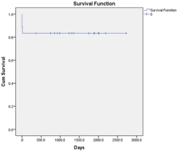
Survival analysis
The mean follow-up duration was 1235.34±815.26, whereas median was 1788.5 (4.9 years) (range 369 – 2730) days. Out of the 18 patients, overall mortality was observed in 3 (16.66%). The survival analysis was presented as Kaplan-Meier analysis and is shown in Figure 3.
DISCUSSION
TGA is a common congenital heart disease seen in paediatric cardiac clinics. The ASO is the treatment of choice for patients with TGA. The term “intramural coronary artery” refers to the coronary patterns in which there is an intimate relationship between the aortic and coronary arterial walls; histologically, the aortic and coronary medial walls are attached without interposed
adventitia[2]. The incidence of intramural coronary artery varies
from 1.4% to 5% in various series[2,3]. Successful relocation of
the coronary arteries is the most important step in ASO. Almost all kinds of coronary patterns can be relocated, however, the presence of an intramural coronary artery offers several surgical
challenges and carries high mortality and morbidity[4-8]. A
meta-analysis by Pasquali et al.[9] reported that the presence
of an intramural coronary artery is associated with the highest mortality compared to any other coronary artery pattern, with more than a six-fold increase in mortality compared to the usual coronary arrangement.
Diagnosis of intramural coronary artery in TGA using
two-dimensional echocardiography is explained by Pasquini et al.[10].
In our experience, intramural coronary artery is always seen as an intraoperative surprise after aortic transection. However, the absence of conical projection of the coronary artery should always raise suspicion in the surgeon’s mind. Furthermore, if there is no left coronary artery ostium in sinus one after aortotomy, the surgeon should strongly suspect there is an intramural left
coronary artery[11]. In our series of patients, we have come across
eight patients with a single ostial opening from Sinus 2 (one of them had kissing edges) with intramural course of both left
and right coronary arteries, which is very rare[2,12,13]. None of the
patients in our series had supra commisural origin of intramural coronary artery.
The single button aortocoronary flap technique for intramural coronary artery is associated with a high incidence of coronary complication, especially when the arrangement of the great arteries is anterior and posterior. In that case, the neo pulmonary artery may compress the coronary button and it can cause myocardial ischemia in the immediate postoperative period and, in the long term, there is a possibility of coronary arterial stenosis
when the covering tissue does not grow or shrink[2,11]. We have
no practical experience with the single button technique. We believe that the creation of two buttons and the transfer of the coronary button to the neo aorta in usual fashion is more physiological, preventing any early or late coronary artery compression and related complications.
The successful surgical treatment for bilateral intramural
coronary arteries has been reported earlier[12,13]. Our technique
of creating two buttons and transferring them in usual fashion is a technical modification of the technique described by Asou
et al.[14], and we believe that the incidence of accidental injury to
coronary artery is least with our technique.
Our technique of using coronary shunts for identifying the course of the intramural coronary artery and its unroofing is very useful, especially when there is stenosis of the ostium of the left intramural coronary artery. The color of the shunt (usually blue) can be seen (Figure 2C) through the aortic intima and the entire course of the intramural portion of the coronary artery can be
delineated. The stay stitch (Figure 2D) provides full control over intimal tissue flap and helps in the unroofing up to the distal portion of the intramural segment. As the coronary shunts are atraumatic and are easily available in various sizes, probing of the intramural coronary artery with metallic coronary probes should be avoided since they may cause trauma to intima as well as dissection and the possibility of late ostial stenosis. In the past, we have also reported the use of coronary shunts for giving
ostial cardioplegia[15]. The bulbous end of the shunt obliterates
the internal opening of the coronary artery and prevents it from any accidental trauma during unroofing while the entire length of the coronary shunt prevents any accidental injury to the tissue around the coronary ostia. We have observed that, with our modified technique, the distal portion of the intramural segment (where the coronary artery actually leaves the aortic wall) can also be opened, which remains potentially stenotic.
CONCLUSION
ASO is a preferred operation for TGA. However, performing arterial switch in the presence of intramural coronary artery is still challenging in pediatric cardiac surgery, even in experienced hands. Our technique is simple, atraumatic, and it helps to outline the course of intramural coronary arteries, to unroof the intramural coronary artery and to create two buttons. We have observed that, with our technique, ASO in the presence of intramural coronary artery can be performed in routine fashion with acceptable results.
ACKNOWLEDGEMENT
Authors acknowledge Mrs. Jyotsna Bhatnagar, Dr. Komal Shah, Ms. Himani Pandya, Mr. Sanjay Patel and Mr. Pratik Shah for assistance.
Authors’ roles & responsibilities
AM AJ MH VW RP NV DR HP
Conception and study design; execution of operations and/or trials; analysis and/or data interpretation; final manuscript approval
Conception and study design; analysis and/or data interpretation; final manuscript approval
Conception and study design; execution of operations and/or trials; statistical analysis; final manuscript approval
Conception and study design; execution of operations and/or trials; final manuscript approval
Conception and study design; execution of operations and/or trials; final manuscript approval
Execution of operations and/or trials; final manuscript approval
Execution of operations and/or trials; final manuscript approval
REFERENCES
1. Abbott ME. Congenital cardiac disease. In: Osler W, McCrae T, eds. Modern medicine. Vol.4. 3rd ed. Philadelphia: Lea & Febiger; 1927. 2. Metton O, Calvaruso D, Gaudin R, Mussa S, Raisky O, Bonnet D, et al.
Intramural coronary arteries and outcome of neonatal arterial switch operation. Eur J Cardiothorac Surg. 2010;37(6):1246-53.
3. Sachweh JS, Tiete AR, Jockenhoevel S, Mühler EG, von Bernuth G, Messmer BJ, et al. Fate of intramural coronary arteries after arterial switch operation. Thorac Cardiovasc Surg. 2002;50(1):40-4.
4. Kirklin JW, Blackstone EH, Tchervenkov CI, Castaneda AR. Congenital Heart Surgeons Society. Clinical outcomes after the arterial switch operation for transposition. Patient, support, procedural and institutional risk factors. Circulation. 1992;86(5):1501-15.
5. Prêtre R, Tamisier D, Bonhoeffer P, Mauriat P, Pouard P, Sidi D, et al. Results of the arterial switch operation in neonates with transposed great arteries. Lancet. 2001;357(9271):1826-30.
6. Legendre A, Losay J, Touchot-Koné A, Serraf A, Belli E, Piot JD, et al. Coronary events after arterial switch operation for transposition of the great arteries. Circulation. 2003;108(Suppl 1):II186-90.
7. García Hernández JA, Montero Valladares C, Martínez López AI, Romero Parreño A, Grueso Montero J, Gil-Fournier Carazo M, et al. Risk factors associated with arterial switch operation for transposition of the great arteries. Rev Esp Cardiol. 2005;58(7):815-21.
8. Wong SH, Finucane K, Kerr AR, O’Donnell C, West T, Gentles TL.
Cardiac outcome up to 15 years after the arterial switch operation. Heart Lung Circ. 2008;17(1):48-53.
9. Pasquali SK, Hasselblad V, Li JS, Kong DF, Sanders SP. Coronary artery pattern and outcome of arterial switch operation for transposition of the great arteries: a meta-analysis. Circulation. 2002;106(20):2575-80. 10. Pasquini L, Parness IA, Colan SD, Wernovsky G, Mayer JE, Sanders
SP. Diagnosis of intramural coronary artery in transposition of the great arteries using two-dimensional echocardiography. Circulation. 1993;88(3):1136-41.
11. Kim H, Sung SC, Kim SH, Chang YH, Ahn HY, Lee HD. Arterial switch operation in patients with intramural coronary artery: early and mid-term results. Korean J Thorac Cardiovasc Surg. 2011;44(2):115-22. 12. Padalino MA, Ohye RG, Devaney EJ, Bove EL. Double intramural
coronary arteries in D-transposition of the great arteries. Ann Thorac Surg. 2004;78(6):2181-3.
13. Cetrano E, Carotti A. Surgical treatment of transposition of the great arteries with bilateral intramural coronary arteries. Ann Thorac Surg. 2012;93(3):986-7.
14. Asou T, Karl TR, Pawade A, Mee RB. Arterial switch: translocation of the intramural coronary artery. Ann Thorac Surg. 1994;57(2):461-5. 15. Mishra A, Hinduja M, Solanki A. Novel technique for pediatric