1
Arquivos Brasileiros de Cardiologia - Volume 84, Nº 1, Janeiro 2005
Case Report
Single Coronary Artery: Angioplasty with Stent
Implantation
Ian C. D. Teixeira, Alexandre X. Brant, Alexandre Ares, Bruno Sarbi, Isaac Moscoso,
Adnan A. Salman, Salvador A. B. Cristóvão, João B. Oliveira Neto, Maria Fernanda Z. Mauro,
José A. Mangione
São Paulo, SP - Brazil
We report the case of a rare anomaly of the right coronary artery originated from the middle third of the left anterior des-cending (LAD) coronary artery with a proximal atheromatous obstructive lesion immediately before the emergence of the right coronary artery (RCA). The patient underwent successful angio-plasty with stent implantation in the left anterior descending coronary artery. Only 7 cases of this anomaly of distribution have been reported in the literature, but none of them was treated with percutaneous revascularization.
Anomalies of the coronary arteries occur in 1 to 2% of the population, constituting a rare, but important, type of nonatheros-clerotic coronary artery disease. These anomalies may cause sudden death on exertion and increase the risk of coronary arterial trauma during surgical procedures. In addition, certain types of anomaly may lead to myocardial ischemia 1.
The single coronary artery (SCA), described as an isolated coronary artery, has no gender predominance, being a rare entity with an incidence of 0.024% when isolated, and of 0.03 to 0.04% when associated with other cardiac abnormalities 2-4. Its main
characte-ristic is being originated from the aortic root through a single ostium, with no evidence of a second ostium, being then responsible for the irrigation of the entire heart, independently of its distribution 2.
In these cases, proximal atherosclerotic obstructions may have severe consequences for the patient 4.
We report the case of a patient who underwent percutaneous coronary intervention with stent implantation and whose RCA originated from the LAD. The cases of only 7 patients with that anomaly have so far been reported in the literature, but none of them was treated with coronary angioplasty 5.
Case Report
The patient is a 73-year-old, white, male, retired lumber jack, bornt and residing in the town of Maria da Fé, in the state of Minas Gerais. The patient has type II diabetes, hypertension and quit smoking 20 years earlier. He was referred to our service for
Hospital Beneficência Portuguesa de São Paulo and Discipline of Cardiology of the Faculdade de Medicina de Mogi das Cruzes Mailing address: Ian C. D. Teixeira - Rua Santa Madalena, 220/84A Cep 01322-020 – São Paulo, SP, Brazil – E-mail: [email protected] Received for publication: 12/13/2003
Accepted for publication: 01/26/2004 English version by Stela Maris Costalonga
coronary angiography due to a one-year history of oppressive chest pain on exertion, which irradiated to the left upper limb, was relieved with rest, and had no aggravating factors. The patient reported that in the preceding 2 months the pattern of the chest pain evolved progressively until being triggered at rest.
On physical examination, the patient had no alterations, except for his cardiac auscultation, which had a regular cardiac rhythm with the presence of S4. The chest teleradiography was normal. The rest electrocardiogram (fig. 1) showed a sinus rhythm, SÂQRS +70º, heart rate of 75 bpm, with no ventricular repolarization alterations. During the exercise test (fig. 2), a significant 3-mm depression of the ST segment was observed on the MC5 lead with a load of 1.7 mph 10% at 3 minutes, characterizing an ischemic myocardial response to exercise.
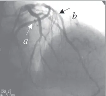
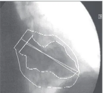
The patient underwent coronary angiography according to the Sones technique on July 14th, 2003, which revealed a LAD originating from the left Valsalva sinus, and dividing into the 2 following arteries: the circumflex artery, which reached the distal third of the left atrioventricular sulcus with obstructive lesions of 90% in the distal third and of 80% in the proximal third of a small posterior ventricular branch; and the LAD, which reached the middle third of the posterior interventricular sulcus with an obstructive lesion of 90% in the proximal third. The RCA originated right after that obstruction and had smooth walls with no obstruc-tive lesions (fig. 3 and 4). The left ventriculography showed an increase in the end-systolic volume due to moderate anteroapical hypokinesia (fig. 5).
Because the lesion in the LAD jeopardized a large myocardial area, the patient was medicated with acetylsalicylic acid (200 mg/day) and clopidogrel (300 mg in an attack dose). Twenty-four hours after coronary angiography, percutaneous coronary inter-vention (PCI) in the lesion of the LAD was performed through the femoral access, using a 2.5x20 mm balloon catheter for predilation of the lesion, followed by implantation of a 3.5x12mm AVE S7 stent with a final deployment pressure of 12 atmospheres. The procedure was successfully performed, resulting in TIMI III coro-nary flow, and 0% residual lesion (fig. 6).
The in-hospital evolution was uneventful, and the patient was discharged 24 hours after the intervention.
Discussion
2
Arquivos Brasileiros de Cardiologia - Volume 84, Nº 1, Janeiro 2005
Single Coronary Artery: Angioplasty with Stent Implantation
no difference in its incidence is observed between genders. It may be classified into 3 types: anomalies of origin, anomalies of termi-nation, and anomalies of distribution (the case reported) 6.
Coronary anomalies of origin are those in which the coronary system is at least biarterial, with one coronary artery emerging from a large vessel or from another coronary. The most common anomaly, corresponding to 90% of the cases of this anatomical alteration, is the circulation pattern in which the LAD emerges from the pulmonary trunk 6.
One modification of that pattern is the emergence of the left coronary artery from the pulmonary trunk (2.5 to 4.6% of congenital heart diseases). Although reported by Brooks in 1886, Bland,
Fig. 1 - Electrocardiogram at rest.
Fig. 2 - Positive exercise test for ischemia.
Fig. 3 - Coronary angiography on cranial RAO projection, showing RC (a) originating from the AD (b).
Fig. 4 - Coronary angiography on caudal LAO projection, showing RC (a) originating from the AD (b).
White, and Garland, in 1933, reported for the first time the clinical and hemodynamic syndrome. In more than 90% of the cases, the left coronary artery emerges from the posterior sinus of the pulmo-nary artery 7. Its physiopathology was established by Edwards 8,
who reported the existence of RCA collaterals to the left coronary artery, and the occurrence of a possible left-right shunt. The most common symptom is angina pectoris, due either to the lack of appropriate collaterals or to the phenomenon of “flow steal” to the pulmonary trunk. If the individual lives beyond childhood, the risk of sudden death persists in up to 80 to 90% of the cases, occurring around the age of 35 years 6.
Most cases of anomaly of origin cause a reduction in survival, except for the origin of the RC from the pulmonary trunk, a more rare form, which usually has benign prognosis 6.
3
Arquivos Brasileiros de Cardiologia - Volume 84, Nº 1, Janeiro 2005
Single Coronary Artery: Angioplasty with Stent Implantation
presenting worser prognosis in the short and long run, because it results in a single coronary system 6.
Coronary anomalies of termination are the most common he-modynamically significant coronary anomalies, the arteriovenous coronary fistulae (AVCF) being one example.
The AVCF, reported for the first time by Krause in 1865, is defined as a direct precapillary anastomosis between a larger coro-nary artery and a cardiac chamber or another larger vessel, such as the coronary sinus, superior vena cava, or pulmonary trunk 7.
The right coronary artery and right ventricle are more commonly involved. Drainage to the left chambers is rare (less than 10% of cases), the coronary sinus and bronchial veins being the most rare sites of drainage 6.
Angina pectoris is the most common symptom, resulting from the left-right “flow steal”, which deviates blood from the high resistance system of the myocardial capillary bed to the low-pressure system of the fistulae 7.
Indication for surgical correction depends on the degree of the shunt, and aims at occluding the AVCF with no hindrance to the
coronary flow. The most used techniques are as follows: inner closure of the fistulae from the recipient cavity; laterolateral tan-gential arteriorrhaphy (Cooley and Ellis, 1962); simple distal nary ligature (Bjork and Crafoord, 1947); proximal and distal coro-nary ligature; and ligature with the use of bypass.
If the surgical treatment is not performed when indicated, the probability of complications, such as congestive heart failure, bac-terial endocarditis, and, rarely, moderate pulmonary hypertension, increases with age. Cases of aneurysmatic dilation of the involved vessel and rupture of the fistulae have also been reported 9.
The single coronary artery (SCA), is an extremely rare anomaly, with an incidence of 0.04% 2. It is associated with a congenital
heart defect (as tetralogy of Fallot, transposition of the great vessels, truncus arteriosus) in 40% of cases 6.
The single coronary artery was considered a variation of the normal until the 18th century. In 1716, Thebesius reported for the first time a case of SCA, which was finally considered an anomaly in 1761, when Morgagni recognized as normal the presence of 2 major coronary arteries (2 ostia). Only 45 cases of single coronary artery had been reported until 1950 10.
The incidences of the anomaly of the right coronary artery RCA and of the left coronary artery LCA are similar 6. However, the even
rarer pattern of occurrence of SCA is that of the RCA originating from the LAD, which has only 7 cases reported in the literature 5.
In all cases, the RCA originated from the LAD after the first septal perforating branch, with an anterior trajectory to the right ventricular outflow tract and to the pulmonary trunk. Four cases had evidence of atheromatous coronary artery disease, but percutaneous revas-cularization was not performed in any of them10.
The prognosis of SCA ranges from excellent (without a decrease in survival) to poor, according to its anatomical distribution, inclu-ding the risk of sudden death, because 15% of the individuals with that anomaly develop severe heart disease before the age of 40 years. This risk is justified by the trajectory of the anomalous coronary artery on the cardiac base to reach its territory of distri-bution, which may expose the coronary artery to compression by other structures or angulation of its origin. In regard to developing atherosclerosis, a single coronary artery is considered at higher risk than normal coronaries are 2,4,6,8,9.
When atheromatous coronary artery disease develops, the con-sequences are significant, and the prognosis is severe if the obs-truction is proximal, because the ostium is single and no possibility of collateral circulation exists 6.
The indication of revascularization should be considered in the presence of symptoms (ie, angina), even in the absence of atheromatous coronary artery disease, due to the considerable chance of acute myocardial infarction and sudden death 7.
In conclusion, we report the case of an anomaly of coronary distribution with RCA originating from the proximal third of the LAD, the rarest type reported in the literature, right after a signi-ficant atheromatous lesion, jeopardizing a large area of myocardial musculature. It is worth emphasizing the significant role played by coronary angiography in this context, accurately defining the coronary artery anatomy and allowing a successful percutaneous treatment with stent implantation and excellent in-hospital evolu-tion, despite the severity of the case of this rare anomaly. Fig. 5 - Left ventriculography on RAO projection.
4
Arquivos Brasileiros de Cardiologia - Volume 84, Nº 1, Janeiro 2005
Single Coronary Artery: Angioplasty with Stent Implantation
1. Engel HJ, Torres C, Page L Jr. Major variations in anatomical origin of the coronary arteries: angiographic observations in 4,250 patients without associated conge-nital heart disease. Cathet Cardiovasc Diagn 1975; 1:157-69.
2. Sharbaugh MJAH, White RS. Single coronary artery: analysis of the anatomic va-riation, clinical importance, and report of five cases. JAMA 1974; 230:243-6. 3. Click RL, Holmes Jr DR, Vliestra RE, et al, and participants of the CASS –
Ano-malous coronary arteries: location, degree of atherosclerosis and effect on survival – a report from the Coronary Artery Surgery Study. J Am Coll Cardiol 1989; 13:531-7.
4. Lipton MJ, Barry WH, Obrez I, et al. Isolated single coronary artery: Diagnosis, angiographic classification, and clinical significance. Radiology 1979; 130:39-47. 5. Iyisoy A, Kursaklioglu H, Barcin C, et al. Single coronary artery with anomalous
origin of the rigth coronary artery as a branch from the left anterior descending ar-tery: a very rare coronary anomaly. Heart Vessels 2002; 16:161-3.
6. Fernandes ED, Kadivar H, Hallman GL, et al. Congenital malformations of the co-ronary arteries: the Texas Heart Institute Experience. Ann Thorac Surg 1992; 54:732-40.
7. Levin DC, Fellows KE, Abrams HL. Hemodynamically significant primary anomalies of the coronary arteries: angiographic aspects. Circulation 1978;58:25-34. 8. Edwards JE. Symposium on cardiovascular diseases: functional pathology of
congenital cardiac disease. Pediatr Clin North Am 1954; 1:13-49.
9. Ogden JA. Congenital anomalies of the coronary arteries. Am J Cardiol 1970; 25: 474-9.
10. Moreira AELC, Meireles GCX, Silva MVB, et al. Artéria coronária única e infarto agudo do miocárdio. Arq Bras Cardiol 1996; v 66:225-28.