1 PhD in Sciences by the Medicine School of the University of São Paulo - Medical doctor Assistant Surgery Division of the Heart Institute of the Hospital das Clínicas Medicine School of the University of São Paulo
2. Livre Docente Professor of the Thoracic Surgery and Cardiovascular Department of the Medicine School of the University of São Paulo - Director of the Research Surgery Unit of the Heart Institute of the Hospital das Clínicas Medicine School of the University of São Paulo
3. Medical doctor graduated in the Medicine School of the University of São Paulo - Medical doctor of Santa Casa de Misericórdia in São Bernardo do Campo.
4. PhD in Sciences by the Medicine School of the University of São Paulo - Assistant physician of the Pathologic Anatomy Department of the Heart Institute of the Hospital das Clínicas Medicine School of the University of São Paulo
5. Master in Sciences by the Veterinary College of the University of São Paulo Veterinarian.
6. Full professor of Cardiovascular Surgery in the Medicine School of the University of São Paulo - Director of the Surgery Division of the Heart Institute of the Hospital das Clínicas Medicine School of the University of São Paulo
Work performed in the Heart Institute of the Hospital das Clínicas Medicine School, University of São Paulo.
Support: FAPESP - Fundação de Amparo à Pesquisa do Estado de São Paulo.
Correspondence address:
Anderson Benício. Av. Dr. Enéas de Carvalho Aguiar, 44 / 2 andar -Bloco II / Sala 13 Cerqueira César - São Paulo – SP - CEP: 05403-000. E-mail: [email protected]
Anderson BENÍCIO1, Luiz Felipe Pinho MOREIRA2, Bernardo Assumpção de MÔNACO3, Jussara B. CASTELLI4, Larissa Eckmann MINGRONE5, Noedir Antônio Groppo STOLF6
Article received in October 16th, 2006 Article accepted in February 21st, 2007
RBCCV 44205-864
Estudo comparativo entre o pré-condicionamento isquêmico e a drenagem liquórica como métodos de
proteção medular em cães
Comparative study between ischemic
preconditioning and cerebrospinal fluid drainage as
methods of spinal cord protection in dogs
Abstract
Objective: This study compares the effects of immediate
ischemic preconditioning based on somatosensory evoked potentials (SSEP) monitoring, with those of cerebrospinal fluid drainage in a model of descending thoracic aorta occlusion in dogs
Method: Eighteen dogs were submitted to spinal cord
ischemia induced by descending thoracic aorta cross-clamping
INTRODUCTION
Paraplegia after the repair of dissections and aneurysms is an unpredictable complication that can occur in 0.2% of elective surgeries for abdominal aortic aneurysms or in up to 40% of the patients treated for acute aortic dissections or descending thoracic aortic or thoracoabdominal aneurysms [1,2].
Some techniques are widely utilized in the clinical practice to prevent paraplegia with satisfactory results [1,8]. Among them, the left atrial-femoral artery shuntand control of cerebrospinal fluid pressure by cerebrospinal fluid drainage should be mentioned. However, these methods do not provide definitive protection, specifically in more complex cases when a rate of paraplegia of from 15% to 20% is observed. On the other hand, other techniques such as the utilization of new drugs, the application of chronic ischemic preconditioning, as well as the introduction of somatosensory evoked potentials (SSEP) and motor induced
Results: Aortic pressure before and after the occluded
segment was similar in the three groups. Seven days after the procedure, Tarlov scores were significantly higher in comparison to the Control Group only in Drainage Group (p<0.05). Lower values of SSEP recovery times were also observed with the cerebrospinal fluid drainage during the final reperfusion period (p<0.01). Histopathologic study stain showed less important neuronal necrosis in the thoracic and lumbar gray matter in animals submitted to both methods of spinal cord protection, being more pronounced in IPC Group (p<0.001).
Conclusion: Cerebrospinal fluid drainage and immediate
ischemic preconditioning seems to protect spinal cord during descending thoracic aorta cross clamping. Nevertheless, the obtained level of spinal cord protection seems to be more significant with cerebrospinal fluid drainage.
Descriptors: Aorta, surgery. Spinal cord. Cerebrospinal
fluid. Somatosensory evoked potentials. Dogs.
Resumo
Objetivo: Este estudo compara os efeitos do
pré-condicionamento isquêmico imediato, baseado na monitorização do potencial evocado somatossensitivo (PESS), com aqueles da drenagem do líquido cefalorraquidiano, em um modelo de oclusão da aorta torácica descendente em cães.
Método: Dezoito cães foram submetidos à isquemia medular
induzida pela oclusão da aorta torácica descendente por 60
minutos. O Grupo Controle foi submetido à oclusão da aorta (n=6), o Grupo Pré-Condicionamento Isquêmico (PCI), ao pré-condicionamento isquêmico (n=6) e o grupo drenagem, à drenagem do líquido cefalorraquidiano (n=6), imediatamente antes da oclusão da aorta. A condição neurológica foi acessada por um observador independente, de acordo com a escala de Tarlov. Os animais foram sacrificados e as medulas retiradas para exame histopatológico.
Resultados: Pressões da aorta proximal e distal à oclusão
foram semelhantes nos três grupos. Sete dias após o procedimento, o índice de Tarlov foi significativamente maior em comparação ao Grupo Controle, somente no Grupo PCI (p<0,05). Foram observados valores menores no tempo de recuperação do PESS com o uso da drenagem liquórica durante a fase final de reperfusão (p<0,01). Exame histopatológico evidenciou necrose menos grave na substância cinzenta torácica e lombar, nos animais submetidos aos dois métodos de proteção medular, sendo mais pronunciada no Grupo PCI (p<0,001).
Conclusão: A drenagem do líquor e o pré-condicionamento
isquêmico parecem proteger a medula espinhal, durante a oclusão da aorta torácica descendente. Entretanto, o nível de proteção medular obtido parece ser mais significativo com a drenagem do líquido cefalorraquidiano.
Descritores: Aorta, cirurgia. Medula espinhal. Líquido
cefalorraquidiano. Potenciais somatosensoriais evocados. Cães.
potentials to identify spinal cord ischemia in the intraoperative period are being developed aiming at providing complete spinal cord protection during aortic surgery [1-8]. The application of acute ischemic preconditioning seems to be associated with some spinal cord protection, however, the methodology is not well established and its effectiveness is still debatable.
The goal of this study is to assess the efficacy of acute ischemic preconditioning compared to cerebrospinal fluid drainage using SSEP monitoring, as complementary methods of spinal cord protection in descending thoracic aortic surgery of dogs.
METHOD
measurements were controlled by continuous arterial gas analysis (Nova Medical, Stat Profile Ultra, USA).
Monitoring was performed with continuous ECG, invasive measurement of proximal and distal systemic arterial pressure using catheters (Gelco 20) introduced into the left common carotid artery and right femoral artery, respectively. Central venous pressure measurements and infusion of drugs were achieved by a central catheter introduced into the left internal jugular vein. The body temperature was controlled between 35.5ºC and 37ºC using an esophageal thermometer. To monitor and drain the cerebrospinal fluid (CSF) the cistern magna was punctured and a nº 16 peridural catheter was inserted. The CSF was drained before aortic occlusion. Hemodynamical measurements were stored using the Acknowledge 3.0 computer program. Hydration was maintained by administering 50 mL/kg/h of simple ringer solution aiming at maintaining the central venous pressure between 3 mmHg and 5 mmHg.
For all dogs, during the aortic occlusion phase, the proximal systemic arterial pressure was controlled by the intravenous administration of sodium nitroprusside using the central catheter.
In all groups, to control of the acid-base balance, arterial gas analysis was performed before, during and after aortic occlusion; in cases in which there was a necessity of metabolic acidosis correction, sodium bicarbonate was administered depending on the result of gas analysis.
SSEP monitoring was performed by lumbar puncture at L2-L3 to introduce a silver electrode. Another electrode for stimulation was placed in the right tibial nerve and a third in the occipital region as a central reference. Recording of SSEP readings was achieved using a 4ME Medleck Saphire device. The experiments were performed in sterile conditions. With the dogs in the right lateral decubitus position, left lateral thoracotomy was performed in the fourth left intercostal space. The aorta was dissected and isolated at the root of the left subclavian artery. Heparin was administrated at a dose of 100 IU/kg of weight. When the procedure had been completed, the effect of the heparin was reverted by administering protamine sulphate at a rate of 1:1 in relation to the administered heparin. Continuous blood sampling to assess arterial gases and serum electrolytes was performed during the surgery. At the end of the experiment, an intramuscular injection of 1200000 IU of benzathine penicillin and an endovenous dose of 15 mg/ kg cephalotin were administered.
The animals were clinically and anatomopathologically assessed for seven days after the surgery. An evaluation of the sensory-motor recovery of the hind paws was made according to clinical criteria, using the Tarlov method* (Apud Matsuyama et al.) [10]:
0 - Without movements of the hind paws; principles described for animal experimentation (COBEA)
and the Guide for the Care and Use of Laboratory Animals (Institute of Laboratory Animal Resources, National Academy of Sciences, Washington, D.C., 1996). The study protocol was approved by the Ethics Committee for the Analysis of Research Projects – CAPPesq of the Hospital das Clínicas of the Medical School of Sao Paulo University. Eighteen crossbred dogs of both genders, weighting between 15 and 20 kg were utilized and divided into the Control Group (six), Ischemic Preconditioning Group (IPC -six) and Drainage Group (-six).
Control group
In this group, the dogs were submitted to simple occlusion of the descending thoracic aorta for a period of 60 minutes. The aorta was occluded in the portion adjacent to the root of the left subclavian artery. During aortic occlusion, the SSEP was monitored and the time for the potential to drop was observed. Sixty minutes of reperfusion followed the occlusion phase. The time of potential recovery was observed. Potential monitoring was performed in this way for all groups.
Ischemic preconditioning group
In this group, the dogs were submitted to ischemic preconditioning, with aortic occlusion (as in the Control Group), causing ischemic injury so that there was an evoked potential drop of up to 60% [9]. The potential was measured at one-minute intervals until the planned drop was achieved. After spinal cord ischemia, the aorta was released, and spinal cord reperfusion followed until complete recovery of the induced potential drop. Then, respective times of ischemia and potential recovery were determined. Immediately after recovery of the potential, the aorta was occluded and reperfused at two other predetermined times (for ischemia and reperfusion), completing the preconditioning phase. Subsequently, a final occlusion for 60 minutes followed by 60 minutes of reperfusion was performed.
Drainage group
In this group, the procedure was similar to the Control Group. However, cerebrospinal fluid drainage was performed immediately before aortic occlusion. The drainage aimed at maintaining cerebrospinal fluid pressure close to zero mmHg.
Surgical procedure
All dogs were submitted to general anesthesia using 20 mg/kg sodium pentobarbital and 4 µg/kg fentanyl citrate.
1 - Perceptible movements of the hind paws; 2- Good movements, but with static instability; 3 - Ability to stand and walk;
4 - Complete recovery.
* Tarlov IM. Spinal cord compression: mechanisms of paralysis and treatment. Springfield, III: Charles C. Thomas; 1957. pg. l47.
Anatomopathological study
Collection and processing of the material
After seven postoperative days the animals were sacrificed by the intravenous administration of 20 mg/kg sodium pentobarbital followed by 10 mL potassium chloride solution. Then the spinal column was removed in one piece from its thoracolumbar transition. Laminectomy was performed exposing the spinal cord along its complete length. The spinal cord was removed sectioning the peripheral nerve roots. After removal of the spinal cord, the dura-mater was opened lengthwise exposing it for better fixation. It was placed for 24 hours in a buffered solution of 10% formaldehyde for fixation. Three cross-sectional samples of approximately 0.5 cm were taken from thoracolumbar T13, L1 and L2 segments and conditioned in 70º alcohol for another 24-hour period. Subsequently, they were placed in an automatic histologic tissue processor (Leica TP 1020 or Shandon-Citadel 1000) overnight and embedded in paraffin. From the paraffin blocks 3-micron histological sections were made using a Leica RM2145 microtome and placed on slides previously prepared with organosilane.
Conventional histological study using hematoxylin and eosin
The histological sections of the spinal cord dully identified and stained using hematoxylin and eosin (HE) were examined at light-microscopy blind to the treatment group.
The number of neurons in half the grey matter was estimated. Detected alterations were morphometrically treated so that a comparative analysis among the groups would be possible in respect to the numbers of viable and necrotic neurons.
Statistical analysis
To analyze the hemodynamical data and number of neurons, two-way analysis of variance was used complemented by the Bonferroni test. To assess the time for SSEP loss and recovery, as well as for the Tarlov rate and neuron count, the non-parametrical Kruskal-Wallis test for non-related variables was utilized, complemented by the multiple comparison Dunn test. The central values are presented as averages, with the spread presented as a standard deviation or 95% confidence interval. The level of significance established for this study was 5%.
RESULTS
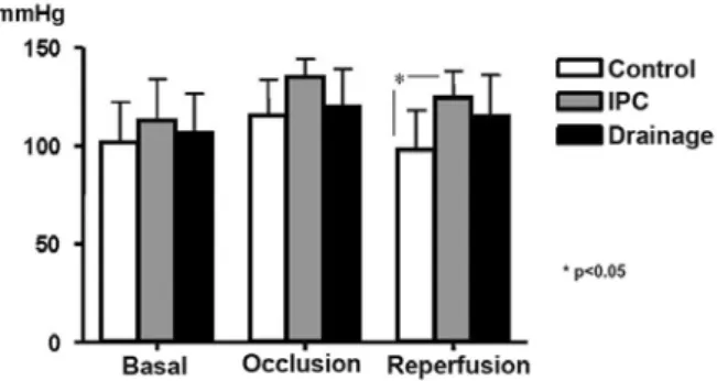
There were no significant differences among the weight and genders of the dogs in the three groups. Arterial gases, esophageal temperatures and hematocrit levels did not present significant differences among the groups too, although some of the dogs presented with significantly different values. The mean proximal (Figure 1) and distal (Figure 2) arterial pressure measurements were performed at baseline, during aortic occlusion and after 60 minutes of reperfusion. When the proximal mean systemic arterial pressure was compared among the three groups, a statistically significant difference was observed among the observed values (p=0.01). When the groups were compared, a statistically significant difference was identified only between the values of the Control and IPC Groups in the reperfusion phase (p<0.05). On comparing the distal mean systemic arterial pressure among the three groups, the pressure in the aortic occlusion phase was significantly lower compared to the basal and reperfusion phases.
Fig. 1 – Mean proximal systemic arterial pressure of the three groups, before, during and after aortic occlusion. The values are shown as means and standard deviation
However, there was no significant difference among the groups.
The cerebrospinal fluid pressure was analysed in the three groups before and during aortic occlusion. In the reperfusion phase, there was a great loss of cerebrospinal fluid pressure, a fact that made a study in this variable unviable. Before aortic occlusion, there was no statistical difference in the cerebrospinal fluid pressures among the three groups. However, a significant difference was observed in the occlusion phase when the Drainage and Control Groups (p<0.001) and the IPC and Drainage Groups (p<0.001) were compared – Figure 3.
In the analysis of SSEP recovery during reperfusion, a statistically significant difference was seen among the groups (p=0.0056). With the comparative Dunn test, a statistically significant difference was identified between the Control and Drainage Groups (p<0.001) – Figure 5.
The clinical evaluation of the Tarlov rate also demonstrated variations among the groups. When the Tarlov rate of the groups was compared, a statistical difference was detected giving a p-value = 0.0185. When the Control group was compared with the Drainage Group, a statistically significant difference was identified with a p-value < 0.05 (Table 1).
IPC - Ischemic preconditioning
Table 1. Tarlov rate of the six dogs in each group
Tarlov
0 1 2 3 4
Control
5 0 1 0 0
IPC
1 2 0 0 3
Drainage*
1 0 0 0 5 Fig. 3 – Cerebrospinal fluid pressure of the three groups, before
and during aortic occlusion. The values are shown as means and standard deviation
Fig. 4 – Time of SSEP drop of the three groups, in the aortic occlusion phase
Fig. 5 –Time of SSEP recovery of the three groups, in the spinal cord reperfusion phase
The time of SSEP loss during the aortic occlusion period and the recovery time during the reperfusion period were studied. There was a statistically significant difference among the groups in respect to SSEP loss during aortic occlusion (p=0.0014). Comparing the groups using the comparative Dunn test a statistically significant difference was identified only when the time of SSEP loss during aortic occlusion between the Control and Drainage Groups was evaluated (p<0.001). However, an early loss in the IPC Group was also observed (Figure 4).
Histological analysis
When the necrotic neurons were analysed the difference was greater. On comparing the groups a statistically significant difference was found giving a p-value < 0.0001. With further investigation using the Bonferroni test, a statistically significant difference was identified between the Control and IPC groups at L2 (p<0.001). However, on comparing the Control and Drainage Groups, a statistically significant difference was seen at T13 and L1, with p-values < 0.05, and at L2, with a p-value < 0.001. On the other hand, comparing the IPC and Drainage Groups no statistically significant differences were found at any point, even though there was a trend of better preservation of the spinal cord histological structure in the Drainage Group (Figure 7).
contribute to the high morbimortality: Type III acute aortic dissections, preoperative renal insufficiency, advanced ages, symptomatic aneurysms, Crawford Type II aortic aneurysms [4], coronary artery disease, chronic obstructive pulmonary disease (COPD), total occlusion time [5] and diabetes [6].
Neurological complications after surgical aneurysm corrections may be presented as immediate and late deficits. Immediate neurological deficits are directly due to the results of hypoxia related to acute deprivation of the blood flow during prolonged aortic occlusion. Late neurological deficits may develop between the first and 21st postoperative day,
and are responsible for approximately one third of all neurological injuries. Late injuries are generally the consequence of subclinical ischemia, reperfusion hyperemia with cell injury mediated by the release of free radicals in the formation of medullary edema or regional hypotension, restricting blood flow by high vascular resistance in the medullary plexus [7].
The physiopathology of spinal cord ischemia, associated to acute interruption of the blood flow during surgery of descending thoracic aortic aneurysms, involves a series of progressive interdependent events that, if allowed to prevail, lead to irreversible neurological injury. This group includes proximal hypertension, increase in the left ventricular post-load, increase in the cerebrospinal fluid pressure, compromise of perfusion of the intercostal branches or lumbar arteries during aortic occlusion and, finally, the extent of the injuries [8]. The risk of paraplegia during aortic surgery is determined by the interaction of four independent processes: reduction in spinal cord blood flow, metabolism rate of neural tissue, reperfusion injury after ischemia and blood flow after reperfusion [2]. Several techniques have been utilized to avoid these mechanisms in an attempt to counteract or minimize their effects. Deep hypothermia with total circulatory arrest [11,12], angiographic identification of the intercostal arteries in the preoperative period for later surgical implantation [13], the use of SSEP [14,15], cerebrospinal fluid drainage [7,16], utilization of pharmacological agents [8,15] and distal aorta perfusion (aorta-aorta or left atrium-femoral artery shunts)[17-19] are widely utilized strategies of spinal cord protection in the clinical practice. However, none of them protect the marrow totally.
Deep hypothermia decreases oxygen demand of the neural tissue and, thus, increases its tolerance to hypoxia. Despite the harmful effects of hypothermia in cardiovascular surgery, this is an important coadjuvant technique to protect the spinal cord in thoracoabdominal aortic surgeries. Although this technique provides some benefit in respect to a drop in the metabolic rate due to the reduction in temperature there are some limitations. The main disadvantage is the elevation in the CSF pressure after infusion of iced saline solution which is necessary to Fig. 6 – Number of viable neurons, in the three groups, at T13, L1
and L2. The values are shown as means and standard deviation
Fig. 7 – Number of necrotic neurons in the three groups at T13, L1 and L2. The values are shown as means and standard deviation
DISCUSSION
decrease the CSF temperature to between 23ºC and 25ºC. Moreover, the CSF temperature increases soon after cessation of the intrathecal infusion of saline solution. Additionally, an epidural catheter is not used for drainage as the fluid quickly spreads out of the epidural space along the nerve roots. Nevertheless, Rokkas et al. [11] and Kouchoukos et al. [12] showed more encouraging results with the association of distal aortic perfusion and deep hypothermia.
SSEP is used to assess the integrity of central somatosensory pathways. It also detects and identifies the location of injuries in afferent pathways of the central nervous system. In cases of spinal cord injury monitoring may determine the magnitude of the injury and also the existence of a cortical response, and therefore, a better prognosis for functional recovery [2,15]. In the clinical practice, some groups have utilized this technique for the intraoperative identification of spinal cord ischemia [15]. However, this technique does not guarantee prevention of paraplegia, even after detecting intraoperative situations in which further ischemic compromise would have a great probability of leading to irreversible injury.
The great limitation of this technique is the possibility of false-negative results, as only lateral and posterior horn conductions are monitored and motor injury is caused by ischemia of the anterior horn of the marrow. Another fact is that cortical dysfunction of the peripheral nerve due to ischemia or anesthetic agents can also result in false-positive results. However, techniques to directly detect the anterior horn function of the spinal cord (motor induced potential) in the intraoperative period are being developed with the purpose of eliminating possible false-positive interpretations that occur in sensory induced potentials [15].
The occlusion of the aorta invariably causes proximal arterial hypertension and increases in the left ventricular post-load, promoting an elevation in the CSF pressure with distal hypotension. Spinal cord perfusion is measured by the spinal cord perfusion pressure that is determined by the gradient between the distal aortic pressure and the CSF pressure [5,20]. Randomized clinical trials [7,16] support the clinical use of drainage and monitoring of the SCF during resection of ascending aortic aneurysms, as a method of spinal cord protection.
Other techniques also utilized are perfusion of the distal aorta and left atrium-femoral artery shunt. The essential premise is that the increase in the perfusion pressure of the distal aorta (by the shunt) will result in an increase in the spinal cord blood flow and thus, a reduction in spinal cord ischemia during aortic occlusion. Safi et al. [17] reported their experience of 45 consecutive patients who underwent the surgical correction of thoracoabdominal aortic aneurysms. Comparing the results of these patients with the initial unpublished results of the same author (112
patients), we observed that in the group of 45 patients, who were submitted to CSF drainage and distal aorta perfusion, there was a lower incidence of paraplegia in the postoperative period than with the early experience of the author. With similar results, Robertazzi et al. [15] confirmed the efficacy of distal aortic perfusion either associated with other methods of spinal cord protection or in isolation to prevent spinal cord ischemia. On the other hand, Svensson et al. [5], in their experience with 1509 patients who underwent thoracoabdominal aortic surgery, proved that, in spite of a reduction in the mortality rate with the new operative techniques, the incidence of postoperative paraplegia was still a severe and unpredictable complication.
At the end of the last decade, the physiopathologic consequences of human myocardial ischemia received special attention. With the description of stunned myocardium and myocardial hibernation, it was believed, until recently, that intermittent ischemic events may cause cumulative injury of the myocardium. Murray et al. [20] demonstrated that endogenous protective mechanisms of the myocardium really exist. Ironically, short intervals of ischemia followed by reperfusion, increase resistance against greater ischemic compromise, making the tissue more resistant to subsequent more prolonged ischemic aggression. This response is called ischemic preconditioning (IPC) [20,21].
Subsequently, the protective effect of IPC was demonstrated in several subsystems. In the last decade, several publications, including a national study [22], tried to demonstrate a beneficial effect of IPC on the medulla, although all were performed under chronic conditions. Munyao et al. [23] studied two groups of rabbits that were submitted to thirty minutes of aortic occlusion: one group with (12 or 48 hours before occlusion) and the other without IPC of the spinal cord. The rabbits that were submitted to IPC 12 hours before ischemia had significantly better motor functions compared to the control group. The rabbits that were submitted to 48 hours of IPC before aortic occlusion presented variable recovery, with a slight anatomopathological correlation in respect to the posterior spinal cord.
REFERENCES
1. Albuquerque LC, Palma JH, Braile D, Gomes W, Guimarães JI. Diretrizes para a cirurgia das doenças da aorta. Arq Bras Cardiol. 2004;82(suppl.5):35-50.
2. Gharagozloo F, Neville RF Jr, Cox JL. Spinal cord protection during surgical procedures on the descending thoracic and thoracoabdominal aorta: a critical overview. Semin Thorac Cardiovasc Surg. 1998;10(1):73-86.
the mechanism of the appearance of HSP, with a positive correlation between its appearance and neurological protection. These results suggest that chronic IPC offers protection to the marrow and that HSP is a protein that, possibly, offers an important additional protection to the marrow.
However, in relation to the immediate IPC, the conclusion is still controversial, as some reports, apart from not presenting this association with HSP, did not demonstrate its protective effect, probably due to the lack of standardization in the technique [25]. Contreras et al. [9] demonstrated a novel form of standardizing the immediate IPC, confirming its benefits. The utilization of SSEP, to determine the ischemia time and spinal cord recovery time, optimizes the IPC effect on the spinal cord, making the method really efficient. Similar to the work of Contreras et al. [9], in our work, we observed a very satisfactory level of spinal cord protection with IPC as was also shown by Tampoulis et al. [26] although they did not utilize SSEP during IPC.
In spite of there being several techniques of spinal cord protection described in publications, none totally and efficiently protect the spinal cord, even though the mechanisms of spinal cord injury are well established [27]. On the other hand, there has been much effort to combine several techniques to find the best way possible to protect against all the mechanisms responsible for spinal cord injury. However, the surgical treatment of aortic diseases still produces tragic complications, including paraplegia for many patients.
CONCLUSIONS
Considering these results, we conclude that both methods (IPC and cerebrospinal fluid drainage) are efficient from clinical and histopathological points of view, to protect the spinal cord during acute aortic ischemia in this experimental model. However, cerebrospinal fluid drainage proved to be better in this experimental model.
3. Coselli JS, LeMaire SA, Figueiredo LP, Kirby RP. Paraplegia after thoracoabdominal aortic aneurysm repair: is dissection a risk factor? Ann Thorac Surg. 1997;63(1):28-36.
4. LeMaire SA, Miller CC 3rd, Conklin LD, Schmittling ZC, Köksoy C, Coselli JS. A new predictive model for adverse outcomes after elective thoracoabdominal aortic aneurysm repair. Ann Thorac Surg. 2001;71(4):1233-8.
5. Svensson LG, Crawford ES, Hess KR, Coselli JS, Safi HJ. Experience with 1509 patients undergoing thoracoabdominal aortic operations. J Vasc Surg. 1993;17(2):357-70.
6. Coselli JS, LeMaire SA, Miller CC 3rd, Schmittling ZC, Köksoy C, Pagan J, et al. Mortality and paraplegia after thoracoabdominal aortic aneurysm repair: a risk factor analysis. Ann Thorac Surg. 2000;69(2):409-14.
7. Coselli JS, LeMaire SA, Köksoy C, Schmittling ZC, Curling PE. Cerebrospinal fluid drainage reduces paraplegia after thoracoabdominal aortic aneurysm repair: results of a randomized clinical trial. J Vasc Surg. 2002;35(4):631-9.
8. Laschinger JC, Izumoto H, Kouchoukos NT. Evolving concepts in prevention of spinal cord injury during operations on the descending thoracic and thoracoabdominal aorta. Ann Thorac Surg. 1987;44(6):667-74.
9. Contreras IS, Moreira LF, Ballester G, Mônaco BA, Lancellotti CL, Dias AR, et al. Immediate ischemic preconditioning based on somatosensory evoked potentials seems to prevent spinal cord injury following descending thoracic aorta cross-clamping. Eur J Cardiothoracic Surg. 2005;28(2):274-9.
10. Matsuyama K, Chiba Y, Ihaya A, Kimura T, Tanigawa N, Muraoka R. Effect of spinal cord preconditioning on paraplegia during cross-clamping of the thoracic aorta. Ann Thorac Surg. 1997;63(5):1315-20.
11. Rokkas CK, Kouchoukos NT. Profound hypothermia for spinal cord protection in operations on the descending thoracic and thoracoabdominal aorta. Semin Thorac Cardiovasc Surg. 1998;10(1):57-60.
12. Kouchoukos NT, Wareing TH, Izumoto H, Klausing W, Abboud N. Elective hypothermic cardiopulmonary bypass and circulatory arrest for spinal cord protection during operations on the thoracoabdominal aorta. J Thorac Cardiovasc Surg. 1990;99(4):659-64.
13. Griepp RB, Ergin MA, Galla JD, Lansman S, Khan N, Quintana C, et al. Looking for the artery of Adamkiewicz: a quest to minimize paraplegia after operations for aneurysms of the descending thoracic and thoracoabdominal aorta. J Thorac Cardiovasc Surg. 1996;112(5):1202-15.
15. Robertazzi RR, Cunningham JN Jr. Intraoperative adjuncts of spinal cord protection. Semin Thorac Cardiovasc Surg. 1998;10(1):29-34.
16. Svensson LG, Stewart RW, Cosgrove DM 3rd, Lytle BW, Antunes MD, Beven EG, et al. Intrathecal papaverine for the prevention of paraplegia after operation on the thoracic or thoracoabdominal aorta. J Thorac Cardiovasc Surg. 1988;96(5):823-9.
17. Safi HJ, Bartoli S, Hess KR, Shenaq SS, Viets JR, Butt GR, et al. Neurologic deficit in patients at high risk with thoracoabdominal aortic aneurysms: the role of cerebral spinal fluid drainage and distal aortic perfusion. J Vasc Surg. 1994;20(3):434-44.
18. Svensson LG, Hess KR, Coselli JS, Safi HJ. Influence of segmental arteries, extent, and atriofemoral bypass on postoperative paraplegia after thoracoabdominal aortic operations. J Vasc Surg. 1994;20(2):255-62.
19. Safi HJ, Campbell MP, Ferreira ML, Azizzadeh A, Miller CC. Spinal cord protection in descending thoracic and thoracoabdominal aortic aneurysm repair. Semin Thorac Cardiovasc Surg. 1998;10(1):41-4.
20. Murray CE, Jennings RB, Reimer KA. Preconditioning with ischemia: a delay of lethal cell injury in ischemic myocardium. Circulation. 1986;74(5):1124-36.
21. Castro e Silva O Jr, Centurion S, Pacheco EG, Brisotti JL, Oliveira AF, Sasso K. Aspectos básicos da lesão de isquemia e
reperfusão e do pré-condicionamento isquêmico. Acta Cir Bras. 2002,17(supl 3):96-100.
22. Sader AA, Chimelli LMC, Sader SL, Barbieri Neto J, Coutinho Neto J, Roselino JES, et al. Pré-condicionamento precoce da medula espinhal isquêmica: pesquisa em coelhos. Rev Bras Cir Cardiovasc. 1998;13(2):146-51.
23. Munyao N, Kaste M, Lindsberg PJ. Tolerization against loss of neuronal function after ischemia-reperfusion injury. Neuroreport. 1998;9(2):321-5.
24. Perdrizet GA, Lena CJ, Shapiro DS, Rewinski MJ. Preoperative stress conditioning prevents paralysis after experimental aortic surgery: increased heat shock protein content is associated with ischemic tolerance of the spinal cord. J Thorac Cardiovasc Surg. 2002;124(1):162-70.
25. Ondrejcak T, Vanicky I, Galik J. Ischemic preconditioning does not improve neurological recovery after spinal cord compression injury in the rat. Brain Research. 2004;995(2):267-73.
26. Toumpoulis IK, Anagnostopoulos CE, Drossos GE, Malamou-Mitsi VD, Pappa LS, Katritsis DG. Early ischemic preconditioning without hypotension prevents spinal cord injury caused by descending thoracic aortic occlusion. J Thorac Cardiovasc Surg. 2003;125(5):1030-6.