Serum and cerebrospinal fluid
concentrations of E-selectin in
patients with aneurysmal
subarachnoid hemorrhage
1Department of Neurosurgery, 2Department of Anesthesiology, 3Department of Biochemistry, Cerrahpasa Medical Faculty,
Istanbul University, Istanbul, Turkey T. Tanriverdi1, G.Z. Sanus1,
M.O. Ulu1, E. Tureci2,
H. Uzun3, S. Aydin3
and M.Y. Kaynar1
Abstract
The goal of the present study was to determine concentrations of E-selectin in both cerebrospinal fluid (CSF) and serum of patients with aneurysmal subarachnoid hemorrhage (SAH) and to evaluate the correlation between the clinical parameters and E-selectin levels. Both CSF and serum samples obtained from 12 patients with aneurysmal SAH and 8 patients with hydrocephalus (control group) without any other known central nervous system disease were assayed for E-selectin by quantitative enzyme-linked immunosorbent assay and the results were compared between the two groups. Mean levels of soluble forms of E-selectin within the first 3 days and on the 5th and 7th days of SAH were 4.0 ± 7.9, 2.8 ± 5.2, and 3.1 ± 4.9 ng/ml in the patient’s CSF, and 33.7 ± 9.2, 35.1 ± 7.0, and 35.2 ± 8.7 ng/ml in serum, respectively. In contrast, mean E-selectin levels were 0.1 ± 0.2 ng/ml in CSF and 8.7 ± 5.0 ng/ml in serum of control patients. The difference between groups was statistically significant regarding both CSF and serum E-selectin levels (P < 0.05). Thus, we have demonstrated a marked increase of E-selectin concentration in both CSF and serum of patients with aneurysmal SAH compared with control and suggest that blocking the interaction between E-selectin and vascular endothe-lium may have a beneficial effect on vasospasms.
Correspondence
T. Tanriverdi P.K: 4, Cerrahpasa 34301, Istanbul Turkey
Fax: +90-212-414-3427 E-mail: tanerato2000@yahoo.com
Received January 21, 2005 Accepted August 11, 2005
Key words
•Adhesion molecules •E-selectin
•Selectin
•Subarachnoid hemorrhage •Vasospasm
Introduction
The contribution of vascular endothe-lium (VE) to inflammation after a pathologi-cal condition has been demonstrated and the recruitment of leukocytes from circulating blood and their interaction with endothelium is crucial in inflammatory reactions (1-3). This interaction occurs through a multistep
Ap-pearance or up-regulation of CAMs has been observed in a variety of central nervous sys-tem (CNS) diseases (5-8) and the therapeu-tic approaches blocking leukocyte-endothe-lium interactions have provided encourag-ing results, suggestencourag-ing that these molecules have a pivotal role (9-13).
Over the last decade, the bulk of the evidence from both experimental and hu-man studies has shown that CAMs may par-ticipate in the pathogenesis of vasospasm (VS) after subarachnoid hemorrhage (SAH) following an aneurysm rupture, but few stud-ies have investigated the role of selectins in aneurysmal SAH.
In this report, we focus on the response of one member of the selectin family, E-selec-tin, that has been proved to participate in the early step of inflammation, namely, rolling of the leukocytes on the VE. We determined the concentrations of E-selectin in both the cerebrospinal fluid (CSF) and serum of pa-tients with aneurysmal SAH and compared them to those of patients with hydroceph-alus.
Patients and Methods
Patients
Ethical approval for this study was ob-tained from the Human Investigations Com-mittee at Istanbul University and all patients or the next of kin if the patient was uncon-scious, provided written informed consent. We studied the patients referred to our neu-rosurgical unit from January to June 2003 with SAH established by computed tomog-raphy (CT). We excluded patients who had any kind of infection at the time of CSF and serum collection, in which CAMs may play a part. The sole inclusion criterion was the admission of the patients to our unit within the first three days of SAH. Demographic and clinical data, i.e., sex, age, initial Glasgow coma scale score, history of chronic illness and smoking, Hunt-Hess (HH) neurological
grade, amount of blood within the cisterns determined by Fisher grade on initial CT, initial leukocytosis (white blood cell count more than 11,000 x 103/ml), symptomatic
VS, and clinical outcome at 6 months as assessed using the Glasgow outcome scale were recorded on standardized data collec-tion charts. A patient was considered to have symptomatic VS if neurological deteriora-tion (confusion or decreased level of con-sciousness with focal neurological deficits, such as speech or motor deficits) occurred after day 3 and was managed by the induc-tion of a hyperdynamic state included hyper-tension, hypervolemia, and hemodilution. If the patients showed clinical signs of symp-tomatic VS, we performed a CT scan to confirm VS and the scan showed a focal ischemic zone in these patients. In this study, we also tried to determine whether there was a correlation between the levels of E-selec-tin and the above clinical parameters.
Demographic data of patients and controls
surgery (≥10-14 days post-SAH) in 2 pa-tients with grade IV HH and 1 patient with grade V HH. One patient with grade II HH underwent endovascular coiling. A summary of the demographic data for the patients with SAH and the control group is provided in Table 1.
Specimen handling
Eighty-eight samples (44 each of CSF and serum) were assayed for E-selectin. For each patient, serial blood and CSF samples were collected at the same time within 3 days, and on the 5th and 7th days of SAH. Blood and CSF samples were collected by venipuncture and lumbar puncture, respec-tively. In the control hydrocephalus group, blood samples were collected by venipunc-ture, and CSF samples were obtained during
the execution of ventriculo-peritoneal shunt-ing. The samples from the control group were obtained on one occasion. As soon as possible, each 10-ml CSF and blood sample was centrifuged at 10,000 rpm for 15 min and the supernatant was stored at -70ºC until assayed.
E-selectin measurement
CSF and serum soluble E-selectin (sE-selectin) concentrations were measured quan-titatively and are reported as ng/ml using a commercially available enzyme-linked im-munosorbent assay kit (R&D Systems, Min-neapolis, MN, USA, and Abingdon, UK). A dilution of 1:10 was used for E-selectin as-says and absorption measurements were car-ried out at 450 nm using a microtiter plate reader (Automated Microplate Reader,
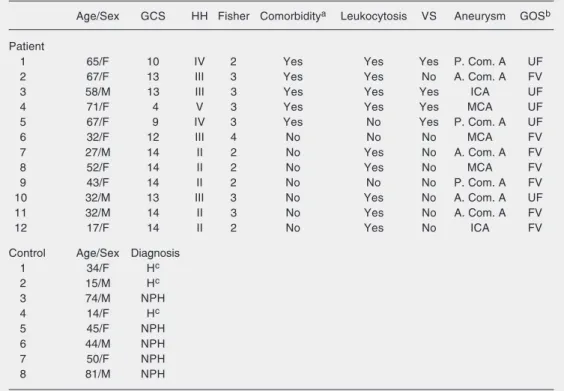
Mo-Table 1. Clinical data of the patients with subarachnoid hemorrhage and hydrocephalus controls.
Age/Sex GCS HH Fisher Comorbiditya Leukocytosis VS Aneurysm GOSb
Patient
1 65/F 10 IV 2 Yes Yes Yes P. Com. A UF
2 67/F 13 III 3 Yes Yes No A. Com. A FV
3 58/M 13 III 3 Yes Yes Yes ICA UF
4 71/F 4 V 3 Yes Yes Yes MCA UF
5 67/F 9 IV 3 Yes No Yes P. Com. A UF
6 32/F 12 III 4 No No No MCA FV
7 27/M 14 II 2 No Yes No A. Com. A FV
8 52/F 14 II 2 No Yes No MCA FV
9 43/F 14 II 2 No No No P. Com. A FV
10 32/M 13 III 3 No Yes No A. Com. A UF
11 32/M 14 II 3 No Yes No A. Com. A FV
12 17/F 14 II 2 No Yes No ICA FV
Control Age/Sex Diagnosis
1 34/F Hc
2 15/M Hc
3 74/M NPH
4 14/F Hc
5 45/F NPH
6 44/M NPH
7 50/F NPH
8 81/M NPH
GCS = Glasgow coma scale; HH = Hunt-Hess grade; VS = symptomatic vasospasm; GOS = Glasgow outcome scale; P. Com. A. = posterior communicating artery; A. Com. A = anterior communicating artery; ICA = internal carotid artery; MCA = middle cerebral artery; UF = unfavorable; FV = favorable; H = hydrocephalus;
NPH = normal pressure hydrocephalus. aComorbidity includes hypertension and/or diabetes mellitus. bAt 6
del EC311sx; Biotech Instruments, Wi-nooski, VT, USA). All samples and stan-dards were run in duplicate.
Statistical analysis
Data were analyzed using the SPSS sta-tistical program (SPSS, Chicago, IL, USA). Statistical analyses were performed using the nonparametric Mann-Whitney U-test. Results are reported as means ± standard deviation (SD). A probability value of less than 0.05 was considered to be statistically significant.
Results
Thirty-six CSF and 36 serum samples
from the patients with SAH and 8 CSF and 8 serum samples from the control group were obtained for this prospective clinical study. The samples were used for the deter-mination of E-selectin. A summary of the statistical data is provided in Table 2 and Figure 1.
Concentrations of E-selectin in CSF
Concentrations of E-selectin were mark-edly different in patients with SAH and the hydrocephalus controls. In the control group, 3 patients had no detectable E-selectin, with a mean concentration of 0.16 ± 0.21 ng/ml. In contrast, all patients with SAH had quan-tifiable E-selectin in their CSF. The average values were 4 ± 7.95, 2.84 ± 5.21, and 3.10 ± 4.91 ng/ml within 3 days of SAH, on day 5, and on day 7 after SAH, respectively. The difference was statistically significant (P values: 0.002, 0.001, and 0.001 related to days, respectively). Although there appeared to be a trend to a decrease in E-selectin concentrations, the mean levels of E-selec-tin measured post-SAH at the three time points were not statistically significant when compared with each other (P > 0.05).
Serum E-selectin concentrations
The mean serum E-selectin concentra-tion for the control group was 8.78 ± 5.06 ng/ml, compared with 33.7 ± 9.28, 35.11 ± 7.07, and 35.23 ± 8.75 ng/ml after 3, 5 and 7 days of SAH, respectively. There was a sta-tistically significant difference between con-trol and SAH patients (P = 0.00001 for each time point). The patients with SAH showed a tendency to a decrease in serum E-selectin levels during post-SAH days, although the differences were not statistically significant.
Association between sE-selectin concentra-tions and clinical and demographic variables
Of the 12 SAH patients studied, 4
(pa-Figure 1. Comparison of soluble E-selectin concentrations in the CSF of SAH patients and con-trols with hydrocephalus and in the serum of SAH patients and controls with hydrocephalus. The squares represent the means ± SD and the bars de-note the range of values (min/ max). The differences in the con-centrations of E-selectin be-tween the two groups at the three time points were statisti-cally significant (P = 0.001 and 0.00001 for CSF and serum, re-spectively (Mann-Whitney U-test).
Table 2. Cerebrospinal fluid and serum concentrations of E-selectin in 12 patients with subarachnoid hemorrhage and 8 hydrocephalus controls.
Post-SAH days
Control First 3 days Day 5 Day 7
E-selectin (CSF) 0.1 ± 0.2 4.0 ± 7.9* 2.8 ± 5.2* 3.1 ± 4.9*
E-selectin (serum) 8.7 ± 5.0 33.7 ± 9.2** 35.1 ± 7.0** 35.2 ± 8.7**
tients 1, 3, 4, and 5) had symptomatic VS. The E-selectin levels in the CSF of these patients were one order of magnitude higher than the average for all other SAH patients at each time point. The mean (± SD) sE-selectin levels in the CSF of the 4 patients with symptomatic VS measured post-SAH at the three time points were 10.8 ± 1.7, 7.1 ± 7.8, and 7.7 ± 6.7 ng/ml. In contrast, the mean (± SD) levels detected at the same time points in the 8 SAH patients without symptomatic VS were 0.5 ± 0.2, 0.6 ± 0.2, and 0.7 ± 0.4 ng/ml. However, among these patients symptomatic VS had no effect on E-selectin levels (P > 0.05), a fact probably due to the small number of patients with SAH included in this study. In addition, no difference in E-selectin levels was observed according to age, sex, initial Glasgow coma scale, HH grade on presentation, Fisher grade on CT, smoking, co-morbidity, initial leu-kocytosis, and Glasgow outcome scale at 6 months after SAH. These findings are con-sistent with the literature but future clinical trials including more patients are needed in order to provide more accurate results.
Discussion
Recruitment of leukocytes during an in-flammatory reaction is a crucial episode, which occurs through a multistep process. The leukocyte-endothelium interaction, the fundamental event of inflammation, is or-chestrated by some CAMs found on both leukocytes and endothelial cells and differ-ent subsets of these molecules are respon-sible for the different steps of inflammation (3,4). The capture, rolling along, firm adhe-sion to, and finally transmigration through the microvascular endothelium of leuko-cytes are executed step-by-step by the ac-tion of CAMs, which are now divided into three families with dissimilar structural ar-chitecture: the selectins, the integrins, and some glycoproteins incorporated into the immunoglobulin super-family (2,4,14-16).
For the purpose of this study, we will focus on the selectins.
Selectins and inflammation
The selectin family of CAMs consists of three members, P-, E-, and L-selectins, all of which mediate rolling of leukocytes along the VE, the early step of the inflammatory process (17,18). P-selectin is found in the granules of endothelium and platelets and comes to the cell surface rapidly in response to several inflammatory stimuli (17). E-se-lectin, on the other hand, is exclusively ex-pressed on endothelial cells and up-regu-lated after stimulation by inflammatory cy-tokines, such as tumor necrosis factor-α and interleukin-1 (17,18). L-selectin is expressed in many subclasses of leukocytes and is rapidly shed from the surface of leukocytes after stimulation (19). It seems that the first contact between leukocytes and VE during inflammation is provided by the induced expression of E- and P-selectin (1,4,14,15). When endothelial cells are activated they first release/express P-selectin and then E-selectin whose adhesion force is sufficiently strong to permit rolling and a reduction in velocity to occur (1,4,14). P-selectin is re-sponsible for the major part of the rolling process of leukocytes, whereas E-selectin mediates slow rolling along the inflamed endothelium (20,21).The interaction between selectin molecules and VE is possible by the action of integrins, a group of macromol-ecules which are expressed constitutively in leukocytes and in several other types of cells (19). By the end of leukocyte rolling, glycoproteins of the immunoglobulin super-family cause firm adhesion in order to allow transmigration of leukocytes through the intercellular junction of endothelial cells (22).
some pathological conditions of the CNS including VS after SAH(9-13).
Selectins in cerebral ischemia
Evidence for a pathological role of selec-tin molecules has been obtained in both ani-mal and human studies. In a primate model of focal ischemia-reperfusion injury, per-sistent up-regulation of P-selectin in both postcapillary microvascular endothelium and platelets has been shown (23). Wang et al. (24) demonstrated elevation and up-regula-tion of E-selectin mRNA up to 2 days after middle cerebral artery occlusion in ischemic rat cortex compared with non-ischemic cor-tex. Up-regulation of E-selectin has been reportedin rats to occur in the early period (2 to 46 h) of reperfusion in rats (25), as well as in middle cerebral artery occlusion and re-perfusion injury in non-human primates af-ter 24 h of reperfusion(26).
Human studies trying to demonstrate the role of selectin molecules in the pathogen-esis of cerebral ischemic stroke have provid-ed conflicting data. Shyu et al. (27) found normal levels of E-selectin within 24 h of ischemic stroke in adults. A fall in E-selectin over 5 days in ischemic stroke patients has also been reported (28). In contrast, signifi-cant elevations of P- and E-selectin mol-ecules in the acute stage of ischemic stroke have been demonstrated (29-31). Taken to-gether, these findings and encouraging re-sults of studies with different therapeutic options targeting the interaction between selectins and VE strongly support the notion that selectins may play an important role in the acute stages of cerebral ischemia.
Selectins in subarachnoid hemorrhage
It has been demonstrated that activation of VE of large cerebral arteries in the sub-arachnoid space is initiated once the blood contacts the outermost layer of the vessel after SAH(32), leading to a cascade of
changes in both the morphology and vaso-motor regulation of the exposed vessel, a process that often leads to the clinical condi-tion recognized as VS, with peaks occurring on the 7th day of SAH(12,33,34). The role of CAMs, principally selectins, in SAH is less clear and few studies have investigated the changes in selectin molecules in aneu-rysmal SAH (35,36). Polin et al. (35) showed marked elevation of soluble forms of E-selectin in the CSF of 17 patients with rup-tured SAH compared with levels in the CSF of 16 controls, patients with unruptured an-eurysms, and patients tested months after the occurrence of SAH. In addition, 3 pa-tients with angiographically demonstrated VS showed higher levels of E-selectin than the average for all other SAH patients. More recently, in a prospective study, Nissen et al. (36) compared the serum concentrations of three selectin molecules in patients with and without delayed ischemic neurological defi-cit (DIND) after grade 1 or 2 SAH. The E-selectin levels were similar for the two groups. On the other hand, P-selectin con-centration was significantly higher in pa-tients with DIND compared with papa-tients without DIND. By contrast, L-selectin con-centration was lower in patients with DIND. The authors concluded that P- and L-selectin might be involved in the pathophysiology of DIND after aneurysmal SAH.
VS, which is typically possible between 4 and 14 days after the onset of SAH. This may provide an opportunity for prophylactic anticipatory therapy. Our findings are con-sistent with those in which serum selectin molecules were detected in human stroke, but not with the study by Nissen et al. (36). There was no statistically significant cor-relation between E-selectin concentrations and the clinical profile of the patients, con-sistent with previous human SAH studies related to E-selectin (35,36). Nevertheless, we think that this may be due to the small number of patients studied here.
Our findings suggest that there may be an association between elevated sE-selectin lev-els in the CSF and serum of patients with aneurysmal SAH and the relevance of our findings for the treatment of VS in SAH remains to be investigated. The importance of our findings is that they suggest that block-age of the E-selectin pathway may be of potential therapeutic benefit to these patients. Additional clinical studies involving a larger number of patients are required in order to determine whether there is a correlation be-tween VS and the elevation of E-selectin molecules.
References
1. Imhof BA & Dunon D (1995). Leukocyte migration and adhesion.
Advances in Immunology,58: 345-416.
2. Luscinskas FW & Gimbrone Jr MA (1996). Endothelial-dependent
mechanisms in chronic inflammatory leukocyte recruitment. Annual
Review of Medicine, 47: 413-421.
3. Muller WA (2002). Leukocyte-endothelial cell interactions in the inflammatory response. Laboratory Investigation, 82: 521-533. 4. Carlos TM & Harlan JM (1994). Leukocyte-endothelial adhesion
molecules. Blood, 84: 2068-2101.
5. Buhrer C, Herold R, Stibenz D et al. (1996). Cerebrospinal fluid
soluble L-selectin (sCD62L) in meningoencephalitis. Archives of
Disease in Childhood, 74: 288-292.
6. Jander S, Heidenreich F & Stoll G (1993). Serum and CSF levels of soluble intracellular adhesion molecule-1 (ICAM-1) in inflammatory neurological diseases. Neurology, 43: 1809-1813.
7. Rossler K, Neuchrist C, Kitz K et al. (1992). Expression of leucocyte
adhesion molecules at the human blood-barrier (BBB). Journal of
Neuroscience Research, 31: 365-374.
8. Tsukada N, Matsuda M, Miyagi K et al. (1993). Increased levels of intercellular adhesion molecule-1 (ICAM-1) and tumor necrosis fac-tor recepfac-tor in the cerebrospinal fluid of patients with multiple sclero-sis. Neurology, 43: 2679-2682.
9. Aydt E & Wolf G (2002-3). Development of synthetic pan-selectin antagonists: a new treatment strategy for chronic inflammation in asthma. Pathobiology, 70: 297-301.
10. Bavbek M, Polin R, Kwan A et al. (1998). Monoclonal antibodies against ICAM-1 and CD18 attenuate cerebral vasospasm after ex-perimental subarachnoid haemorrhage in rabbits. Stroke, 29: 1930-1936.
11. Clark WM, Madden KP & Rothlein R (1991). Reduction of central nervous system ischaemic injury by monoclonal antibody to inter-cellular adhesion molecule. Journal of Neurosurgery, 75: 623-627. 12. Oshiro EM, Hoffman PA, Dietsch GN et al. (1997). Inhibition of
experimental vasospasm with anti-intercellular adhesion molecule-1 monoclonal antibody in rats. Stroke, 28: 2031-2038.
13. Romano SJ & Slee DH (2001). Targeting selectins for the treatment
of respiratory diseases. Current Opinion in Investigational Drugs, 2: 907-913.
14. Kansas GS (1996). Selectins and their ligands: current concepts and controversies. Blood, 88: 3259-3287.
15. Springer TA (1994). Traffic signals for lymphocyte recirculation and leukocyte emigration: the multistep paradigm. Cell, 76: 301-314. 16. Zimmerman GA, Prescott SM & McIntyre TM (1992). Endothelial cell
interactions with granulocytes: tethering and signalling molecules.
Immunology Today, 13: 93-100.
17. Ley K (2001). Functions of selectins. Results and Problems in Cell
Differentiaton, 33: 177-200.
18. Vestweber D & Blanks JE (1999). Mechanisms that regulate the function of the selectins and their ligands. Physiological Reviews, 79: 181-213.
19. Takagi J & Springer TA (2002). Integrin activation and structural
rearrangement. Immunological Reviews, 186: 141-163.
20. Xia L, Sperandia M, Yago T et al. (2001). P-selectin glycoprotein ligand-1-deficient mice have impaired leukocyte tethering to E-se-lectin under flow. Journal of Clinical Investigation, 109: 939-950. 21. Yang J, Hirata T, Croce K et al. (1999). Targeted gene disruption
demonstrates that P-selectin glycoprotein ligand 1 (PSGL-1) is re-quired for P-selectin-mediated but not E-selectin-mediated neutro-phil rolling and migration. Journal of Experimental Medicine,190: 1769-1782.
22. Hogg N, Henderson R, Leitinger B et al. (2002). Mechanisms con-tributing to the activity of integrins on leukocytes. Immunological Reviews, 186: 164-171.
23. Pitzalis C, Pipitone N & Perreti M (2002). Regulation of leukocyte-endothelial interactions by glucocorticoids. Annals of the New York Academy of Sciences,966: 108-118.
24. Wang X, Yue TL, Barone FC et al. (1995). Demonstration of in-creased endothelial-leukocyte adhesion molecule-1 mRNA expres-sion in rat ischemic cortex. Stroke, 26: 1665-1668.
2094-2107.
26. Haring HP, Berg EL, Tsurushita N et al. (1996). E-selectin appears in non-ischemic tissue during experimental focal cerebral ischemia.
Stroke, 27: 1386-1391.
27. Shyu KG, Chang H & Linn CC (1997). Serum levels of intercellular adhesion molecule-1 and E-selectin in patients with acute ischae-mic stroke. Journal of Neurology, 244: 90-93.
28. Bitsch A, Klene W, Murtada L et al. (1998). A longitudinal prospec-tive study of soluble adhesion molecules in acute stroke. Stroke, 29: 2129-2135.
29. Fassbender K, Mossner R, Motsch L et al. (1995). Circulating selec-tin- and immunoglobulin-type adhesion molecules in acute ischemic stroke. Stroke, 26: 1361-1364.
30. Frijins CJM, Kappelle LJ, van Gijin J et al. (1997). Soluble adhesion molecules reflect endothelial cell activation in ischemic stroke and in carotid atherosclerosis. Stroke, 28: 2214-2218.
31. Wu G, Li F, Li P et al. (1993). Detection of plasma alpha-granule membrane protein GMP-140 using radiolabelled monoclonal anti-bodies in thrombotic disease. Haemostasis, 23: 121-128.
32. Sonobe M & Suzuki J (1978). Vasospasmogenic substance pro-duced following subarachnoid hemorrhage, and its fate. Acta Neuro-chirurgica,44: 97-106.
33. Handa Y, Kubota T, Kaneko M et al. (1995). Expression of intercellu-lar adhesion molecule 1 [ICAM-1] on the cerebral artery following
subarachnoid haemorrhage in rats. Acta Neurochirurgica, 132:
92-97.
34. Sills A, Clatterbuck RE, Thompson RC et al. (1997). Endothelial cell expression of intercellular adhesion molecule 1 in experimental
posthaemorrhagic vasospasm. Neurosurgery, 41: 453-460.
35. Polin RS, Bavbek M, Shaffrey ME et al. (1998). Detection of soluble E-selectin, ICAM-1, VCAM-1, and L-selectin in the cerebrospinal fluid of patients after subarachnoid hemorrhage. Journal of Neuro-surgery, 89: 559-567.