• Nenhum resultado encontrado

Braz. Dent. J. vol.25 número3

N/A
N/A
Protected

Academic year: 2018

Share "Braz. Dent. J. vol.25 número3"

Copied!
7
0
0

Texto

(1)

The aim of this study was to evaluate the bond strength to the dentin of an adhesive material used for root reinforcement light activated with different sources. Roots were divided into 4 groups (n=15) according to the light source used to activate the resin reinforcement: GI, non-weakened roots (control); GII, halogen light (H) 600 mW/cm2;

GIII, LED 800 mW/cm2 and GIV, LED 1500 mW/cm2. The reinforcement was done with

adhesive, composite resin and fiberglass posts. After 24 h, the specimens were sectioned and the first slice of each post region was used in the push out test in a universal testing machine with a crosshead speed of 0.5 mm/min. Failure modes of the debonded specimens were examined. Data (MPa) were analyzed by ANOVA and Holm-Sidak test (α=0.05). The second slice from each region was analyzed by scanning electron microscopy (SEM). LED-1500 (4.69 ± 1.74) provided bond strength similar to the control group (5.05 ± 2.63) and statistically different from H-600 (1.96 ± 0.94) and LED-800 (2.75 ± 1.90), which were similar to each other (p<0.05). Cervical (4.16 ± 2.32) and middle (4.43 ± 2.32) regions showed higher bond strength than the apical (2.25 ± 1.50) (p<0.05). There was a prevalence of adhesive failures in H-600 and LED-800 and cohesive failures in LED-1500. SEM showed the formation of long, numerous and fine tags. It was concluded that LED-1500provided higher bond strength of resin reinforcement to the dentin.

E f f e c t o f L i g h t S o u r c e s o n

t h e B o n d S t r e n g t h o f R e s i n

M a t e r i a l t o T h i n - w a l l e d R o o t s

Rosângela Paniago Machado1, Carolina de Andrade Lima Chaves1, Fuad Jacob Abi Rached-Junior1, Cassio José de Souza2, Danielle Cristine Messias1, Yara Corrêa Silva-Sousa1

1School of Dentistry,

University of Ribeirão Preto, Ribeirão Preto, SP, Brazil

2School of Dentistry, UFU -

Federal University of Uberlândia, Uberlândia, MG, Brazil

Correspondence: Profa. Dra. Yara T. C. Silva-Sousa, Rua Célia de Oliveira Meireles, 350, Jardim Canadá, 14024-070 Ribeirão Preto, SP, Brasil. Tel: +55-16-3623-6002. e-mail: ysousa@unaerp.br

Key Words: bond strength, resin, light source, weakened roots.

Introduction

Endodontically treated teeth often exhibit excessive loss of dental structure and become fragile and prone to fractures (1). In these cases, restorative techniques may reinforce the weakened roots (2).

Satisfactory results have been found when materials with mechanical properties similar to dentin and bond capacity to the dental tissue are employed to restore endodontically treated teeth (3).Reinforcement of the intraradicular dentin with composite resin and fiber posts has been considered an effective technique to restore weakened teeth (1,2).

Despite advances of adhesive materials and techniques, the difficulty of obtaining optimal bond to root dentin, especially in deep regions of root canals, remains as the main cause of failure of these restorations (2). Among the factors that can result in lower bond strength of adhesive material to root canal walls failure it should be highlighted contamination during or after etching and failure in adhesive system application and light-activation of composite resin (2,4). Roberts et al. (2004) (5) reported curing light difficulty to reach light- or dual-cured resin-based materials in the most apical areas of root canals that may cause restoration failures (6). This is a critical aspect even with the use of light-transmitting posts (5,6), which

could aid in the transmission of the curing light and enable adequate polymerization of resin materials at deeper levels within the root canal (7).

The intensity of light emitted for the light-curing units may also influence the polymerization and properties of the resin-based materials (8,9). However, the influence of light-activation parameters, such as light source and power density, on the polymerization of composite resin along the root reinforcement is unclear.

The hypothesis tested in this study was that light-curing units with different power densities may affect the bond strength of composite resins used in root reinforcement. The objectives of this study were: (1) to analyze the bond strength of resin-based material to the dentin in the canal-post regions after root reinforcement with composite resin and glass fiber post as function of the light-curing unit used: one unit of quartz-tungsten-halogen and two units of light-emitting diode (LED) with power density of 600, 800 and 1500 mW/cm2, respectively; (2) to assess the dentin/composite resin interface by SEM.

Material and Methods

(2)

R

.P

. Machado et al.

product. Subsequently, the teeth were radiographically examined to verify absence of calcification or resorptions in the canals and inspected with a stereomicroscope at ×4 magnification (Leica Microsystems, Wetzlar, Germany) to exclude those with fractures or fissures.

Teeth were cut transversally at the cementoenamel junction to separate crowns and roots using a diamond disc (Brasseler Dental Products, Savannah, GA, USA) at low speed with air/water spray coolant (Dabi Atlante Ltda, Ribeirão Preto, SP, Brazil) to obtain 17-mm-long roots.

The working length was determined visually by subtracting 1 mm from the length of a size 15 K-file (Dentsply Maillefer, Ballaigues, Switzerland) at the apical foramen. The canals were instrumented with the Profile rotary system (Dentsply Maillefer, Tulsa, OK, USA) according to a crown-down technique using 2 mL of 1% NaOCl between each file size instrumentation. All canals were enlarged to a size 40.06 taper to the working length. After preparation, the canals were irrigated with 5 mL of 17% EDTA (pH=7.7) for 5 min followed by a final 1-min 2-mL rinse with deionized water. The canals were dried with paper points.

The roots in the control group (n=15) were not weakened. In the experimental groups, the roots were weakened by reducing the thickness of the dentin canal walls using high-speed diamond burs #4137 (Vortex Ind. e Com., São Paulo, SP, Brazil) and KG 717 (KG Sorensen, São Paulo, SP, Brazil) with air/water spray coolant up to 12 mm from the root canal foramen.

Fiber posts (White Post DC #2; FGM, Joinville, SC, Brazil) were individually tested in the root canal to ensure the presence of a 1.0 mm space between the post and intraradicular dentin surface. Roots were irrigated with 10 mL of 1% NaOCl followed by a final rinse with 2 mL deionized water.

All roots were filled with gutta-percha and AH Plus (Dentsply De Trey, Konstanz, Germany) using Tagger´s hybrid technique with #45 Mc Spadden compactors (Moyco Union Broach, York, PA, USA). After completion of these procedures, the specimens of the experimental group were radiographed from the buccal and the proximal view to confirm the quality of endodontic treatment. The roots were sealed with a non-eugenol temporary filling (Cotosol; Coltene, Mahwah, NJ, USA) and the specimens were stored in relative humidity at 37 ºC.

After 24 h, the temporary coronal seal was removed and the canals were prepared for post placement by removing the gutta-percha up to a depth of 12 mm using the preparation drill of the specific fiber post system (White Post DC; FGM) and electric heated pluggers (System B; Sybron Dental Specialties, Orange, CA, USA).

The weakened roots were randomly assigned to three

groups (n=15) according to the light-curing unit used for curing the composite resin reinforcement: GI, quartz-tungsten-halogen (QTH) lamp with 600 mW/cm2 (Ultralux; Dabi Atlante,); GII, light-emitting diode (LED) with 800 mW/ cm2 (Spaceled; Ecel, Ribeirão Preto, SP, Brazil); and GIII, LED with 1500 mW/cm2 (Radii Plus; SDI Ltd., Bayswater, Vic, Australia).

Prior to root reinforcement, the canals were irrigated with 10 mL deionized water and dried with absorbent paper points. Intracanal dentin was etched with 35% phosphoric acid for 15 s, rinsed with deionized water for 30 s, and dried with absorbent paper points.

A three-step etch-and-rinse adhesive system (Adper Scotchbond Multipurpose; 3M/ESPE) was applied to the slightly moist dentin with disposable microbrush tips (3M/ ESPE). A coat of primer followed by prebond resin were applied and gently dried with absorbent points. Light curing was performed by placing the light tip perpendicular through the post for 20 s with the light-curing unit chosen for each experimental group.

The composite resin (Z250; 3M/ESPE) was filled into the dowel space. After receiving a thin coat of petroleum jelly on its surface, the post was centrally inserted into the resin mass along the entire post space extension. The post was seated to full depth in the prepared space with a slight finger pressure while excess of the composite resin was removed with a small brush and then light-activated with the allocated light-curing unit for 40 s by placing the light tip on the remaining coronary post. Power density of the light-curing units was checked using a radiometer (Ecel) prior to activating each specimen. After that, the fiber-reinforced posts were sectioned horizontally with a water-cooled diamond disc (KG Sorensen) 4 mm above the coronal border of the root.

The fiber posts were removed from the root canal and a thin layer of silane coupling agent (3M ESPE) was applied on the post surfaces with a brush, gently dried with air and light-cured for 10 s. The dowel space was cleaned with alcohol and air-dried as recommended by the resin cement manufacturer. Equal amounts of base and catalyst pastes of RelyX U100 (3M ESPE) were mixed for 20 s. The silanized fiber-reinforced post was coated with the resin cement and seated into the post space with slight finger pressure while light-curing for 40 s using the light source of each group.

In non-weakened roots, the canals were washed with deionized water and dried with absorbent paper. The fiber posts were luted as described above sequence. The roots were stored in a dark container at 37 °C for 24 h.

(3)

Bond of light-activated resin to root dentin

region - coronal, middle and apical -, using a low-speed saw (Isomet 1000; Buehler, Lake Forest, IL, USA) with water coolant. The first section from each post-root region was selected for the push out test, which was performed in a universal testing machine (Instron 4444; Instron, Canton, MA, USA) operating at a cross-head speed of 0.5 mm/min using a 0.6-mm diameter cylindrical stainless steel plunger until bond failure. A stainless steel support was used to hold the specimens in a way that the side with the smaller diameter of the root canal faced upward and was aligned to the shaft that would exert pressure on the cement in the apex-crown direction until dislodgement occurred.

The force needed to dislodge the set of post-adhesive cement-composite resin (in kN) was transformed into tension (r; in MPa) by dividing the force by the adhesive area of the resin (SL; in mm2), using the following equation: r = F/SL; SL was calculated using the following equation: SL = π (R + r) g; where SL = resin adhesion area; π = 3.14; R = mean radius of the coronal canal, in mm; r = mean radius of the apical canal, in mm; g = height of the tapered inverted cone, in mm.

After the push out test, the slices were examined with a stereomicroscope (Leica Microsystem) at·×25 magnification to determine the failure modes that occurred due to displacement of the post-luting-composite resin from

the specimen. Failures were classified as follows: adhesive between post and resin cement, adhesive between dentin and composite resin, mixed of the types above, cohesive within dentin, and cohesive within resin.

Statistical analysis

Parametric statistical analysis was performed by two-way ANOVA and Holm-Sidak test at 5% significance level, considering light-curing unit and root-post region as independent variables, using SPSS software (Statistical Package for the Social Sciences; SPSS Inc., IL, USA)

SEM analysis

The second slice obtained from each root-post region was prepared for SEM analysis of the resin material/root dentin interface. The sliced surfaces were polished with wet silicon carbide paper of decreasing abrasiveness (up to 1,200 grit) and were sequentially dehydrated in ascending grades of ethanol (25%, 50%, 75%, and 95% for 20 min each, and 100% for 60 min). After that, the samples were demineralized in HCl 6 mol/L for 2 min and deproteinized in 2.5% NaOCl for 10 min. Then samples were dried, mounted on aluminum stubs, placed in a vacuum chamber, and sputter-coated with a gold layer of 300 Å (Bal-Tec SCD 005, Bal-Tec, Liechtenstein) and examined with a scanning electron microscope (JEOL, JSM 5410, Tokyo, Japan) operating at 15 kV. SEM micrographs of the representative areas were obtained at ×500 magnification.

Results

Table 1 summarizes the means and standard deviations of push out bond strength (in MPa) for the displacement of reinforcement material from the root dentin, light-activated with QTH, LED-800 and LED-1500, in the cervical, middle and apical root-post regions. Table 2 presents the distribution of failure modes in the root thirds after the push out test.

Two-way ANOVA showed significant difference for light-curing units (p<0.001), canal/post regions (p<0.001) and interaction between the factors (p=0.006).

Table 1. Push out bond strength (in MPa) recorded for the different light-curing units and root/post regions

Light-curing unit

Root/post regions

Coronal Middle Apical

Control* 4.95 ± 2.18 Ba 6.83 ± 1.40 Aa 3.40 ± 1.80 Ca

QTH-600 2.55 ± 0.99 Ab 2.15 ± 0.77 Ab 1.21 ± 0.46 Ab

LED-800 3.92 ± 2.25 Aab 2.94 ± 1.31 Ab 1.33 ± 0.93 Bb

LED-1500 5.24 ± 1.35 Aa 5.79 ± 1.51 Aa 3.06 ± 0.98 Ba

*Control refers to non-weakened/non-restored roots. Values are mean ± standard deviation. Different uppercase letters in rows and lowercase letters in columns indicate statistically significant difference (p>0.05).

Table 2.Distribution of failure modes (%) in the cervical (C), middle (M) and apical (A) thirds of each group after the push out test

Failure mode

Non-weakened QTH-600 LED-800 LED-1500

C M A C M A C M A C M A

Adhesive/ post 53.33 80.00 93.33 7.69 - 38.46 20.00 20.00 33.33 20.00 18.18 9.09

Adhesive/ resin - - - 30.77 92.31 53.85 13.33 46.67 66.67 20.00 18.18 72.73

Mixed - - - 20.00 6.67 - - 9.09

-Cohesive/ dentin 46.67 20.00 6.67 53.85 7.69 7.69 40.00 6.67 - 50.00 36.36 18.18

(4)

-R.P

. Machado et al.

Holm-Sidak test evidenced that the non-weakened group presented the highest bond strength values (5.05 ± 2.63), similar to LED-1500 (4.69 ± 1.74) (p>0.05). QTH-600 (1.96 ± 0.94) and LED-800 (2.73 ± 1.90) had the lowest values of bond strength and did not differ from each other (p>0.05).

The cervical (4.16 ± 2.32) and middle (4.43 ± 2.32) root/post regions were presented statistically similar bond strength (p>0.05) to each other and both presented significantly higher (p<0.05) bond strength than the apical region (2.25 ± 1.50).

In all root/post regions, the non-weakened and

LED-1500 groups presented greater bond strength than QTH and LED-800. In the non-weakened, LED-800 and LED-1500 groups, the apical region showed higher bond strength values than the cervical and middle areas. There was no difference among root/post regions for specimens light activated with QTH (Table 1).

The analysis of failure modes revealed that in the non-weakened group the most frequent type of failure was adhesive between post and resin cement, regardless of the root/post region. In the QTH-600 and LED-800 groups, adhesive between dentin and composite resin were predominantly found in the middle and apical root/

(5)

Bond of light-activated resin to root dentin

post regions, whilst in the cervical area most failures were cohesive within dentin. In the group light-activated with LED-1500, the most frequent type of failure was cohesive within dentin in the cervical and middle regions, and adhesive between dentin and composite dentin in the apical root/post region (Table 2).

The SEM analysis of the resin material/root dentin interface exhibited long and numerous resin tags, distributed in a non-homogenous manner in the tubules. This irregular distribution of tags was observed on the entire interface and was constant in different regions of the dowel space (Figs. 1 and 2). In the non-weakened group,

in which specimens were not reinforced with composite resin, resin tags were formed from the penetration of resin cement in the root dentin. In other groups, the resin tags derived from the restorative system (adhesive system and composite resin).

Discussion

Endodontically treated and weakened teeth have been restored with resin materials and intraradicular posts, reestablishing form and thickness of dentin walls and reinforcing the root structure (1). The loss of adhesion in the dentin/resin material interface is the most common

(6)

R.P

. Machado et al.

failure cause of this procedure (10).

The relationship among light intensity, composite polymerization level, polymer quality and bonding between resin reinforcement and dentin has been discussed in the literature (15). Previous studies have shown that the power density of the light-curing unit can affect the conversion degree of a composite (8,9). The present investigation assessed the bond strength to root dentin of composite resin reinforcement light activated with different light-curing units. .

Considering that the distance between the curing light and the resin material can influence the power density (12-14),this parameter was standardized during light-activation of the adhesive system, composite resin and adhesive cement by placing the tip end of the light output over the cervical root region or the remaining crown of the post, respectively.

The results of this study evidenced bond strength values similar between non-weakened and non-restored specimens and those weakened and restored with light-activated resin by LED-1500. Additionally, both presented higher bond strength the roots light-activated with QTH-600 and LED-800. A possible explanation is that the spectral emission of high-power LED is equal to the camphorquinone absorption peak, which is the major photo-initiator in most resin materials (15,16), improving the composite resin polymerization. Additionally, in the third-generation LED lights, such as LED-1500 selected for this study, there is an association with one or more low power density chips that emit light wavelengths able to activate photo-initiators other than camphorquinone (15).

For composite resins, the intensity of light emitted by light-curing units affects the degree of conversion of monomers (5), the complete material cure and, consequently, their mechanical properties (17). In quartz tungsten halogen lamps part of the energy is used to convert monomers into polymers during light-activation (21), and the rest is lost as heat, which can affect the dental structure and adjacent soft tissues of the oral cavity (21). This may be associated with the low bond strength values of specimens light-activated with QTH.

Regarding the root/post regions, the greater bond strength in cervical and middle regions than in apical part may be partially explained by the differences in density and distribution of dentinal tubules along the canal walls, which decrease from the cervical to apical area (22, 23). Additionally, the difficulty of curing lights to reach the most apical areas of roots canals (6,24) can impair the material polymerization, causing failure in the composite resin-dentin adhesive bond (25).

Although translucent fiber-reinforced posts were used to transmit light in attempt to enhance the cure in the

deeper area, this was not enough to increase the bond strength in the apical root/post region. The reduced bond in the apical area can also be ascribed to the difficulty of inserting the adhesive material and sealer in a narrow apical region with very high C-factor. In root canals, it contributes to increase the polymerization stress of resin-based materials along the root canal walls, particularly in the light-cured resin cements, affecting the retention of fiber post (21).

The findings of this study may be corroborated by the analysis of failure modes, which showed prevalence of adhesive failures in QTH-600 and LED-800 groups in the middle and apical root/post regions, and cohesive in dentin in the cervical part. On the other hand, in the LED-1500 group, most failures were cohesive in dentin in the cervical and middle regions and adhesive in the apical root/post region.

High-power LED curing devices include a plurality of micro diodes that enable a high degree of conversion of monomers into polymers increasing the polymerization rate of resin material in deeper regions (20). However, a high contraction stress in the initial stages of polymerization may cause formation of gaps at the dentin interface (22). This disadvantage may be counterbalanced by the soft-start polymerization technique, which employs an initially low irradiance followed by a final cure with high irradiance. Further studies should be conducted to assess different high-power LED units, especially for root canals.

In conclusion, LED-1500 provided greater bond strength of resin material to the dentin than LED-800 and QTH-600. In addition, the bond strength of the resin reinforcement to the dentin was superior in the cervical and middle areas than in the apical region.

Resumo

O objetivo deste estudo foi avaliar a resistência de união à dentina de uma resina utilizada para reforço de raiz, ativada com diferentes fontes de luz. De acordo com a fonte de luz utilizada as raízes foram divididas em 4 grupos (n = 15): GI, raízes não fragilizadas (controle); GII, luz halógena (H) 600 mW/cm2; GIII, LED 800 mW/cm2 e GIV, LED

1500 mW/cm2. O reforço foi feito com adesivo, resina composta e

(7)

Bond of light-activated resin to root dentin

References

1. Gonçalves LAA, Vansan LP, Paulino SM, Sousa-Neto MD. Fracture resistance of weakened roots restored with a transilluminating post and adhesive restorative materials. J Prosthet Dent 2006;96:344-399. 2. Teixeira CS, Felippe MCS, Silva-Sousa YT, Sousa-Neto MD. Interfacial

evaluation of experimentally weakened roots restored with adhesive materials and fibre posts: an SEM analysis. J Dent 2008;36:672-682. 3. Qualtrough AJE, Chandler NP, Purton DG. A comparison of the

retention of tooth-colored posts. Quintessence Int 2003;34:199-201. 4. Vichi A, Grandini S, Davidson CL, Ferrai M. An evaluation of several

adhesive systems used for bonding fiber posts under clinical conditions. Dent Mater 2002;18:495-502.

5. Roberts HW, Leonardo DL, Vandewale KS, Cohen ME, Charlon DG. The effect of translucent posts on resin composite depth of cure. Dent Mater 2004;20:617-622.

6. Yoldas O, Alaçam T. Microhardness of composites in simulated root canals cured with light transmitting posts and glass-fiber reinforced composite posts. J Endod 2005;31:104-106.

7. Zorba YO, Erdemir A, Ahmetoglu F, Yoldas O. Effect of different light sources in combination with a light-transmitting post on the degree of conversion of resin composite at different depths of simulated root canals. Dent Traumatol 2011;27:195-198.

8. Miyazaki M, Oshida Y, Moore BK, Onose H. Effect of light exposure on fracture toughness and flexural strength of light cured composites. Dent Mater 1996; 12:328-332.

9. Halvorson RH, Erickson RL, Davidson CL. An energy conversion relationship predictive of conversion profiles and depth of cure for resin-base composites. Oper Dent 2003;28:307-314.

10. Mannocci F, Bertelli E, Watson TF, Ford TP. Resin- dentin interfaces of endodontically -treated restored teeth. Am J Dent 2003;16:28-32. 11. Miguel-Almeida ME, Azevedo MLC, Rached-Júnior FA, Oliveira CF,

Silva RG, Messias DC. Effect of activation with different light-curing units and time intervals on resin cement bond strength to intraradicular dentin. Braz Dent J 2012;23:1-5.

12. Bennett AW, Watts DC. Performance of two blue light-emitting-diode dental light curing units with distance and irradiation-time. Dent Mater 2004;20:72-79.

13. De Paula AB, Tango RN, Sinhoreti MA, Alves MC, Puppin-Rontani RM. Effect of thickness of indirect restoration and distance from the light-curing unit tip on the hardness of a dual-cured resin cement. Braz Dent J 2010;21:117-122.

14. Price RB, Labrie D, Whalen JM, Felix CM. Effect of distance on irradiance and beam homogeneity from 4 light-emitting diode curing units. J Can Dent Assoc 2011;77:b9.

15. Rueggeberg FA, Blalock JS, Callan RS. LED curing lights – what’s new? Compend Contin Educ Dent Suppl 2005;26:586-588.

16. Santos MJ, Passos SP, Encarnação MO, Santos-Junior GC, Bottino MA. Hardening of a dual-cure resin cement using QTH and LED curing units. J Appl Oral Sci 2010;18:110-115.

17. Yoshida K, Meng X. Influence of light-exposure methods and depths of cavity on the microhardness of dual-cured core build-up resin composites. J Appl Oral Sci 2014;22:44-51.

18. Asmussen E, Peutzfeldt A. Temperature rise induced by some light emitting diode and quartz-tungsten-halogen curing units. Eur J Oral Sci 2005;113:96-98.

19. Ferrari M, Mannocci F, Cagidiaco MC, Mjor IA. Bonding to root canal: structural characteristics of the substrate. Am J Dent 2000;13:255-260. 20. Zorba YO, Erdemir A, Turkyilmaz A, Eldeniz AU. Effects of different

curing units and luting agents on push-out bond strength of translucent posts. J Endod 2010;36:1521-1525.

21. Akgungor G, Akkayan B. Influence of dentin bonding agents and polymerization modes on the bond strength between translucent fiber posts and three dentin regions within a post space. J Prosthet Dent 2006;95:368-378.

22. Cunha LG, Alonso RC, de Souza-Junior EJ, Neves AC, Correr-Sobrinho L, Sinhoreti MA. Influence of the curing method on the post-polymerization shrinkage stress of a composite resin. J Appl Oral Sci 2008;16:266-270.

23. Visvanathan A, Ilie N, Hickel R, Kunzelmann KH. The influence of curing times and light curing methods on the polymerization shrinkage stress of a shrinkage-optimized composite with hybrid-type prepolymer fillers. Dent Mater 2007;23:777-784.

24. Matsumoto M1, Miura J, Takeshige F, Yatani H. Mechanical and morphological evaluation of the bond-dentin interface in direct resin core build-up method. Dent Mater 2013;29:287-293.

25. Yoshida K, Meng X. Microhardness of dual-polymerizing resin cements and foundation composite resins for luting fiber-reinforced posts. J Prosthet Dent 2014;111:505-511.

Imagem

Table 1. Push out bond strength (in MPa) recorded for the different  light-curing units and root/post regions
Figure 1. Scanning electron micrographs of the surface of the non-weakened specimens (A, B, C) and specimens reinforced with composite resin light  activated with QTH-600 (D, E, F)
Figure 2. Scanning electron micrographs of the surface of the specimens reinforced with composite resin light activated with LED-800 (A, B, C) and  with LED-1500 (D, E, F) that presented long and thin tags in the interface (500× magnification).

Referências

Documentos relacionados

Desta forma, diante da importância do tema para a saúde pública, delineou-se como objetivos do es- tudo identificar, entre pessoas com hipertensão arterial, os

Therefore, the purpose of this in vitro study was to evaluate the color change and the microtensile bond strength of bulk fill resin composites in dentin after the immersion

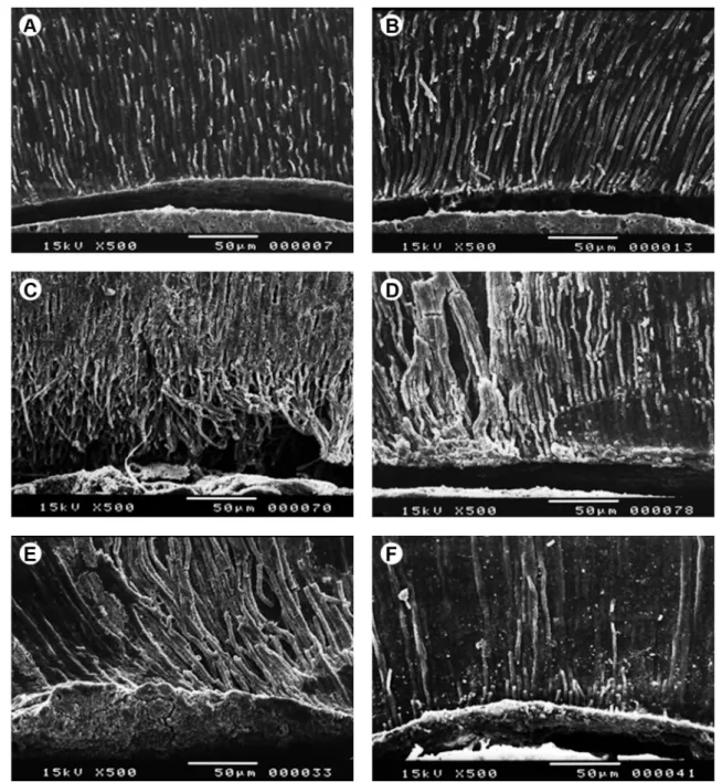
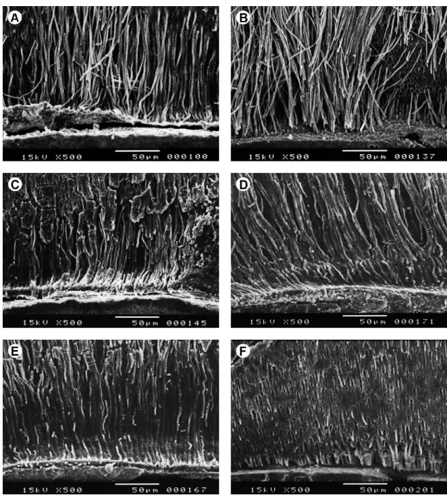
This study evaluated the effect of flowable composite resin application on the microtensile bond strength (µTBS) of adhesive systems to dentin.. Occlusal surfaces of human third

The purpose of this study was to evaluate the influence of the cementation system on the regional push-out bond strength of fiber post to root canal dentin..

The unset endodontic sealer had an influence on the adhesion of the luting resin cements to root canal dentin since the bond strength values were lower when the post

The tested hypotheses were as follows: (1) when FRCs are cemented immediately after root canal filling, the salicylate resin-based sealer presents a bond strength similar to that

The aim of this study was to evaluate the microtensile bond strength (µTBS) of two substrates (enamel and dentin) considering two study factors: type of composite resin

The objective of this study was to evaluate the influence of resin cement thickness on the push-out bond strength of prefabricated and customized glass fiber posts to dentin,