The aim of this study was to evaluate the microtensile bond strength (µTBS) of two substrates (enamel and dentin) considering two study factors: type of composite resin [methacrylate-based (Filtek Supreme) or silorane-based (Filtek LS)] and aging time (24 h or 3 months). Twenty human molars were selected and divided into 2 groups (n=10) considering two dental substrates, enamel or dentin. The enamel and dentin of each tooth was divided into two halves separated by a glass plate. Each tooth was restored using both tested composite resins following the manufacturer’s instructions. The samples were sectioned, producing 4 sticks for each composite resin. Half of them were tested after 24 h and half after 3 months. µTBS testing was carried out at 0.05 mm/s. Data were analyzed by three-way ANOVA and Tukey's HSD tests at α=0.05. Significant differences between composite resins and substrates were found (p<0.05), but no statistically significant difference was found for aging time and interactions among study factors. The methacrylate-based resin showed higher µTBS than the silorane-based resin. The µTBS for enamel was significantly higher than for dentin, irrespective of the composite resin and storage time. Three months of storage was not sufficient time to cause degradation of the bonding interaction of either of the composite resins to enamel and dentin.
M i c r o t e n s i l e B o n d S t r e n g t h
o f M e t h a c r y l a t e a n d S i l o r a n e
R e s i n s t o E n a m e l a n d D e n t i n
Gisele Rodrigues da Silva1, Isabela Sousa Araújo2, Rodrigo Dantas Pereira1, Bruno de Castro Ferreira Barreto1, Célio Jesus do Prado2, Carlos José Soares1,
Luís Roberto Marcondes Martins3
1Department of Operative Dentistry and Dental Materials, School of Dentistry, UFU - Federal University of Uberlândia, Uberlândia, MG, Brazil 2Department of Occlusion, Fixed Prosthodontics and Dental Materials, School of Dentistry, UFU - Federal University of Uberlândia, Uberlândia, MG, Brazil 3Department of Operative Dentistry, Piracicaba School of Dentistry, UNICAMP - University of Campinas, Piracicaba, SP, Brazil
Correspondence: Dra. Gisele Rodrigues da Silva, Avenida Pará, 1720, Bloco 4L, Anexo A, Sala 4LA33, Campus Umuarama, 38400-902 Uberlândia, MG, Brasil. Tel: +55-34-3218-2255. e-mail: [email protected]
Key Words:microtensile bond strength, BisGMA methacrylate composite resin, silorane composite resins, enamel, dentin.
Introduction
Resin composites have been widely used in direct adhesive restorations (1,2) due to their excellent physical and mechanical properties (3). However, these composites have inherent shortcomings that are mainly relative to polymerization shrinkage (4). Shrinkage creates stress and compromises restoration integrity (3), which leads to microleakage (1) and failures in the tooth-restoration interface (5,6). Some restorative strategies have been developed in order to decrease the polymerization shrinkage and its effects, such as the incremental filling technique (2,4,5), curing light intensity (1) and photoactivation time (3). Additionally, the manufacturers have worked to improve resin composition by increasing the volume and size of inorganic fillers and developing new monomers (4-6).
New types of monomers, known as low-shrinkage monomers, have been developed with the intention of reducing problems inherent to resins based on methacrylates (6), like monomeric volume reduction during polymerization shrinkage (4,5). Silorane composite resin is filled with a combination of fine quartz particles and radiopaque yttrium fluoride. The quartz surface is modified with a silane layer (7). Silorane technology has afforded a highly hydrophobic restorative material with lower polymerization shrinkage that results in lower residual shrinkage stress (2,4,5). This composite resin presents also better color stability as well as
lower insolubility in biologic fluids and adequate physical and mechanical properties, making it clinically suitable (7,8). Studies confirm that a commercial silorane-based composite accounts for less than 1.0% of total volumetric shrinkage, compared with 2.0–3.5% for BisGMA-based composites and causes less tooth deflection (2,4,5) and microleakage (6). Some studies have shown that this low shrinkage of composites provide clinical longevity (9,10). Composite resin bonding strength to dental substrate varies with the substrate. Tooth structure consists mostly of dentin (11), which is a hard tissue containing approximately 45% mineral, 35% of organic matrix and 20% water, by volume. Dentinal tubules extend radially from the pulp through the dentin and toward the dentin-enamel junction (12). Enamel is a highly mineralized tissue, which covers the entire crown of the tooth and consists of 92-96% of inorganic matrix, 1-2% organic material and 3-4% of water by weight (13). Thus, the physicochemical characteristics of these substrates probably influence the quality of the composite resin adhesion. More studies that make simultaneous comparisons between different resin monomers and tooth structures, concerning microtensile bond strength (µTBS), would be of great value.
328
G.R. da Silva et al.
literature shows that laboratory storage and actual aging do not occur in exactly the same way since oral environment is complex and composed by several interrelated factors such as temperature, pressure, chemical and mechanical phenomena, among others. Nevertheless, artificial aging is important for monitoring restoration, improving the correlation of the in vitro results with clinical outcomes (14).
The aim of this study was to test the effect of the composite resin composition and aging time on the µTBS to different dental substrates. The null hypothesis is that the composite resin composition and the aging time have no effect on µTBS to enamel or dentin substrates.
Material and Methods
After approval from the Federal University of Uberlândia Ethics Committee (CEP / UFU 001/10), 20 intact third molars extracted for orthodontic reasons were collected. The teeth were stored in an aqueous 0.2% thymol solution for no more than 3 months, cleaned with periodontal curettes
and pumice prophylaxis and then maintained in distilled water at 4 °C.
To test the µTBS to dentin, the tooth roots (n=10) were embedded in polyester resin (Aerojet, São Paulo, SP, Brazil) inside a silicone matrix (Aerojet) and their coronal occlusal surfaces were ground wet onto 320- to 600-grit silicon carbide paper. Abrasion continued until the superficial dentin was completely exposed, which was confirmed by surface analysis with a stereomicroscope (Leica, Weztlar, Hesse, Germany). To test the µTBS to enamel, the buccal enamel surface (n=10) was abraded with 600-grit silicon carbide paper for 30 s until the surface was flattened.
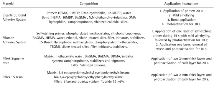
The substrates were separated in the middle by a cover slip and restored with both resins simultaneously (Fig. 1). The substrate was restored using both resins to exclude the influence of possible morphological differences of the specimens caused by intrinsic mineralization. Half of specimens were restored with the methacrylate-based resin (Filtek Supreme A2, 3M ESPE) (FSU) after application of a two-step self-etching adhesive system (Clearfil SE Bond, Kuraray, Okayama, Japan), and half with the silorane-based composite (Filtek LS A2, 3M ESPE, St. Paul, MN, USA) (FLS) after application of its specific adhesive system. All procedures were done following the manufacturers’ protocol (Table 1). Two 2-mm-thick increments were used on enamel and dentin to build composite resin restorations measuring 4x4x4 mm (4). Photoactivation was performed with a halogen light source (Demetron 501, 550 mW/cm2, Kerr, Orange, CA, USA) for 40 s each increment.
The restored teeth were stored for 24 h at 37 °C and then cut into sticks with 1 mm2 cross section area using a precision saw (Isomet 2000, Buehler, Lake Bluff, IL, USA) with speed of 300 rpm. Four sticks were obtained for each Figure 1. Schematic illustration describing the study design and
specimen preparation. A: Restorations on enamel: buccal surface of the tooth. B: Restorations on dentin: occlusal surface of the tooth.
Table 1. Materials, compositions, manufacturers and protocols of usage
Material Composition Application instructions
Clearfil SE Bond Adhesive System
Primer: HEMA, 10MDP, DMA hydrophilic, 12-MDBP, water Bond: HEMA, 10MDP, BisGMA , N,N-diethanol-p-toluidina, DMA
hydrophilic, camphorquinone, silanized colloidal silica.
1. Application of primer: 20 s; 2. Mild air-drying 3. Bond application 4. Photoactivation for 10 s.
Silorane Adhesive System
Self-etching primer: phosphorylated methacrylates, vitrebond copolymer, BisGMA, HEMA, water, ethanol, silane-treated silica filler, initiators, stabilizers.
LS Bond: Hydrophobic methacrylates, phosphorylated methacrylates, TEGM), silane-treated silica filler, initiators, stabilizers.
1. Application of one layer of self-etching primer during 15 s with mild air-drying,
followed by photoactivation for 10 s; 2. Application one layer, removal of excess and photoactivation for 10 s.
Filtek Supreme resin
Matrix: methacrylate resin , BisGMA, BisEMA, UDMA, initiator system: camphorquinone, stabilizers and pigments.
Filler: Silanized zirconia,
Application of two 2-mm-thick layers and photoactivation of each layer for 20 s.
Filtek LS resin
Matrix: 3,4 epoxycyclohexylethyl cyclopolymethylsiloxane, bis-3,4-epoxycyclohexylethylphenylmethylsilane. Filler: Silanized quartz; yttrium fluoride 76 wt%
Application of two 2-mm-thick layers and photoactivation of each layer for 20 s.
329
Bond strength of silorane composite resin
longitudinal restoration. Half the sticks were used for the immediate test and the other half were tested after 3 months of storage in water at 37 °C.
The area of bond interface of each stick was individually measured with a digital caliper (Starrett 727-2001, Itu, SP, Brazil). The specimens were actively gripped onto a Geraldeli’s device (15) with cyanoacrylate adhesive (Superglue Gel, Loctite, Henkel Corp., Avon, OH, USA). Each testing assembly was connected to a universal testing machine (EMIC, São Jose dos Pinhais, PR, Brazil) and the specimens were stressed to failure under tensile force at 0.5 mm/min (15). Final values were expressed in MPa considering the bonded area. The µTBS of each sample was defined as the average of results from two sticks, and assuming the tooth as the experimental unit (16). All tested sticks were checked on the stereomicroscope (Leica) to ensure that the failure occurred at the adhesive interface. Two sticks that had cohesive failure and different failures of the adhesive were excluded from the study.
The Shapiro-Wilk and Levene tests were conducted to test the data for normality and homogeneity. Data were transformed using log10 to fulfill this requirement and three-way ANOVA was used, followed by post hoc Tukey’s HSD (Honestly Significant Difference) to determine whether there was a significant difference in the bond strength (α=0.05). For all analyses was used SigmaPlot V 12.0 software (Systat Software, Inc., San Jose, CA, USA).
Results
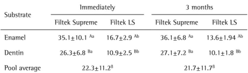
The µTBS results are presented in Table 2. The statistical analysis showed a significant effect for composite resin (p<0.001) and for dental substrates (p<0.001). However no significant effect was found for aging time (p=0.329), for interactions between composite resin and aging time (p=0.083), composite resin and dental substrate (p=0.424), dental substrate and aging time (p=0.622), or for the interaction among all three study factors (p=0.437). Tukey's HSD test showed that µTBS on enamel was higher than
on dentin, for all composite resins and aging times. The methacrylate-based composite resin had higher µTBS values than silorane-based ones, irrespective of dental substrate or aging time (p<0.001).
Discussion
The null hypothesis was rejected. Methacrylate resin showed higher bond strength than silorane regardless of dental substrate and aging time. Additionally, the µTBS on enamel was higher than dentin regardless the composite resin type and aging time. On the other hand, the storage time had no effect on µTBS results.
Although the silorane system is based on cationic polymerization, which occurs by photo cationic ring opening and results in lower polymerization shrinkage compared with methacrylate-based resins (5,17), silorane had lower µTBS values on both substrates. It is important to observe that the low polymerization shrinkage of a composite does not always indicates reduction of the shrinkage stress on the restored tooth (2,5). The silorane-based resin forms a low-viscosity layer and may induce shrinkage stress similar to that produced by methacrylate resins (17). The viscoelastic behavior changes that occur during the polymerization of predominantly elastic-viscous material can make the development of strains an event of significantly complex polymerization. The low initial flow presented by the base resin can restrict the flow of viscoelastic silorane, increasing the stress despite the low shrinkage (17). However, it is worth emphasizing that the polymerization stress is a physical condition that is not solely based on material properties but also on the geometry of the cavity and the boundary conditions (2,5). The other aspect that may explain the results of the present study was the need of using a dedicated adhesive system for the silorane composite resin (18). However, both adhesive systems were two-step self-etching adhesives to avoid the influence of this factor. Clearfil SE Bond was used in combination with Filtek Supreme because it is considered as the “gold standard” (19) for this class of adhesive systems. The primer agent of the silorane restorative system presents different curing method from Clearfil SE Bond.
The silorane primer agent is first light-cured and then the bonding agent is applied. Therefore, the primer agent creates the hybrid layer, in contrast with the conventional self-etching adhesive systems, where the hybrid layer formation is determined by the combination of primer and bonding agent. The silorane adhesive system produces an interface composed by a hybrid layer, produced by the primer agent, an intermediate resin layer with Table 2. Microtensile bond strength of methacrylate and silorane-based resin composites
on the dental substrates, enamel and dentin, according aging time.
Substrate
Immediately 3 months
Filtek Supreme Filtek LS Filtek Supreme Filtek LS
Enamel 35.1±10.1 Aa 16.7±2.9 Ab 36.1±6.8 Aa 13.6±1.94 Ab
Dentin 26.3±6.8 Ba 10.9±2.5 Bb 27.1±7.2 Ba 10.1±1.8 Bb
Pool average 22.3±11.2ß 21.7±11.7ß
330
G.R. da Silva et al.
low viscosity and finally the composite resin. Due to this complex process, a weak bonding interaction between the two substrates may compromise the µTBS (18). Moreover, the pH of the silorane-based self-ethcing primer is less acidic than the methacrylate-basedadhesive used in this study (20). The primer agent of the silorane composite resin is cured before application of the bond; therefore the dentin hybridization may be entirely dependent on the degree of demineralization, penetration, and cross-linking produced by the primer (18). pH and the resulting hydrophilicity of the silorane primer may greatly determine the extent of resin permeation into dentin and enamel (21). Although this in vitro test showed a significant difference
between both composite resins, a one year clinical study that analyzed the performance of Class I and II cavities of three different composite resins, the silorane-based system showed acceptable results (9). The restorations had no advantage over those with methacrylate-based composite combined with etch-and-rinse adhesive. However, silorane restorations tended to degrade in terms of marginal adaptation compared with baseline values. In their two-year follow-up, the three restorative systems showed statistically similar clinical performances (9).
Bond strength of the composites did not differ whether immediately after or three months after manufacture. This was likely because the time interval was insufficient to reduce the strength of the adhesive resins. For example, Martins et al. (22) found that the properties of the adhesive layer start to degrade after 6 months of storage. Dental substrate was another studied factor and it was observed that µTBS on enamel was greater than on dentin. The self-etching adhesives essentially modify the smear layer and provide chemical adhesion to the mineralized component of enamel (23). The effectiveness of some self-etching adhesive on enamel is probably due to a secondary connection caused by calcium affinity (24). Nevertheless, several factors may influence the bond including surface preparation, adhesive thickness, test method, crosshead speed and material type (6). In this study, enamel was abraded with silicon carbide paper increasing its roughness. This method is used in laboratory research to standardize the specimen preparation, simulating clinical dentin/ enamel preparation using a medium-grit diamond bur (16,21). Surface roughness creates an increased surface area and mechanical retention may have enhanced slightly on enamel (21). Moreover, removing the outer aprismatic enamel layer and reaching the inner prismatic enamel may also improve enamel bonding (21). The experimental design using the same tooth to test different materials is advised to minimize the effect of substrate on the composite resin factor (16). The use of only two sticks is a limitation of the tooth size and the number of the variations tested on each
unit sample (15). However, it is advisable to have a smaller number of sticks for one composite but to have the same substrate tested for both study factors (composite resin – 2 sticks each; aging time – 2 sticks each).
The evolution of restorative materials tends to induce the clinicians to expect that new products result directly in a better performance. New technologies of the composite resin manufacturers aim to reduce shrinkage of the composite during polymerization. But the modifications of the composite resins do not always alter only one mechanical property, reflecting in a negative performance of the material because it is dependent of multifactorial aspects (2,5). Despite its low polymerization shrinkage, the silorane-based resin does not appear to be a better alternative then methacrylate resins. In this study was used a flat surface for enamel and dentin. Flat surface does not actually account for all the aspects involved in the residual shrinkage stress generated after resin polymerization. The C factor is much smaller on flat surfaces than in conventional dental cavities. However, using this method to measure the post-gel shrinkage, where the composite is inserted over the strain-gauge, revealed that the shrinkage stress of Filtek LS is significantly lower than that of Filtek Supreme (5,25). Therefore the lower bonding strength performance of silorane composite resulted from the inefficiency of the adhesive procedure and mechanical interaction with the composite resin. Moreover, bonding to enamel was significantly higher than to dentin for both composites and 3 months aging appears to be not long enough to promote changes in dentin and enamel µTBS, given the used restorative systems. Additional in vitro tests that evaluate
wear and fracture resistance and mainly the randomized clinical trials with longer follow-up period should be performed to verify the clinical behavior of this material, since the oral environment has characteristics that cannot be faithfully reproduced in laboratory studies. In addition, due the short storage time and intrinsic limitations of laboratory studies, this research outcomes were limited by the immediate bonding of the restoration, different from what happens usually in the oral environment, as in this study the C factor had little influence because the restored surface was flat.
Resumo
331
Bond strength of silorane composite resin
dos palitos foi testada após 24h e o restante após três meses. O ensaio de microtração (µTBS) foi conduzido numa velocidade de 0,05 mm/s. Os dados foram analisados usando three-way ANOVA e teste de Tukey HSD (α= 0,05). Diferença significante foi encontrada para o fator resina e substratos (p<0,05), porém não houve influência do tempo de envelhecimento e interações entre fatores estudados. A resina à base de metacrilato apresentou maior resistência adesiva do que a silorano. A adesão em esmalte foi significativamente maior do que em dentina, independente da resina e do tempo de envelhecimento. Três meses de armazenamento não foram suficientes para causar degradação da interação adesiva, para ambas as resinas compostas, no esmalte e na dentina.
Acknowledgements
This study was supported by FAPEMIG, CNPq, CAPES and FOUFU. The authors thank 3M/ESPE for providing some of the restorative materials used in this study.
References
1. Guiraldo RD, Consani S, Consani RL, Berger SB, Correr AB, Sinhoreti MA, Correr-Sobrinho L. Comparison of silorane and methacrylate-based composites on the polymerization heat generated with different light-curing units and dentin thicknesses. Braz Dent J 2013;24:258-262. 2. Bicalho A, Valdívia A, Barreto B, Tantbirojn D, Versluis A, Soares
C. Incremental filling technique and composite material-Part II: Shrinkage and shrinkage stresses. Oper Dent 2014;39:E71-E82. 3. Ferracane JL. Resin composite - State of the art. Dent Mater
2011;27:29-38.
4. Soares CJ, Bicalho AA, Tantbirojn D, Versluis A. Polymerization shrinkage stresses in a premolar restored with different composite resins and different incremental techniques. J Adhes Dent 2013;15:341-350. 5. Bicalho A, Pereira R, Zanatta R, Franco S, Tantbirojn D, Versluis A,
et al.. Incremental filling technique and composite material-Part I: Cuspal deformation, bond strength, and physical properties. Oper Dent 2014;39:E83-E92.
6. Yaman BC, Doğruer I, Gümüştaş B, Efes BG. Three-year randomized clinical evaluation of a low-shrinkage silorane-based resin composite in non-carious cervical lesions. Clin Oral Investig 2014;18:1071-1079. 7. Weinmann W, Thalacker C, Guggenberger R. Siloranes in dental
composites. Dent Mater 2005;21:68-74.
8. Schmidt M, Kirkevang LL, Hørsted-Bindslev P, Poulsen S. Marginal adaptation of a low-shrinkage silorane-based composite: 1-year randomized clinical trial. Clin Oral Investig 2011;15:291-295. 9. Baracco B, Perdigão J, Cabrera E, Ceballos L. Two-year clinical
performance of a low-shrinkage composite in posterior restorations. Oper Dent 2013;38:591-600.
10. Gonçalves FS, Leal CD, Bueno AC, Freitas AB, Moreira AN, Magalhães CS. A double-blind randomized clinical trial of a silorane-based resin composite in class 2 restorations: 18-month follow-up. Am J Dent 2013;26:93-98.
11. Giannini M, Soares CJ, Carvalho RM. Ultimate tensile strength of tooth structures. Dental Mater 2004;20:322–329.
12. Arola D, Reprogel RK. Tubule orientation and the fatigue strength of human dentin. Biomat 2006;27:2131–2140.
13. Gwinnett AJ. Structure and composition of enamel. Oper Dent 1992;Suppl5:10-17.
14. Cenci MS, Venturini D, Pereira-Cenci T, Piva E, Demarco FF. The effect of polishing techniques and time on the surface characteristics and sealing ability of resin composite restorations after one-year storage. Oper Dent 2008;33:169-176.
15. Raposo LH, Armstrong SR, Maia RR, Qian F, Geraldeli S, Soares CJ. Effect of specimen gripping device, geometry and fixation method on microtensile bond strength, failure mode and stress distribution: laboratory and finite element analyses. Dent Mater 2012;28:e50-e62. 16. Armstrong S, Geraldeli S, Maia R, Raposo LH, Soares CJ, Yamagawa J. Adhesion to tooth structure: a critical review of "micro" bond strength test methods. Dent Mater 2010;26:e50-e62.
17. Boaro LC, Gonçalves F, Guimarães TC, Ferracane JL, Versluis A, Braga RR. Polymerization stress, shrinkage and elastic modulus of current low-shrinkage restorative composites. Dent Mater 2010;26:1144-1150. 18. Lima AF, Sasaki RT, Araújo LS, Gaglianone LA, Freitas MS, Aguiar FH,
et al.. Effect of tooth bleaching on bond strength of enamel-dentin cavities restored with silorane- and dimethacrylate-based materials. Oper Dent 2011;36:390-396.
19. De Munck J, Shirai K, Yoshida Y, Inoue S, Van Landuyt K, Lambrechts P, et al.. Effect of water storage on the bonding effectiveness of 6 adhesives to Class I cavity dentin. Oper Dent 2006;31:456-465. 20. Tay FR, Pashley DH. Aggressiveness of contemporary self-etching
systems. I: Depth of penetration beyond dentin smear layers. Dent Mater 2001;17:296-308.
21. Adebayo OA, Burrow MF, Tyas MJ, Palamara J. Effect of tooth surface preparation on the bonding of self-etching primer adhesives. Oper Dent 2012;37:137-149.
22. Martins GC, Calixto AL, Gomes OM, Loguercio AD, D'Alpino PH, Reis A. Effect of water storage on resin-dentin bond strengths formed by different bonding approaches. Indian J Dent Res 2009;20:431-436. 23. Sengun A, Orucoglu H, Ipekdal I, Ozer F. Adhesion of two bonding
systems to air-abraded or bur-abraded human enamel surfaces. Eur J Dent 2008;2:167-175.
24. Brackett WW, Tay FR, Looney SW, Ito S, Haisch LD, Pashley DH. Microtensile dentin and enamel bond strengths of recent self-etching resins. Oper Dent 2008;33:89-95.
25. Sakaguchi RL, Versluis A, Douglas WH. Analysis of strain gauge method for measurement of post-gel shrinkage in composite resins. Dent Mater 1997;13:233-239.