INTRODUCTION
The adhesion to dentin is a critical step because of the heterogeneous structure and humidity of this tissue (1). In this sense, self-etch adhesives emerged with the proposal to simplify the adhesive restorative procedure (2), making the technique less critical in controlling dentin moisture.
Self-etch adhesives are aqueous mixtures of hydrophilic and hydrophobic monomers and solvents (3), combining steps of etching and priming (self-etch primers) or etching, priming and bonding in a single bottle (all-in-one adhesives). They do not require a separate etching step; rather, they incorporate the smear layer into the hybrid layer (3). Although theoretically the discrepancy between the depth of demineralization and infiltration of monomers is not present in self-etch systems (4), in vitro studies have shown nanoleakage (3) and phase separation (5) in the adhesive/resin interface
Bond Strength of HEMA-Containing
versus
HEMA-Free Self-Etch Adhesive Systems to Dentin
Klíssia Romero FELIZARDO Letícia Vargas Freire Martins LEMOS
Rodrigo Varella de CARVALHO Alcides GONINI JUNIOR
Murilo Baena LOPES Sandra Kiss MOURA
Dental School, UNOPAR - University North of Paraná, Londrina, PR, Brazil
This study analyzed the bond strength (BS) of HEMA versus HEMA-free self-etch adhesive systems to dentin. The occlusal surface of 20 third molars was removed and the dentin was abraded with 600-grit silicon carbide paper. The teeth were randomly assigned to 4 groups (n=5) and restored with GO, Adper SE Plus, OptiBond All-In-One, and Clearfil 3S Bond adhesive systems and Filtek Z350 composite. After light curing (600 mW/cm²), the teeth were stored in distilled water at 37ºC for 24 h and were sectioned in the mesiodistal and buccolingual directions to obtain sticks (0.8 mm²). The sticks were subjected to tensile force using a universal testing machine (0.5 mm/min), and the modes of failure were analyzed by scanning electron microscopy and classified as adhesive, cohesive, or mixed. The BS data (in MPa) were treated using one-way ANOVA and Tukey’s tests at 5% significance. GO presented the lowest mean bond strength value (10.57 ± 3.72) and differed significantly from the other materials (p=0.001), which, in turn, presented statistically similar results (p>0.05) among themselves: Adper SE Plus (29.08 ± 8.93), OptiBond All-In-One (28.36 ± 6.49), and Clearfil 3S Bond (28.62 ± 6.97). Mixed fractures were the most prevalent. It was concluded that the influence of HEMA on BS to dentin was material dependent.
Key Words: Bonding, dental adhesives, dentin.
of some self-etch systems, which is believed to result from the high water content of solvent formulations. Therefore, the durability of the adhesion has been questioned because the hydrophilic nature of the self-etch primers would make them vulnerable to water sorption (6).
HEMA (2-hydroxyethyl-methacrylate) is the most frequent hydrophilic monomer present in adhesive systems (7). Due to its low molecular weight, HEMA acts as a co-solvent, minimizing phase separation and increasing the miscibility of hydrophobic and hydrophilic components into the solution (5). However, a formation of an aqueous and unstable gel was reported in adhesives containing HEMA; this should be susceptible to hydrolytic degradation (8) and should encourage more water update (9). Some studies have shown that the removal of HEMA from self-etch adhesives (10) would minimize water sorption, while others observed that the 10% of HEMA content would be beneficial for
the adhesive system performance (11).
Surfactant dimethacrylate monomers have been added to adhesive systems to maintain hydrophilicity (12,13). They are solubility enhancers that facilitate penetration of hydrophobic components into wet, demineralized dentin, reducing phase separation (12,13). The advantage of these monomers is that they result in formation of more stable cross-linked polymers facilitated by two polymerizable groups (12,13).
There is still a controversy about the performance of adhesives with or without HEMA. Therefore, the aim of this study was to evaluate the bond strength (BS) to dentin of commercial containing and HEMA-free self-etch adhesive systems. The null hypothesis tested in the study is that the adhesive systems do not differ with respect to BS to dentin regardless of the presence of HEMA.
MATERIAL AND METHODS
This study was conducted after approval of the institutional Ethics Committee (Protocol #PP0104/09). Twenty third molars were disinfected in 0.5% chloramine solution at 4ºC, cleaned and randomly assigned to 4 groups (n=5), which received self-etch adhesive systems with HEMA (Adper SE Plus; 3M ESPE, St Louis, MN, USA and Clearfil 3S Bond; Kuraray Medical Inc, Tokyo, Japan) and without HEMA (GO; SDI, Chicago, IL, USA and OptiBond All-In-One; Kerr Co, Orange, CA, USA).
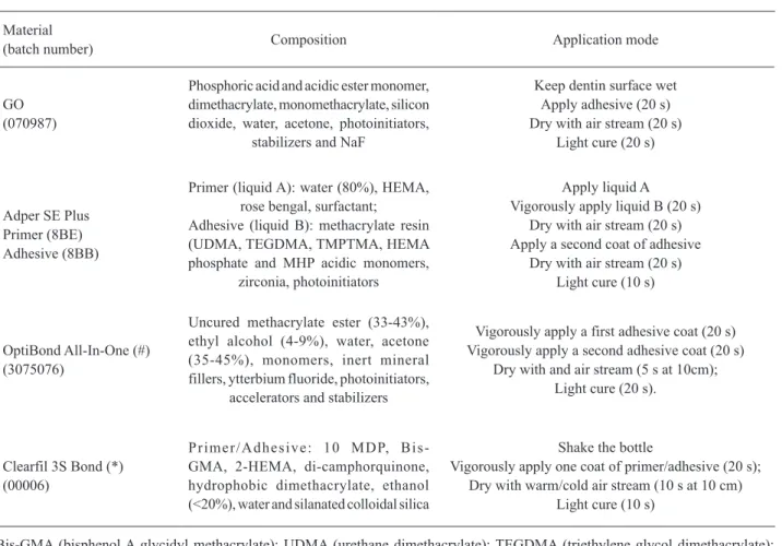
The teeth were ground with 180-grit silicon carbide paper to remove the occlusal surfaces and expose flat coronal dentin, which was polished with 600-grit silicon carbide paper under water cooling for 60 s to produce a standardize smear layer. The adhesive systems were applied as described in Table 1. Light
Table 1. Materials used, composition, and application modes.
Material
(batch number) Composition Application mode
GO (070987)
Phosphoric acid and acidic ester monomer, dimethacrylate, monomethacrylate, silicon dioxide, water, acetone, photoinitiators,
stabilizers and NaF
Keep dentin surface wet Apply adhesive (20 s) Dry with air stream (20 s)
Light cure (20 s)
Adper SE Plus Primer (8BE) Adhesive (8BB)
Primer (liquid A): water (80%), HEMA, rose bengal, surfactant; Adhesive (liquid B): methacrylate resin (UDMA, TEGDMA, TMPTMA, HEMA phosphate and MHP acidic monomers,
zirconia, photoinitiators
Apply liquid A Vigorously apply liquid B (20 s)
Dry with air stream (20 s) Apply a second coat of adhesive
Dry with air stream (20 s) Light cure (10 s)
OptiBond All-In-One (#) (3075076)
Uncured methacrylate ester (33-43%), ethyl alcohol (4-9%), water, acetone (35-45%), monomers, inert mineral fillers, ytterbium fluoride, photoinitiators,
accelerators and stabilizers
Vigorously apply a first adhesive coat (20 s) Vigorously apply a second adhesive coat (20 s)
Dry with and air stream (5 s at 10cm); Light cure (20 s).
Clearfil 3S Bond (*) (00006)
P r i m e r / A d h e s i v e : 1 0 M D P, B i s -GMA, 2-HEMA, di-camphorquinone, hydrophobic dimethacrylate, ethanol (<20%), water and silanated colloidal silica
Shake the bottle
Vigorously apply one coat of primer/adhesive (20 s); Dry with warm/cold air stream (10 s at 10 cm)
Light cure (10 s)
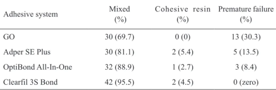
Table 2. Number of specimens (representative percentages) distributed according to the failure mode for each experimental condition.
Adhesive system Mixed
(%)
Cohesive resin (%)
Premature failure (%)
GO 30 (69.7) 0 (0) 13 (30.3)
Adper SE Plus 30 (81.1) 2 (5.4) 5 (13.5)
OptiBond All-In-One 32 (88.9) 1 (2.7) 3 (8.4)
Clearfil 3S Bond 42 (95.5) 2 (4.5) 0 (zero)
curing was carried with a commercial light-curing unit with irradiance of 600 mW/cm² (Ultralux; Dabi Atlante, Rio de Janeiro, RJ, Brazil) and blocks of Filtek Z350 composite (3M ESPE) were build up in five 1-mm-thick increments, each one light cured for 40 s.
The restored teeth were stored in distilled water at 37°C for 24 h and then cut on an Isomet 1000 machine (Buhler, Lake Bluff, IL, USA) in the mesiodistal and buccolingual directions, perpendicular to the bonded interfaces, to obtain sticks. The dimensions of each stick were measured with a digital caliper accurate to the nearest 0.1 mm (Absolute Digimatic; Mitutoyo, Japan) in order to calculate the cross-sectional area (0.8 mm²).
Each sample was fixed in a microtensile testing device (Odeme Biotechnology, Joaçaba, SC, Brazil) with cyanoacrylate-based adhesive (Super Bonder Gel; Loctite, Itapevi, SP, Brazil) and tested for tensile strength in a universal testing machine (EMIC DL 2000; São José dos Pinhais, PR, Brazil 0.5 mm/min). The fragments of fractured specimens were fixed in aluminum stubs with double-faced carbon tape, sputter-coated with gold (SCD 050 Sputter Coater; Bal-Tec, Balzers, Liechtenstein), and the modes of fracture were observed and classified as adhesive, cohesive or mixed using a scanning electron microscope (JSM 5600; JEOL ,Tokyo, Japan) operating at 15 kv. The bond strength values (in N) were divided by the cross-sectional area (in mm2) of each specimen
and the results obtained in MPa were analyzed by one-way analysis of variance and Tukey’s tests at 5% significance level.
RESULTS
The mean values of cross-sectional areas ranged from 0.77 to 0.83 mm2, with no statistically significant
difference among groups (p>0.05). The percentages of specimens tested and fractured before testing and the
modes of failure for each experimental condition are presented in Table 2. Mixed fractures were the most prevalent in all groups.
The means (in MPa) and standard deviations of the experimental groups are described as follows. GO presented the lowest mean bond strength value (10.57 ± 3.72) and differed significantly from the other materials (p=0.001), which, in turn, presented statistically similar results (p>0.05) among themselves: Adper SE Plus (29.08 ± 8.93), OptiBond All-In-One (28.36 ± 6.49), and Clearfil 3S Bond (28.62 ± 6.97).
DISCUSSION
Self-etch systems do not require etching as a separate step, as they run by acidic monomers incorporated into the primer (self-etch primers), and the penetration of monomers occurs simultaneously. Because the acidic monomers increase the wetness of dentin, some authors initially hypothesized that the presence of HEMA in these adhesives would not be necessary (10). Other problems reported due to the presence of HEMA on the adhesive systems were the possibility of allergenic reactions and high hydrophilicity, resulting in water sorption even after the end of the polymerization reaction (7).
50% (14).
The results of OptiBond All-In-One, however, were similar to those of Adper SE Plus and Clearfil 3S Bond, both containing 20% HEMA. One can assume that other factors, such as the solvent type and content, may have influenced the bond strength of these two materials. GO contains acetone, while OptiBond All-In-One contains 35 to 45% acetone and 4-9% ethanol. The solvent evaporation from adhesives is influenced by the vapor pressure (15). As the vapor pressure of acetone is high, it volatilizes rapidly and may dehydrate the dentin. The presence of water in self-etch adhesives is necessary to ensure the ionization of the acidic monomers, but it is not as efficient as acetone or ethanol as a solvent because of its lower vapor pressure (7). Conversely, the presence of acetone and ethanol in OptiBond All-In-One might balance the solvent evaporation without dehydrating dentin, because ethanol ensures the wetness of the substrate, and its vapor pressure is intermediate between acetone and water. The lower percentage of premature failures observed for OptiBond All-In-One compared to those of GO confirms this aspect (Table 2). Another explanation for the good performance of OptiBond All-In-One could be the content of glycerol phosphate dimethacrylate monomer in its formulation, a surfactant monomer that may have facilitated the penetration of hydrophobic components into dentin, reducing the phase separation (12,13).
The disappearance of bubbles in the adhesive layer was observed when a small amount of HEMA was present (5). Adper SE Plus and Clearfil 3S Bond have similar amounts of HEMA, resulting in good bond strength, thus confirming that the presence of HEMA in small amounts might have prevented the phase separation, as suggested.
It was observed the influence of water content, type and amount of solvents in the absence of phase separation, on bond strength, and marginal adaptation in enamel and dentin (16). This was partly reinforced in the present study because although the bond strength values of Clearfil 3S Bond and Adper SE Plus were similar, several premature fractures were observed for Adper SE Plus (Table 2). This may be explained by the 80% of water in its composition and as the vapor pressure of water is low, the solvent evaporation in this adhesive may be decreased in the presence of HEMA (7). No premature fractures were observed for Clearfil 3S Bond, suggesting that its water content and solvent might have optimized solvent evaporation from this adhesive.
Alternative techniques have been presented to improve solvent evaporation and bond strength of HEMA-containing and HEM-free adhesives, such as increasing the air drying time (15). Removal of solvent should be held at the maximum. It has been reported that the type of solvent and the mixture of components including HEMA may influence the solvent evaporation rate of commercial and experimental adhesives (17), with those containing acetone being the fastest ones.
The increased content of HEMA in adhesives decreases the degree of conversion (14) due to its lower polymer rates, which is influenced by the lower concentration of polymerizable groups (18) and it may decrease the mechanical properties of the polymer (19,20). Other studies should be conducted to evaluate the performance of self-etch adhesives with and without HEMA, such as the replacement of HEMA by other surfactant monomers (13) and the evaluation of the durability of bonding. Torkabaki et al. (21) observed a decrease of bond strength to dentin for HEMA-containing adhesives after 1 year of water storage, although this decrease was gradual for the adhesives without HEMA. Recently, Van Landuyt and others (22) evaluated the clinical performance of HEMA-free one-step G-Bond with one three-one-step adhesive in noncarious cervical lesions and they found no statistically significant difference between the materials.
Within the limitations of an in vitro study, it may be concluded that the influence of HEMA on BS to dentin was material dependent and that the presence of HEMA in the self-etch adhesive systems evaluated was not a predominant factor that influenced their bond strength to dentin.
RESUMO
RU foi observada para o adesivo GO (10,57 ± 3,72) (p=0,001). Adper SE Plus (29,08 ± 8,93), OptiBond All-In-One (28,36 ± 6,49) e Clearfil 3S Bond (28,62 ± 6,97) foram estatisticamente semelhantes entre si (p>0.05). Fraturas mistas predominaram nos grupos. Concluiu-se que a influência do HEMA na RU foi material-dependente.
REFERENCES
1. Swift EJ Jr., Perdigão J, Heymann HO. Bonding to enamel and dentin: a brief history and state of the art. Quintessence Int 1995;26:95-110.
2. Van Meerbeek B, De Munck J, Yoshida Y, Inoue S, Vargas M, Vijay P, et al.. Buonocore memorial lecture. Adhesion to enamel and dentin: current status and future challenges. Oper Dent 2003;28:215-235.
3. Tay FR, King NM, Chan KM, Pashley DH. How can nanoleakage occur in self-etching adhesive systems that demineralize and infiltrate simultaneously? J Adhes Dent 2002;4:255-269. 4. Katz JL, Spencer P, Nomura T, Wagh A, Wang Y. Micromechanical
properties of demineralized dentin collagen with and without adhesive infiltration. J Biomed Mater Res A 2003;66:120-128. 5. Van Landuyt KL, De Munck J, Snauwaert J, Coutinho E, Poitevin
A, Yoshida Y, et al.. Monomer-solvent phase separation in one-step self-etch adhesives. J Dent Res 2005;84:183-188.
6. Sideridou I, Tserki V, Papanastasiou G. Study of water sorption, solubility and modulus of elasticity of light-cured dimethacrylate-based dental resins. Biomater 2003;24:655-665.
7. Van Landuyt KL, Snauwaert J, De Munck J, Peumans M, Yoshida Y, Poitevin A, et al.. Systematic review of the chemical composition of contemporary dental adhesives. Biomater 2007;28:3757-3785.
8. Yiu CK, Pashley EL, Hiraishi N, King NM, Goracci C, Ferrari M, et al.. Solvent and water retention in dental adhesive blends after evaporation. Biomater 2005;26:6863-6872.
9. Moszner N, Salz U, Zimmermann J. Chemical aspects of self-etching enamel-dentin adhesives: a systematic review. Dent Mater 2005;21:895-910.
10. Van Meerbeek B, Van Landuyt K, De Munck J, Hashimoto M, Peumans M, Lambrechts P, et al.. Technique-sensitivity of contemporary adhesives. Dent Mater J 2005;24:1-13.
11. Van Landuyt KL, Snauwaert J, Peumans M, De Munck J,
Lambrechts P, Van Meerbeek B. The role of HEMA in one-step self-etch adhesives. Dent Mater 2008;24:1412-1419.
12. Guo X, Spencer P, Wang Y, Ye Q, Yao X, Williams K. Effects of a solubility enhancer on penetration of hydrophobic component in model adhesives into wet demineralized dentin. Dent Mater 2007;23:1473-1481.
13. Zanchi CH, Munchow EA, Ogliari FA, Chersoni S, Prati C, Demarco FF, et al.. Development of experimental HEMA-free three-step adhesive system. J Dent 2010;38:503-508.
14. Collares FM, Ogliari FA, Zanchi CH, Petzhold CL, Piva E, Samuel SM. Influence of 2-hydroxyethyl methacrylate concentration on polymer network of adhesive resin. J Adhes Dent 2010; in press. 15. Ikeda T, De Munck J, Shirai K, Hikita K, Inoue S, Sano H, et al.. Effect of air-drying and solvent evaporation on the strength of HEMA-rich versus HEMA-free one-step adhesives. Dent Mater 2008;24:1316-1323.
16. Furukawa M, Shigetani Y, Finger WJ, Hoffmann M, Kanehira M, Endo T, et al.. All-in-one self-etch model adhesives: HEMA-free and without phase separation. J Dent 2008;36:402-408. 17. Nihi FM, Fabre HSC, Garcia G, Fernandes KBP, Ferreira
FBA, Wang L. In vitro assessment of solvent evaporation from commercial adhesive systems compared to experimental systems. Braz Dent J 2009;20:396-402.
18. Andrzejewska E. Photopolymerization kinetics of multifunctional monomers. Progressive Polymer Sci 2001;26:605-665. 19. Ferracane JL. Hygroscopic and hydrolytic effects in dental
polymer networks. Dent Mater 2006;22:211-222.
20. Zanchi CH, Münchow EA, Ogliari FA, de Carvalho RV, Chersoni S, Prati C, Demarco FF, Piva E. A new approach in self-etching adhesive formulations: replacing HEMA for surfactant dimethacrylate monomers. J Biomed Mater Res B Appl Biomater 2011;99:51-77.
21. Torkabadi S, Nakajima M, Ikeda M, Foxton RM, Tagami J. Bonding durability of HEMA-free and HEMA-containing one-step adhesives to dentine surrounded by bonded enamel. J Dent 2008:36:80-86.
22. Van Landuyt KL, Peumans M, De Munck J, Cardoso MV, Ermis B, Van Meerbeek B. Three-year clinical performance of a HEMA-free one-step self-etch adhesive in non-carious cervical lesions. Eur J Oral Sci 2011;119:511-516.