The aim of this study was to assess the effects of intracanal irrigation with sodium hypochlorite (NaOCl) and ethylenediaminetetracetic acid (EDTA) prior to calcium hydroxide medication on the compressive strength of root dentin. Forty-eight roots with single and straight canals were instrumented with a crown-down technique using Flexofile files and Gates Glidden burs, under 0.9% sodium chloride (NaCl) irrigation. Root canals were randomly separated into 4 groups (n=12), according to the irrigating solution: Control Group, 0.9% NaCl; GroupNaOCl, 1% NaOCl; GroupEDTA, 17% EDTA; and GroupEDTA+NaOCl, 17%
EDTA and 1% NaOCl. Solutions remained for 40 min, renewed every 10 min, except for GroupEDTA+NaOCl, where the solutions were alternated every 10 min. Canals were dried and
filled with calcium hydroxide paste, and radiographed. Each canal was sealed and stored at 37 °C for 30 days. After this period, the roots were cut 1 mm below the cementoenamel junction and 6 mm apically from this point in order to obtain 6 mm-long cylinders. The compressive test was conducted with a crosshead speed of 1 mm/min. The means (±SD) (MPa) of compressive strength were: Control Group, 151.23±94.36; GroupNaOCl,
167.50±57.25; GroupEDTA, 108.79±99.88; GroupEDTA+NaOCl, 60.12±92.10. Significant statistical
differences among groups were found (ANOVA and Tukey, α=5%, p=0.02). Control Group and GroupNaOCl showed higher means of compressive strength than GroupEDTA+NaOCl.
Intracanal irrigation with 17% EDTA and 1% NaOCl produced a significant reduction of dentin compressive strength 30 days after medication with calcium hydroxide.
Intracanal Irrigating Solutions Prior
to Calcium Hydroxide Medication and
Its Effects on Root Dentin Strength
Leila Clarisse Hillesheim, Juliara Bellina Hoffmann, Daniela Peressoni Vieira Schuldt, Maybell Tedesco, Eduardo Antunes Bortoluzzi, Cleonice Silveira Teixeira
Departament of Dentistry, UFSC - Universidade Federal de Santa Catarina, Florianópolis, SC, Brazil
Correspondence: Cleonice Silveira Teixeira, Rua Haroldo Soares Glavan, 929, Condomínio Casa Blanca, casa 16, 88050-005 Florianópolis, SC, Brasil. Tel: +55-048-3721-5842. e-mail: [email protected]. br; [email protected]
Key Words: calcium hydroxide, compressive strength, irrigation, sodium hypochlorite.
Introduction
The endodontic treatment goal is to obtain bacteria-free root canals. The elimination of microorganisms can be achieved using various instrumentation techniques, irrigation solutions and intracanal medicaments (1). Mechanical instrumentation results in the debridement of root canal system, but alone it cannot effectively disinfect root canals, emphasizing the need for an irrigating solution to effectively kill more microorganisms (2,3).
Irrigation is currently the best method for removing pulp tissue remnants and dentin debris during instrumentation (1). Ideally, an irrigating solution should promote debridement, lubrication, dissolve organic tissue and eradicate the microorganisms from the root canal system (4-8).However, sodium hypochlorite is unable to remove inorganic components, and thus, unable to remove the smear layer produced during canal instrumentation (5,6). In an effort to remove the smear layer completely, studies suggest that the canals should be irrigated with ethylenediaminetetracetic acid (EDTA) and NaOCl (6,9). EDTA is a chelate that acts on the inorganic components of the smear layer. Therefore, irrigation with 15% EDTA and 1% NaOCl removes both inorganic and organic smear
layer components (6).
Besides the known importance of employing sodium hypochlorite solution and EDTA during canal instrumentation, and also prior to intracanal medications and canal filling, there has been much discussion over the effects of using these solutions on root dentin strength. In this way, studies have related sodium hypochlorite irrigation with dentin microhardness reduction (10). Other reports correlate sodium hypochlorite with dentin weakening by reduced dentin flexural strength (3,10,11) and elastic modulus (3,11).
Irrigating solutions effects on dentin
min, followed by 2.5% NaOCl irrigation for another minute, provided the best experimental combination when trying to avoid changes to dentin chemical composition and its morphological structures (15).
For necrotic tissue and microbes, intracanal medication with calcium hydroxide is recommended (2). Meanwhile, root canals treated with calcium hydroxide medication, even for short (16) or long periods (17), may be associated with reduced dentin strength. However, there is little scientific knowledge about the effects of irrigating solutions, like NaOCl and EDTA, prior to calcium hydroxide medication on root dentin strength. In view of this, the purpose of this study was to evaluate the effect caused by different irrigating solutions, prior to calcium hydroxide intracanal dressing, on root dentin strength. The null hypothesis of this experiment is that the use of different solutions before calcium hydroxide dressing does not affect the root dentin strength in the experimental groups.
Material and Methods
This study was approved by the institutional Human Ethics Committee (Committee’s approval #2083/2011).
Specimen Selection and Preparation
Forty-eight intact and single-rooted extracted human mandibular premolars were chosen for the study and stored in 0.1% thymol solution. The selected teeth were 22 to 25 mm long. Each tooth was radiographed and carefully examined with a 4× magnifying glass (Illuminated magnifying glass, Tokyo, Japan) to confirm the existence of a single and straight canal, no cracks and fully formed apical foramen. After cleaning the roots with periodontal curettes, the crowns were removed near of the cement-enamel junction with a water-cooled low-speed diamond saw to obtain a standardized root length of 16 mm. The access cavity was made and the roots with large oval canals were replaced in order to avoid canals without round
sections after endodontic preparation. The working length (WL) was defined by introducing a #15 file in the root canal until the tip was observed at the apical foramen, which was subtracted one millimeter from the canal length. Root canals were instrumented with a crown-down technique using Flexofile files (Dentsply Maillefer, Tulsa, OK, USA) until a master apical file size #50, and Gates Glidden burs #4 to #1 (Moyco Union Broach, York, PA, USA). Throughout the preparation, canals were irrigated with 2 mL of 0.9% sodium chloride solution (Dermus, Florianópolis, SC, Brazil) between every file or bur.
The instrumented canals were dried with absorbent paper points (Dentsply Maillefer) and randomly divided into 4 groups (n=12) according to the irrigating solution: Control Group, 0.9% NaCl (Dermus, Florianópolis, SC, Brazil); GroupNaOCl, 1% NaOCl (Dermus); GroupEDTA, 17%
EDTA (Dermus); and GroupEDTA+NaOCl, 17% EDTA (Dermus)
and 1% NaOCl (Dermus).
Irrigation System
The irrigation protocol used for each group was performed with a 22-gauge blunt-end needle (Ibras, São Paulo, SP, Brazil) 2 mm short of the WL. In each group, root canals were irrigated with 3 mL of solution for 10 min. After this time, the solution was removed by aspiration and renewed with the same amount for the same time, totalizing 12 mL of solution (4×3 mL) and 40 min total time (4×10 min) in each group. In GroupEDTA+NaOCl, the solutions
were alternated every 10 min, starting with 17% EDTA.
Intracanal Medication
The canals were dried with absorbent paper points (Dentsply Maillefer) and filled with calcium hydroxide medication (Figs. 1A and 1B). The paste was prepared by mixing 0.712 g Ca(OH)2 powder (Merck KGaA, Frankfurter,
Germany) with 0.4 mL propyleneglycol. The paste was placed into the canal using a spiral Lentulo (Dentsply, Maillefer)
L.C. Hillesheim et al
and radiographs were taken to confirm the complete filling. A provisional 2-mm-thick restoration (Citodur, DoriDent, Vienna, Austria) was placed and the roots were stored in 100% relative humidity at 37 °C for 30 days (Fig. 1C).
Compressive Strength
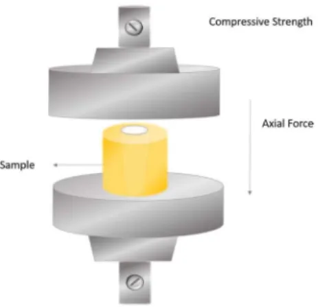
Each root was sectioned (first cut) 1 mm below the cemento-enamel junction using a 0.15 diamond saw at slow speed (Isomet 1000; Buehler, Lake Bluff, IL, USA). A second cut was performed 6 mm apically from the first cut, resulting in 6 mm-long cylinders. The cylinders were polished with smooth sand paper to produce smooth flat ends to prevent any interference with the jaws of the test machine. In order to standardize the cylinder diameter in approximately 3 mm (Fig. 1D), additional wears were performed using diamond burs (2135 model; KG Sorensen, São Paulo, SP, Brazil) in a high-speed handpiece with air/ water coolant. Then, each dentin cylinder was tested for the compressive force required to fracture it by a universal testing machine (Instron Model 4444; Instron, Canton, MA, USA) at a crosshead speed of 1 mm/min (Fig. 2). The samples were placed between the jaws of testing machine and forces were applied from the coronal end of the cylinder and in a vertical direction with the point of application centered in the root canal until the cylinder fractured. Maximum load at fracture was recorded in Newtons and converted to MPa by using the following formula: C=4p/π.d2, where C is the
load in MPa; p=is the maximum applied load in newtons (N); d is the diameter of the specimens in millimeters (mm).
Statistical Analysis
Data were subjected to normality test by Kolmogorov-Smirnov and homogeneity test by Levene’s. The analysis of variance (ANOVA) was performed with SPSS 19.0 for Windows (SPSS Inc., Chicago, IL) statistical software.
Post-hoc tests were calculated with Tukey HSD multiple comparison test at α=0.05.
Results
The results obtained after the compressive strength are given in Table 1. There was a significant difference in the compressive strength between groups (one-way ANOVA, p=0.020). The mean compressive force required to break the samples in the Control Group (151.23±94.36 MPa), which were irrigated with saline (NaCl), was statistically different from GroupEDTA+NaOCl (60.12±92.10 MPa) irrigated with 17%
EDTA and 1% NaOCl (Tukey HSD test, p<0.05). However, no statistical difference was observed between Control Group, GroupNaOCl and GroupEDTA. In addition, GroupNaOCl was also
statistically different (p<0.05) from GroupEDTA+NaOCl and
exhibited a higher mean of compressive strength.
Discussion
The null hypothesis was rejected based on the results of the study. The use of different irrigating solutions, 30 days before the calcium hydroxide intracanal dressing, promoted statistically significant differences among groups. The present study corroborates other studies in the literature, which observed that the dentin exposure to calcium hydroxide for one month or less, has affected negatively some dentin’s mechanical properties (3,11,16,18).
In the present study, the alternate irrigation with EDTA and NaOCl solutions, prior to calcium hydroxide intracanal dressing, decreased dentin compressive strength. However, when the samples were irrigated with NaOCl alone, the results were similar to those obtained with saline irrigation (control group). EDTA irrigation alone did not differ significantly from saline irrigation (control group) either, despite the lower compressive strength means. These findings can be justified by the composition, chemical action, alternation and irrigation time with the solutions, as well as the effect of removing the smear layer over calcium hydroxide dressing action.
Regarding the irrigating solutions, it is well known that NaOCl appears to be the most employed solution due to
Table 1. Compressive strength (MPa, mean±standard deviation) values
Groups and solutions Compressive strength
Control Group: saline 151.23±94.36 a
GroupNaOCl: 1% NaOCl 167.50±57.29 a
GroupEDTA : 17% EDTA 108.79±99.88 ab
GroupEDTA+NaOCl: 17%EDTA, 1% NaOCl 60.12±92.10 b
Different letters indicate statistically significant difference (p<0.05) between groups (Tukey HSD test).
Irrigating solutions effects on dentin
its bactericidal activity (4,5,7,8), excellent lubrication (2), ability to dissolve organic components (1,6), bleaching capacity and low surface tension (1,2). However, adverse effects of NaOCl irrigation on dentin’s mechanical properties (3,10,11,13,19) have been reported, including decrease of microhardness (10), flexural strength (11) and elastic modulus (3). Sodium hypochlorite promoted loss of dentinal organic components (19) because this solution is a non-specific proteolytic agent. Organic phase depletion may change mechanical properties (9), due to the fact that dentin has 22% organic material, especially type I collagen. Collagen plays an important role during dentin structural formation and it is responsible for sustaining dentinal mineral content (19). By losing this organic content, dentinal elastic module and flexural strength decline (3,10).
EDTA is widely used as chelate and cleaning agent during endodontic treatment (1). This solution is able to remove the smear layer and significantly reduce the amount of microorganisms inside the root canal (7). Studies have observed that any irrigating solution is capable of removing both organic and inorganic components from smear layer after canal preparation (1,2,6). Thus, to remove efficiently the smear layer, EDTA is usually recommended, followed by NaOCl (6).
Nevertheless, this association leads to dentinal tubule orifice opening, intertubular dentinal erosion (12,20) and dentin microhardness reduction (21). When NaOCl was used alone as an irrigating agent, the exposed inorganic material prevented further dissolution of dentine. However when the solutions are used alternately, both organic and inorganic contents are removed (6). Niu et al. (20) demonstrated that final irrigation with 6% NaOCl after treatment with 15% EDTA causes erosion of peritubular and intertubular dentin. Surface dentin was neither smooth nor flat and dentinal tubule orifices were irregularly enlarged (20).
In the present study, it may be assumed that irrigation time and the NaOCl-EDTA combination might have resulted in a significant root dentin strength decrease compared with the control group. It is important to emphasize that 20 min irrigation with EDTA may have affected the results. EDTA is mostly used only for final irrigation, after canal preparation and for short periods (1 to 5 min) to remove the smear layer (6). Periods longer than 10 min may cause severe peritubular and intertubular dentinal erosion (12) and dentinal tubule enlargement. When NaOCl was used after irrigation with EDTA, also for 20 min, changing the solutions at every 10 min, there was a major decrease of dentin strength. The use of EDTA for longer periods (20 to 40 min) attempted to simulate its use in atresic canals. In these situations, EDTA is used along all root canal preparation, improving the file actions by lubrication and decalcification of dentin structure (2,19).
Evaluating the impact of using 2.5% NaOCl and 17% EDTA in different sequences on dentin elastic modulus and flexural strength, Marending et al. (11) observed that samples exposed to NaOCl for 24 min had their flexural strength reduced. However, EDTA used for shorter periods, before or after NaOCl irrigation, did not affect the outcomes. It is important to remind that those authors (11) employed dentin bars, which remained totally immersed in hypochlorite, while in our study, only the root canal remained in contact with the solutions, although there was alternate irrigation with EDTA for 20 min. This may have led to higher hypochlorite penetration into dentinal tubules, increasing its action and consequently causing more damage to dentinal collagen matrix. Dentin becomes more fragile and brittle when this matrix is destroyed.
Mineral components in hard tissues contribute to their strength and elastic modulus, whereas the organic component is associated with tissue toughness (22). Collagen matrix destruction in mineralized tissue results in a more brittle and friable substratum that can lead to fatigue crack propagation during cyclic stresses (23).
Finally, a greater dentinal tubule opening by smear layer removal may have improved the effects of calcium hydroxide. Although all groups received intracanal medication for the same time, better effects could be achieved in smear-free canals due to higher hydroxyl ion diffusion in dentin (24). The authors choose to use manual instrumentation in order to minimize the presence of untouched canal walls during preparation, which could result in smear layer formation. Furthermore, canal shaping in the cervical and middle thirds were complemented with Gates Glidden drills, which produced circular sections and standardized the amount of calcium hydroxide dressing inside the canal.
It has been suggested that calcium hydroxide could decrease dentin resistance in smear layer absence. Reports have shown that one week of calcium hydroxide medication can damage dentin resistance (3). Andreasen et al. (17) demonstrated that extracted sheep incisors filled with calcium hydroxide for 1/2, 1, 2, 3, 6, 9, or 12 months had their fracture strength greatly reduced, and this decrease was proportional to the increase of the experimental periods. Sahebi et al.(16) reported that dentin cylinders exposed to calcium hydroxide for 30 days needed a lower compression load to fracture. Zarei et al. (25) observed that under in vitro conditions, teeth with instrumented canals that received calcium hydroxide medication for periods over 1 month had their fracture strength reduced. In shorter periods, resistance also decreased, but it was not statistically significant.
L.C. Hillesheim et al
calcium hydroxide for 30 days. Therefore, the lack of significant difference between groups irrigated with saline and those irrigated with NaOCl could be attributed to the fact that none of those solutions is capable of removing the smear layer (2)
Considering our results, final irrigation with EDTA and NaOCl prior to intracanal medication should be carried out in the shortest irrigation time possible. This could avoid adverse effects over root dentin strength. Furthermore, new studies are needed to observe how irrigation time and amount of solutions, when used in association and prior to intracanal dressing, can affect root dentin mechanical properties.
In conclusion, it was possible to verify that the use of irrigating solutions prior to calcium hydroxide intracanal medication may influence root dentin compressive strength. Canal irrigation with 17% EDTA followed by 1% NaOCl decreased dentin strength significantly, after 30 days on calcium hydroxide dressing.
Resumo
O objetivo desse estudo foi avaliar o efeito da irrigação intracanal com hipoclorito de sódio (NaOCl) e ácido etilenodiamino tetracético (EDTA) previamente à medicação de hidróxido de cálcio na resistência à compressão da dentina radicular. Quarenta e oito raízes com canais únicos e retos tiveram os canais instrumentados pela técnica coroa-ápice com limas Flexofile e brocas Gates Glidden sob irrigação com solução salina (NaCl) a 0,9%. Os canais radiculares foram separados em 4 grupos (n=12), de acordo com a solução irrigadora: Grupo Controle, NaCl 0,9%; GrupoNaOCl, NaOCl 1%; GrupoEDTA, EDTA 17%; e GrupoEDTA+NaOCl, NaOCl 1% e EDTA 17%. As soluções permaneceram por 40 min, sendo renovadas a cada 10 min, exceto para o GrupoEDTA+NaOCl, onde as soluções foram alternadas a cada 10 min. Os canais foram secos, preenchidos com pasta de hidróxido de cálcio e radiografados. Cada canal foi selado e mantido a 37 °C por 30 dias. Após esse período, as raízes foram cortadas 1 mm abaixo da junção cemento-esmalte e 6 mm apicalmente a este corte, a fim de obter cilindros de 6 mm de comprimento. O teste de compressão foi realizado com velocidade de cruzeta de 1 mm/min. As médias (±SD) da resistência à compressão em MPa foram: Grupo Controle, 151.23±94.36; GrupoNaOCl, 167.50±57.25; GrupoEDTA, 108.79±99.88; GrupoEDTA+NaOCl, 60.12±92.10. Diferenças estatisticamente significativas foram encontradas entre os grupos (ANOVA e Tukey; α=0,05; p=0,02). O Grupo Controle e o GrupoNaOCl obtiveram maiores médias de resistência à compressão em relação ao GrupoEDTA+NaOCl. A irrigação intracanal com EDTA 17% e NaOCl 1% promoveu redução significativa da resistência à compressão da dentina, após 30 dias de medicação intracanal com hidróxido de cálcio.
References
1. Haapasalo M, Shen Y, Wang Z, Gao Y. Irrigation in endodontics. Braz Dental J 2014;21:299-303.
2. Good M, El KI, Hussey DL. Endodontic ‘solutions’ Part 1: a literature review on the use of endodontic lubricants, irrigants and medicaments. Dent Update 2012;39:239-246.
3. Grigoratos D, Knowles J, Ng YL, Gulabivala K. Effect of exposing dentine to sodium hypochlorite and calcium hydroxide on its flexural strength and elastic modulus. Int Endod J 2001;34:113-119.
4. Christo JE, Zilm PS, Sullivan T, Cathro PR. Efficacy of low concentrations of sodium hypochlorite and low-powered Er,Cr:YSGG laser activated irrigation against an Enterococcus faecalis biofilm. Int Endod J 2016;49:279-286.
5. Soares ZA, Carvalho MAR de; Santos SMC, Mendonça RMC,
Ribeiro-Sobrinho AP, Brito-Junior M, et al.. Effectiveness of chemomechanical preparation with alternating use of sodium hypochlorite and EDTA in eliminating intracanal Enterococcus faecalis biofilm. J Endod 2010;36:894-898.
6. Teixeira CS, Felippe MCS, Felippe WT. The effect of application time of EDTA and NaOCl on intracanal smear layer removal: an SEM analysis. Int Endod J 2005;38:285-290.
7. Byströn A, Sundqvist G. The antibacterial action of sodium hypochlorite and EDTA in 60 cases of endodontic therapy. Int Endod J 1985;18:35-40.
8. Gonçalves LS, Rodrigues RC, Andrade Junior CV, Soares RG, Vettore MV. The effect of sodium hypochlorite and chlorhexidine as irrigant solutions for root canal disinfection: a systematic review of clinical trials. J Endod 2016;42:527-532.
9. Schmidt TF, Teixeira CS, Felippe MC, Felippe WT, Pashley DH, Bortoluzzi EA. Effect of ultrasonic activation of irrigants on smear layer removal. J Endod 2015;41:1359-1363.
10. Pascon FM, Kantovitz KR, Sacramento PA, Nobre-dos-Santos M, Puppin-Rontani RM (2009). Effect of sodium hypochlorite on dentine mechanical properties. A review. J Dent 2009;37:903-938.
11. Marending M, Paqué F, Fischer J, Zehnder M. Impact of irrigant sequence on mechanical properties of human root dentin. J Endod 2007;33:1325-1328.
12. Çalt S, Serper A. Time-dependent effects of EDTA on dentin structures. J Endod 2002;28:17-19.
13. Ghisi AC, Kopper PM, Baldasso FE, Stürmer CP, Rossi-Fedele G, Steier L, et al.. Effect of super-oxidized water, sodium hypochlorite and EDTA on dentin microhardness. Braz Dent J 2014;25:420-424.
14. Sen BH, Ertürk O, Pişkin B. The effect of different concentrations of EDTA on instrumented root canal walls. Oral Surg Oral Med Oral Pathol Oral 2009;108:622-627.
15. Ozdemir HO, Buzoglu HD, Çalt S, Çehreli ZC, Varol E, Temel A. Chemical and ultramorphologic effects of ethylenediaminetetraacetic acid and sodium hypochlorite in young and old root canal dentin. J Endod 2012;38:204-208.
16. Sahebi S, Moazami F, Abbott P. The effects of short-term calcium hydroxide application on the strength of dentine. Dent Traumatol 2010;26:43-46.
17. Andreasen JO, Farik B, Munksgaard EC. Long-term calcium hydroxide as a root canal dressing may increase risk of root fracture. Dent Traumatol 2002;18:134-137.
18. Yassen GH, Platt, JA. The effect of nonsetting calcium hydroxide on root fracture and mechanical properties of radicular dentine: a systematic review. Int Endod J 2013;46:112-8.
19. Hülsmann M, Effects of mechanical instrumentation and chemical irrigation on the root canal dentin and surrounding tissues. Endodontic Topics 2013;29:55-86.
20. Niu W, Yoshioka T, Suda H. A scanning electron microscopic study of dentinal erosion by final irrigation with EDTA and NaOCl solutions. Int Endod J 2002;35:934-939.
21. Sayin TC, Serper A, Cehreli ZC, Otlu HG. The effect of EDTA, EGTA, EDTAC, and tetracycline-HCl with and without subsequent NaOCl treatment on the microhardness of root canal dentin. Oral Surg Oral Medi Oral Pathol Oral Radiol and Endod 2007;104:418-424. 22. Wang X, Bank RA, Tekoppele JM, Agrawal CM. The role of collagen in
determining bone mechanical properties. J Orthop Res 2001;19:1021-1026.
23. Kruzic JJ, Ritchie RO. Fatigue of mineralized tissues: cortical bone and dentin. J Mech Behav Biomed Mat 2008;1:3-17.
24. Foster KH, Kulild JC, Weller RN. Effect of smear layer removal on the diffusion of calcium hydroxide through radicular dentin. J Endod 1993;19:136-140.
25. Zarei M, Afkhami F, Poor ZM. Fracture resistance of human root dentin exposed to calcium hydroxide inter-visit medication at various time periods: an in vitro study. Dent Traumatol 2013;29:156-160.