Cardiovascular and respiratory changes
during slow-wave sleep in rats are
associated with electrocorticogram
desynchronization
Departamento de Fisiologia, Faculdade de Medicina de Ribeirão Preto, Universidade de São Paulo, Ribeirão Preto, SP, Brasil
J.R. Dias-dos-Santos and B.H. Machado
Abstract
In awake rats a single recurrent larger tidal volume (deep breaths) occurs at regular intervals, followed by oscillations in arterial pressure and heart rate. In the present study we recorded the changes in blood pressure, heart rate and ventilation during the wakefulness-sleep cycle identified by electrocorticographic records in order to determine whether the deep breaths and cardiovascular oscillations were associ-ated with changes in the electrocorticogram. During several episodes of slow-wave sleep (SWS) in 7 rats the deep breaths and oscillations in arterial pressure and heart rate were preceded by SWS desynchroni-zation. The interval between deep breaths during SWS was 71 ± 4 s, the period between initial desynchronization and the generation of deep breaths was 3.98 ± 0.45 s and the duration of SWS desynchroni-zation was 11 ± 0.65 s. Hypotension (-16 ± 1 mmHg) and tachycardia (+15 ± 5 bpm) were observed during deep breaths in the SWS state. These data indicate that the oscillations in arterial pressure and heart rate during SWS are associated with deep breaths, which in turn are preceded by desynchronization of the electrocorticogram in this state of sleep.
Correspondence
B.H. Machado
Departamento de Fisiologia FMRP, USP
14049-900 Ribeirão Preto, SP Brasil
Fax: 55 (016) 633-0017 E-mail: [email protected]
Research supported by FAPESP (Nos. 91/0576-9, 93/2790-3 and 95/4685-8) and CNPq (Nos. 500864/91-8 and 522150/95-0).
Received October 8, 1996 Accepted September 10, 1997
Key words
•Wakefulness-sleep cycle
•Ventilation
•Cardiovascular regulation
•Deep breaths
•Ventilatory patterns
•Arterial pressure oscillations
•Whole body plethysmography
Introduction
Studies on cardiovascular changes in the wakefulness-sleep cycle have shown that short episodes of electrocortical desynchro-nization occur during slow-wave sleep (SWS) in which arterial pressure exhibits rapid os-cillations (1). In previous studies we ob-served that awake normal rats present deep breath events at regular intervals associated with oscillations in arterial blood pressure and heart rate (2,3). These studies also showed that in rats with sino-aortic deafferentation the deep breath events were consistently
In addition to the studies by Junqueira and Krieger (1), other reports have shown that several changes in blood pressure, heart rate and respiratory frequency occur during the sleep-wake cycle (9,10), but no studies have determined whether the cardiovascular and respiratory changes occurring during the deep breath events in rats are associated with electrocorticographic changes during the dif-ferent states of the wakefulness-sleep cycle. For this reason, in the present study we recorded pulsatile arterial pressure, heart rate and ventilation during the wakefulness-sleep cycle identified by the electrocorticogram in normal rats.
Material and Methods
Male Wistar rats weighing 250-300 g were used. Three days before the experi-ments two bipolar electrodes were implanted into the skull of the rats under Nembutal anesthesia (40 mg/kg, ip) by means of two small nickel-chromium screws (100 µm in diameter) fixed into two small holes drilled in the skull. The electrodes were implanted on the dura mater in order to cover a small portion of the parietal cortex (areas 3 and 7). The electrodes were soldered to the two small screws which were fixed 1 mm left to the median suture and 1 mm from the breg-matic suture in the parietal bone and 1 mm from the median suture and 1 mm from the lambda suture on the left side (1,11). The entire system was fixed to the skull with acrylic cement. Electrocorticographic record-ings were obtained three days after electrode implantation. The different steps of the sleep-wake cycle were identified by electrocor-ticographic analysis according to the method of Timo-Iaria et al. (12) but with no meas-urement of the cervical electromyogram. The characterization of desynchronized sleep (REM) was performed by visual observation of the position of the head under the trunk (12) as well as by the increase in respiratory
frequency and reduction in tidal volume, as described by Remmers (13). The electrocor-ticogram was recorded with a Narcotrace 40 physiological recorder using a Universal Coupler (Narco Bio-Systems, Austin, TX).
chamber which was carefully closed and opened at intervals of approximately 7 min. When the chamber was closed PAP, HR, electrocorticogram and ventilation were re-corded continuously.
Despite the limitations of the whole body plethysmographic method for long-term measurement of ventilation, the method was reliable for the purpose of the present study because a) no significant changes in basal respiratory frequency were observed during the period of 5-7 min in which the chamber was closed (2,3); b) the deep breath event was not an artefact of this method because it was also observed in the rats outside the chamber; c) after the rats were trained to stay inside, the chamber could be carefully opened and closed with no major disturbance of the sleep-awake cycle of the rats, and d) the advantage of this method in relation to the diaphragm electromyogram or records of electrical activity of the phrenic nerve is that it can be used in unanesthetized rats without implanting additional electrodes.
For 2 days prior to the experiments the rats used in the present study (N = 7) were trained to stay inside the plethysmographic chamber for periods of 2-3 h. On the day of the recordings the rats were maintained in-side the chamber for at least 2 h before starting the procedure. The recordings were performed between 2:00 and 5:00 p.m. in an acoustically isolated room. The duration of the recordings varied for each rat because it depended on the occurrence of a complete sleep-wake cycle. The cables from the skull for the EKG recordings and the catheter from the femoral artery were exteriorized through a small hole in the plethysmographic chamber to be connected to the transducers and physiological recorder. This small hole was then filled with silicone grease in order to seal the plethysmographic chamber.
The changes in mean arterial pressure and heart rate during the deep breath epi-sodes between different states of the wake-fulness-sleep cycle and the awake state were
compared by the unpaired Student t-test and the level of significance was set at P<0.05.
Results
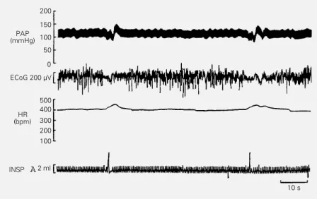
Deep breaths occurred at intervals of 71 ± 4 s during the SWS in the 7 rats studied, in which 29 episodes were analyzed. The time between the initial changes in the electrocor-ticogram and the appearance of the deep breath episodes was 3.98 ± 0.45 s (33 epi-sodes analyzed) and the duration of the changes in the electrocorticogram pattern during SWS was 11 ± 0.65 s (33 episodes analyzed). Figure 1 is a tracing obtained from one rat representative of the group showing 2 episodes of deep breaths with the corresponding changes in electrocorticogram, PAP and HR during the SWS.
Table 1 shows the changes in diastolic arterial pressure and heart rate associated with deep breaths during the awake state (-7 ± 2 mmHg; +4 ± 3 bpm) and SWS (-16 ± 1 mmHg; +15 ± 5 bpm) and in the deep breaths observed during the transition from SWS to the awake state (-10 ± 3 mmHg; +12 ± 6
PAP (mmHg)
200
150
100
50
0
ECoG 200 µV
HR (bpm)
500 400
300 200 100
INSP A2 ml
10 s
bpm) and from desynchronized sleep (REM) to the awake state (-17 ± 7 mmHg; +5 ± 5 bpm).
The transition from the SWS to wakeful-ness in the 4 rats was also associated with deep breaths and hemodynamic changes. The transition episodes from REM to wakeful-ness (5 episodes in 5 different rats) were also recorded and in these cases the transition was also associated with deep breaths and hemodynamic changes. The REM state is characterized by an irregular respiratory rhythm (13), which is normalized after the transition to wakefulness. In 4 rats, the ir-regular respiratory rhythm during the REM state presented reduction in tidal volume and short apnea, which were followed in many cases by tachypnea.
Discussion
The results of the present study indicate that all micro-awake episodes during the SWS are associated with deep breaths and that the transition from SWS to wakefulness as well as from desynchronized sleep (REM) to wakefulness is associated with deep breaths and cardiovascular changes. The tran-sition from REM sleep to wakefulness is also associated with a deep breath episode that seems to be a marker for the normaliza-tion of the frequency and tidal volume of the ventilatory cycle. The cardiovascular changes
observed during the SWS desynchronization and simultaneous to the deep breaths were not greater probably because of the buffer-ing role played by the arterial baroreceptors (2,3).
Studies by Junqueira and Krieger (1) have shown the occurrence of some episodes of desynchronization in the electrocorticogram during the SWS which seem to be coincident with a short period of animal wakefulness. These investigators also observed that im-mediately after this short desynchronization of the electrocorticogram the rat was again in the SWS state of the sleep-wake cycle. In the present study we observed that this de-synchronization is a consistent phenomenon occurring at regular intervals of 71 ± 4 s and all the episodes recorded were associated with deep breaths and with changes in PAP and HR. The deep breaths and cardiovascu-lar changes occurred at regucardiovascu-lar intervals in all phases of the sleep-wake cycle, but the changes in the electrocorticogram were ob-served only in the SWS. It is important to note that deep breaths occurred 3.98 ± 0.45 s after the initial desynchronization of the elec-trocorticogram. The total period of desyn-chronization during the SWS was 11 ± 0.65 s. We may suggest different possibilities to explain these findings: a) deep breaths may be generated by cortical mechanisms, b) the micro-awake episodes may be related to neu-rovegetative adjustments activated by a pos-sible reduction in pO2 during the SWS and c)
the short period of SWS desynchronization may be the electrophysiological expression of micro-awake episodes and the cardiovas-cular and respiratory changes observed are part of this event. The micro-awake episodes observed in the present study are similar to the arousal episodes that follow synchro-nized and desynchrosynchro-nized sleep, in which rostrum and eye movements, vibrissal twitches and tachycardia were observed (15). Since we did not record the cervical elec-tromyogram or eye movements to determine the different steps of the wakefulness-sleep Table 1 - Peak changes in diastolic arterial pressure (∆ DAP) and heart rate (∆ HR) during
episodes of deep breaths in the awake state (deep breaths/wake), in the slow-wave sleep (SWS) state, in the transition from slow-wave state to awake state (SWS/wake) and in the transition from desynchronized to the awake state (REM/wake).
*P<0.05 compared to the changes in deep breaths in the awake state (deep breaths/ wake) (Student t-test).
∆ DAP ∆ HR Number Number of
(mmHg) (bpm) of rats episodes
Deep breaths wake -7 ± 2 +4 ± 3 6 6
Deep breaths SWS -16 ± 1* +15 ± 5* 7 21
Deep breaths SWS/wake -10 ± 3 +12 ± 6 4 10
cycle, we cannot rule out the possibility that desynchronization of the SWS (micro-awake episodes) observed in our study is related to episodes of relaxed wakefulness (15).
Deep breaths induce a 4-5-fold increase in tidal volume (2) and this large expansion of the chest wall may induce cardiovascular changes such as hypotension and tachycar-dia that may be counterbalanced by cardio-vascular reflexes. We have reported that the fall in blood pressure of rats submitted to removal of the arterial baroreceptors was greater than in control rats (2,3). Thus, on the basis of a previous study (2), we may suggest two mechanisms to explain the fall in pressure during deep breaths: 1) activa-tion of cardiopulmonary receptors in response to hemodynamic changes secondary to the large expansion of the chest wall and 2) interaction of neurons in the brain stem asso-ciated with the central neural control of the circulation and ventilation, considering that ventilatory and cardiovascular changes oc-curred at the same time. The activation of cardiopulmonary receptors may result from an increased end-diastolic filling pressure. However, this mechanism may not be im-portant in this case because the fall in pres-sure seems to be too fast for the characteris-tics of this reflex mechanism. In addition, the tachycardia observed is not typical of the cardiopulmonary reflex (Bezold-Jarisch re-flex), which is characterized by an intense bradycardic response. The second possibil-ity, despite the complex neural mechanisms involved, is plausible if we consider the studies by St. John et al. (5-8) showing that gasping, a deep breath that occurs in decer-ebrated cats, is generated by neural circuits in the brain stem.
The hypothesis of respiratory and cardio-vascular neuron interaction in the brain stem is supported by evidence showing that neu-rons generating the breathing rhythm in the pre-Botzinger complex (16) are located in the vicinity of the sympathetic vasomotor neurons in the rostral ventrolateral medulla
(RVLM) (17) and also that neurons of the central respiratory generator may have an excitatory as well as an inhibitory effect on the sympathetic neurons located in the RVLM (18). Therefore, we suggest that the hypo-tension that follows the large deep breath seems to be more closely related to a pos-sible sympatho-inhibitory mechanism at the RVLM level than to hemodynamic changes secondary to the large expansion of the chest wall. However, the interaction of these neu-ral mechanisms in the brain stem to generate the respiratory and cardiovascular changes described in the present study requires fur-ther experiments to be better understood.
barore-ceptor reflex integrity plays a key role in counterbalancing the cardiovascular oscilla-tions during deep breaths and the present data suggest that during SWS desynchroni-zation the sensitivity of the baroreflex may be reduced since the fall in pressure at that time was greater than in the awake state.
The cortical desynchronization during SWS indicates micro-awake episodes, which may induce cardiovascular and respiratory changes. The interaction of rostral brain ar-eas involved in the generation of SWS
de-synchronization with brain stem structures involved in the autonomic regulation of cir-culation and ventilation is still a matter for further investigation.
Acknowledgments
The authors thank Mauro de Oliveira and Leni G.H. Bonagamba for excellent techni-cal assistance, and Dr. Raul Laguzzi and Dr. Patrice G. Guyenet for helpful suggestions and critical comments about the data.
References
1. Junqueira LF & Krieger EM (1976). Blood pressure and sleep in the rat in normoten-sion and in neurogenic hypertennormoten-sion.
Journal of Physiology, 259: 725-735. 2. Machado BH, Mauad H & Glass ML
(1992). Transient changes in blood pres-sure during spontaneous deep breaths in rats with sinoaortic deafferentation. Jour-nal of Applied Physiology, 72: 920-924. 3. Mauad H, Glass ML & Machado BH
(1992). Effect of selective denervation of baroreceptors on pulmonary ventilation and arterial pressure lability in rat. Hyper-tension, 19 (Suppl II): II.182-II.186. 4. Bartlett Jr D (1971). Origin and regulation
of spontaneous deep breaths. Respiratory Physiology, 12: 230-238.
5. St. John WM, Bartlett Jr D, Knuth KV & Hwang JC (1981). Brain stem genesis of automatic ventilatory patterns independ-ent of spinal mechanisms. Journal of Ap-plied Physiology, 51: 204-210.
6. St. John WM, Bledsoe TA & Sokol HW (1984). Identification of medullary loci criti-cal for neurogenesis of gasping. Journal of Applied Physiology, 56: 1008-1019. 7. St. John WM, Bledsoe TA & Tenney SM
(1985). Characterization by stimulation of medullary mechanisms underlying gasp-ing neurogenesis. Journal of Applied Physiology, 58: 121-128.
8. St. John WM (1989). Neurogenesis, con-trol, and functional significance of gasp-ing. Journal of Applied Physiology, 68: 1305-1315.
9. Harper RM (1991). Brain mechanisms un-derlying cardiorespiratory control during sleep. In: Gaultier C, Escourrou P & Curzi-Dascalova L (Editors), Sleep and Cardio-respiratory Control. Colloque INSERM/ John Libbey Eurotext Ltd., Paris, 217: 55-60.
10. Laguzzi R (1991). Cardiovascular changes during the sleep-wake cycle. In: Gaultier C, Escourrou P & Curzi-Dascalova L (Edi-tors), Sleep and Cardiorespiratory Control. Colloque INSERM/John Libbey Eurotext Ltd., Paris, 217: 9-13.
11. Lacombe J, Nosjean A, Meunier JM & Laguzzi R (1988). Computer analysis of cardiovascular changes during the sleep-wake cycle in Sprague-Dawley rats.
American Journal of Physiology, 254: H217-H222.
12. Timo-Iaria C, Negrão N, Schmidek WR, Hoshino K, Menezes CEL & Rocha TL (1970). Phases and states of sleep in the rat. Physiology and Behavior, 5: 1057-1062.
13. Remmers JE (1981). Control of breathing during sleep. In: Hornbein TR (Editor),
Regulation of Respiration. Part II. Marcel Dekker, New York, 1197-1249.
14. Malan A (1973). Ventilation measured by body plethysmography in hibernating mammals and poikilotherm. Respiratory Physiology, 17: 32-44.
15. Valle AC, Timo-Iaria C, Fraga JL, Sameshima K & Yamashita R (1992). Theta waves and behavioral manifesta-tion of alertness and dreaming activity in the rat. Brazilian Journal of Medical and Biological Research, 25: 745-749. 16. Smith JC, Ellenberger HH, Ballanyi K,
Richter DW & Feldman JL (1991). Pre-Botzinger complex: A brainstem region that may generate respiratory rhythm in mammals. Science, 254: 726-729. 17. Guyenet PG (1990). Role of the ventral
medulla oblongata in blood pressure regu-lation. In: Loewy AD & Spyer KM (Edi-tors), Central Regulation of Autonomic Functions. Oxford University Press, New York, 145-167.