online | memorias.ioc.fiocruz.br
Schistosomiasis affects 200 million people world-wide (Lambertucci 1993). Hepatosplenic schistosomiasis mansoni is a severe form of the disease and develops into periportal fibrosis and portal hypertension in nearly 10% of infected individuals (Prata 1987, Pinto-Silva et al. 1994, Lambertucci et al. 2000, Andrade 2004, Maia et al. 2007).
Abdominal ultrasound (US) is considered a simple, safe and inexpensive imaging technique in the evaluation of schistosomal liver involvement and is often used as the surrogate for the gold-standard in the diagnosis of liver fibrosis (LF) and classification of its intensity (Andrade 2005, Lambertucci et al. 2008). The sensitivity and spe-cificity of US in the recognition of LF is comparable to histological examination of liver fragments (Cerri et al. 1984, Homeida et al. 1988a, Abdel-Wahab et al. 1989). The development of portable US devices is considered an advance in the investigation of morbidity in field-based studies (Marinho et al. 2006). US may also be used to
evaluate the dynamic changes produced by portal hyper-tension and, accordingly, may suggest the presence of oesophageal varices (Abdel-Latif et al. 1981, Davidson et al. 1991, Abdel-Wahab et al. 1993, Richter et al. 1998). However, this diagnostic tool requires well-trained phy-sicians and its standardization is still a matter of debate after two World Health Organization (WHO) workshops (Cairo Working Group 1992, Niamey Working Group 1996, Lambertucci et al. 2001).
Magnetic resonance (MR) is a very sensitive imaging technique that has been used to evaluate liver involvement in a variety of diseases. In schistosomiasis, MR altera-tions have already been described in case reports and in series of cases (Patel et al. 1993, Willemsen et al. 1995, Lambertucci et al. 2002, 2004, Silva et al. 2006, Bezerra et al. 2007, Silva 2007). However, to our knowledge, no study has compared US and MR to liver histology in pa-tients with hepatosplenic schistosomiasis.
The aim of the present study was to compare abdom-inal US and MR to liver histology in the diagnosis and evaluation of Symmers’ fibrosis intensity in schistoso-miasis mansoni.
PATIENTS, MATERIALS AND METHODS
Patients - The study was approved by the Ethical Board of the Federal University of Minas Gerais, Brazil.
Imaging techniques and histology in the evaluation
of liver fibrosis in hepatosplenic schistosomiasis mansoni
in Brazil: a comparative study
Izabela Voieta1, Leonardo C de Queiroz2, Luciene M Andrade1,3, Luciana Cristina S Silva1,
Vitor F Fontes1, Aryon Barbosa Jr4, Vivian Resende5, Andy Petroianu5, Zilton Andrade4,
Carlos Mauricio Antunes1, José Roberto Lambertucci1/+
2Serviço de Radiologia, Hospital das Clínicas 5Serviço de Cirurgia de Fígado e Vias Biliares 1Programa de Pós-Graduação
em Ciências da Saúde: Infectologia e Medicina Tropical, Faculdade de Medicina, Universidade Federal de Minas Gerais, Av. Alfredo Balena 190 sala 169, 30.130-100 Belo Horizonte, MG, Brasil 3Laboratórios Hermes Pardini,
Belo Horizonte, MG, Brasil 4Fundação Gonçalo Moniz-Fiocruz, Salvador, BA, Brasil
Few publications have compared ultrasound (US) to histology in diagnosing schistosomiasis-induced liver fibrosis (LF); none has used magnetic resonance (MR). The aim of this study was to evaluate schistosomal LF using these three methods. Fourteen patients with hepatosplenic schistosomiasis admitted to hospital for surgical treatment of variceal bleeding were investigated. They were submitted to upper digestive endoscopy, US, MR and wedge liver biopsy. The World Health Organization protocol for US in schistosomiasis was used. Hepatic fibrosis was classified as absent, slight, moderate or intense. Histology and MR confirmed Symmers’ fibrosis in all cases. US failed to detect it in one patient. Moderate agreement was found comparing US to MR; poor agreement was found when US or MR were compared to histology. Re-classifying LF as only slight or intense created moderate agreement between imaging techniques and histology. Histomorphometry did not separate slight from intense LF. Two patients with advanced hepatosplenic schistosomiasis presented slight LF. Our data suggest that the pres-ence of the characteristic periportal fibrosis, diagnosed by US, MR or histology, associated with a sign of portal hypertension, defines the severity of the disease. We conclude that imaging techniques are reliable to define the presence of LF but fail in grading its intensity.
Key words: schistosomiasis - ultrasound - magnetic resonance - liver fibrosis - Symmers’ fibrosis - splenectomy - portal hypertension
Financial support: CNPq
+ Corresponding author: [email protected] Received 9 January 2009
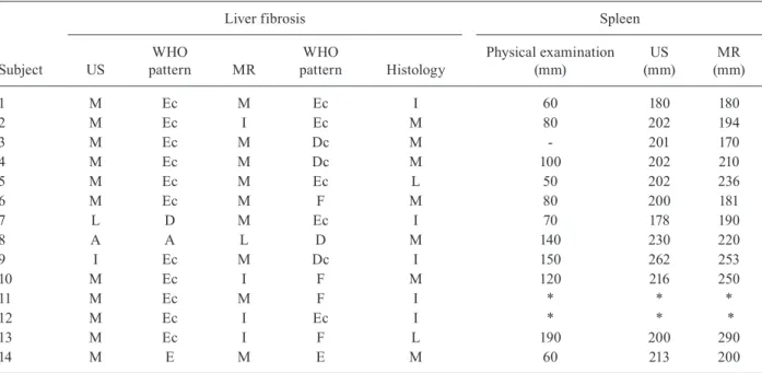
All study subjects signed an informed consent for par-ticipation. Fourteen patients (10 males) referred to the Tropical Diseases Outpatient Clinic with a diagnosis of hepatosplenic schistosomiasis, portal hypertension and indication for surgical intervention (splenectomy and/or suturing of oesophago-gastric varices) were selected for this study. Schistosomiasis was defined by microscopic evidence of active infection (positive parasitological stool examination or eggs in rectal biopsy) and a history of contact with stream waters of an endemic area. Patients included in the study did not have other causes of chronic liver disease, such as cirrhosis, congestive heart failure or toxic or viral hepatitis. Age ranged from 20-57 years (39 ± 10.4). Baseline characteristics are presented in Table I.
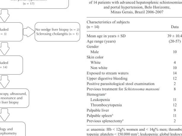
Methods - Outpatient assessment included clinical examination, abdominal US, upper digestive endoscopy, serum markers of autoimmune hepatitis, hepatitis B and C serology, blood cell count and liver function evalua-tion (serum albumin and prothrombin time). Seventeen patients fulfilled the criteria for inclusion, but three were excluded because wedge liver biopsy was not available or because of sclerosing cholangitis (Fig. 1).
US examination of the abdomen was performed using a real-time ALOKA SSD device 1700 (Japan) with electronic convex 3.5 MHz transducer, according to the WHO
proto-TABLE I
Demographic characteristics, clinical and laboratory aspects of 14 patients with advanced hepatosplenic schistosomiasis
and portal hypertension, Belo Horizonte, Minas Gerais, Brazil 2006-2007
Characteristics of subjects
(n = 14) Data
Mean age in years ± SD 39 ± 10.4
Age range (years) (20-57)
Gender
Male 10
Skin color
White 4
Non white 10
Exposed to stream waters 14
Upper digestive bleeding 12
Positive parasitological stool examination 2 Previous treatment for Schistosoma mansoni 8 Hemograma
Leukopenia 11
Thrombocytopenia 12
Palpable liver 9
Palpable spleenb 11
Previous splenectomyb 2
a: anaemia: Hb < 12g% women and < 14g% men; thrombocy-topenia: platelets < 150.000 mm3; leukopenia: global leukocytes < 4.300 cels/mm3; b: two were submitted to previous splenecto-my and one was obese, two subjects had only palpable spleen. Upper endoscopy, ultrasound,
magnetic resonance and wedge liver biopsy
Histology and histomorphometry
No wedge liver biopsy (n = 2) Sclerosing cholangitis (n = 1) Advanced hepatosplenic schistosomiasis
with portal hypertension (n = 17)
Excluded (n = 3)
Included (n = 14)
col for US assessment of schistosomiasis-related morbidity (Niamey Working Group 1996). US hepatic fibrosis was classified as absent (pattern A), slight (B, C, D and Dc), moderate (E and Ec) or intense (F). The presence of collat-eral vessels and the spleen length were evaluated. MR was obtained using a GE 1.5 T Sigma unit (Milwaukee, USA). Axial and coronal 7-mm-thick slices were performed in T1 and T2-weighted sequences, before and after gadopentetate dimeglumine (Gd-DTPA) administration. MR analysis was guided by an adaptation of the WHO protocol for US. US and MR were blindly performed (Fig. 2). Radiologists and pathologists knew that all patients had schistosomiasis, but they were not informed of the clinical form of the disease.
Radiology was the specialty of both observers. The US examiner had seven years of experience in radiol-ogy; he was trained to use the WHO protocol for US-related morbidity in schistosomiasis. The MR examiner had 10 years of experience in radiology. One observer viewed the MR and other the US images. The inter-ob-server agreement for US has been reported to be moderate (kappa 0.45) (Doehringer et al. 1992). For MR the intra and inter-observer agreement has been shown to be substantial (kappa 0.65 and 0.66, respectively) (Bezerra et al. 2007).
Wedge liver biopsy fragments of ~3 cm3 were
ob-tained from the left liver lobe during splenectomy, fixed
in 10% formalin and embedded in paraffin (Fig. 3). The section preparations were stained with hematoxylin and eosin (H&E) and were examined under light
microsco-py. Randomly sampled 5-μm-thick liver histological sec -tions stained with picrosirius red for interstitial collagen were examined by semiautomatic morphometry using the LEICA QWIN-3.1 (Microsystem Imagina Solutions LTD, Cambridge, UK) (Fig. 4). For morphometric meas-urements a total sectional area of 4,296,920 m�� per pa-m�� per pa-per pa-tient was evaluated. The sectional area of the red-stained fibrous tissue was directly measured and calculated as a percentage of the total area examined, as previously described (Barbosa 2001, Coutinho et al. 2003).
Fig. 2: World Health Organization patterns for ultrasound (US) as-sessment of schistosomiasis-related morbidity (middle column) (US on the left and magnetic resonance on the right).
Fig. 3: wedge liver biopsy being performed during surgery on the left and a fragment of 3 cm3 on the right.
Statistical analysis - Data were stored using EpiDa-ta 3.1 software [JM Lauristen (ed.), Odense Denmark, EpiData Association, 2000-2006 (available from: http:// www.epidata.dk)]. Statistical analysis was performed us-ing EpiInfo 3.3.2 [Centers for Disease Control and Pre-vention, USA, 2005 (available from: http://www.cdc.gov/ EpiInfo/epiinfo.htm)] and SPSS 12.0 for Windows (SPSS Incorporation, Chicago, Illinois, USA, 2005). Compari-sons of quantitative and qualitative variables among US, MR and liver biopsy were made using Wilcoxon and McNemar tests. Agreement between image methods was evaluated by kappa index or Spearman’s test. The degree of agreement for kappa was classified according to Lan-dis and Koch (1977) (Fig. 5). Other associations between the three groups (bleeding frequency, spleen length and LF) were appraised by the nonparametric Mann-Whitney test or Fisher’s exact test, as appropriate.
RESULTS
All patients had oesophageal varices confirmed by endoscopy and 12 reported some form of previous diges-tive bleeding. Only four patients underwent sclerothera-py of oesophageal varices. Two patients excreting Schis-tosoma mansoni eggs were treated with praziquantel.
In 13 out of 14 (92.8%) patients, US showed the char-acteristic features of schistosomal liver fibrosis, whereas MR identified periportal thickening in all 14.
Patients with hepatosplenic schistosomiasis are shown in Fig. 5. Symmers’ fibrosis was confirmed by histology. The white clay pipe-stem aspect was pres-ent in all cases, with variable degrees of septal fibrosis maintaining the acinar architecture of the hepatic paren-chyma. Periovular granulomas were rare, small, fibrotic and sometimes with calcified ova.
During splenectomy, the liver surface presented a wide range of patterns (Fig. 6). The surface varied from smooth to pseudo-nodular with macroscopic whitish bands. Liver wedges also varied from sharp to blunt and consistency varied from soft to hard.
of one degree, e.g., from moderate to intense (patients 1, 2, 9, 10, 11 and 12). The three methods completely disagreed in four patients (28.5%; patients 5, 7, 8 and 13). These results are summarised in Table II. Agree-ment between imaging methods, as evaluated by kappa index, was 0.41 (moderate); after re-grouping grades absent and slight together (labelled slight) and grades moderate and intense together (labelled intense), sub-stantial concordance was observed (kappa = 0.63).
Agreement between US and histology was poor (kap-pa = 0.06) and remained so even after re-grouping into slight and intense (kappa = -0.17). Similar results were observed comparing MR to histology. Excluding one pa-tient (papa-tient 7, Table II) who presented absent fibrosis in US and moderate fibrosis in MR, the agreement was moderate between imaging techniques and histology (kappa = 0.41). If this patient was kept in the compari-son, the obtained agreement was poor (kappa = 0.10).
There was no agreement (kappa < 0) between histo-morphometric classification of fibrosis and the subjec-tive histological classification.
No correlation was observed between spleen size or upper digestive bleeding and the intensity of LF.
DISCUSSION
Histology and MR confirmed Symmers’ fibrosis in all cases; US failed to identify one patient. Imaging tech-niques presented moderate agreement with histology to
rank LF. Histomorphometry did not agree with the his-tological classification of fibrosis. We also observed two patients with advanced hepatosplenic schistosomiasis with light LF.
Surgical wedge liver biopsy is considered the gold-standard procedure for diagnosing Symmers’ fibrosis. In our study, MR identified LF with the same precision as histology. A marked divergence, though, occurred during classification of fibrosis intensity. Other studies using MR contained case reports or series of cases and they did not include liver biopsy to confirm the diagnosis and intensity of Symmers’ fibrosis. However, using US, Homeida et al. (1988b) found results comparable to ours: the diagnosis of LF by US coincided with histology, but they presented poor agreement on fibrosis intensity.
Other studies used US to diagnosis the presence and grade the intensity of LF, but liver biopsy was only em-ployed to confirm Symmers’ fibrosis and to exclude other liver diseases (Abdel-Wahab et al. 1978, 1989, 1992, Cerri et al. 1984, Homeida et al. 1988a, Pinto-Silva et al. 1994).
In one of our patients, US failed to diagnosis LF. This deficiency has been previously described by Abdel-Wahab et al. (1989). Of their 18 patients with histological diagno-sis of Symmers’ fibrodiagno-sis, two did not present US evidence of periportal thickening. It is also important to comment on one patient with sclerosing cholangitis excluded from our study: US images were indistinguishable from what is seen in patients with Symmers’ fibrosis.
Needle liver biopsy frequently overlooks periportal fibrosis because it retrieves insufficient and fragment-ed tissue samples with a small number of portal tracts (Bogliolo 1957b). Taking this into consideration, our study was designed to obtain a surgical biopsy of the liver during splenectomy. Some authors criticise the wedge liver biopsy because fragments come from the periphery of the organ and fibrosis is not expected to be uniformly distributed (Brandt et al. 2002). Nevertheless, Dusek et al. (1965) states that a large enough specimen for histological assessment is obtainable only by surgical wedge biopsy and that the specimens are often sufficient to make an ac-curate diagnosis. In our opinion, the fragments obtained in our study (~3 cm3) were large enough to permit a
de-finitive diagnosis of Symmers’ fibrosis.
Abdel-Wahab et al. (1992) and Richter et al. (1992) found that increases in portal and splenic vein diameters were significantly correlated with the degree of hepatic periportal fibrosis and the frequency of bleeding from en-doscopically proven oesophageal varices. Here, we found no correlation between spleen size or a history of oesoph-ageal bleeding and LF intensity. Interestingly, Andrade and Bina (1983), describing an autopsy series of 232 ca-davers, stated that they found no correlation between the intensity of LF and evidence of portal hypertension and that, in fact, the latter resulted from intra-hepatic vessel obstruction rather than from LF (Prata & Andrade 1963).
Histomorphometry is a method used to measure LF as a percentage of the hepatic tissue. No correlation be-tween histological findings and morphometric measure-ments was observed in the present investigation. The reason for this is not clear, but a few points may be noted: (i) the usual histological examination by a pathologist is subjective and therefore difficult to evaluate (for exam-Fig. 4: liver histological slide stained with picrosirius-red for
inter-stitial collagens.
Values of kappa Interpretation
< 0 No agreement
0-0.19 Poor agreement
0.20-0.39 Fair agreement
0.40-0.59 Moderate agreement
0.60-0.79 Substantial agreement
0.80-1.00 Almost perfect agreement
Fig. 6: clinical aspect of patients in the first row; surface of the liver in the second; ultrasound (US) in the third; magnetic resonance (MR) in the fourth and histology in the fifth. On the left column is the case of a patient with severe hepatosplenic schistosomiasis, but with a nearly normal liver, US without fibrosis, MR with light fibrosis and histology with moderate. In the middle column there is an example of moderate fibrosis and of intense fibrosis on the right.
ple, the classification of the intensity of fibrosis in hepa-titis C is also subjective and a significant error among different examiners has been reported) (de Paula Farah et al. 2007) and (ii) LF is not homogeneously dispersed in the liver and even in the same fragment, fibrosis may be unevenly distributed. The same results have been de-scribed by others (Domingues 1998, Brandt et al. 2002).
TABLE II
Liver fibrosis and spleen size of 14 patients with advanced hepatosplenic schistosomiasis evaluated by physical examination, ultrasound (US), magnetic resonance (MR) and histology, Belo Horizonte, Minas Gerais, Brazil 2006-2007
Liver fibrosis Spleen
Subject US
WHO
pattern MR
WHO
pattern Histology
Physical examination (mm)
US (mm)
MR (mm)
1 M Ec M Ec I 60 180 180
2 M Ec I Ec M 80 202 194
3 M Ec M Dc M - 201 170
4 M Ec M Dc M 100 202 210
5 M Ec M Ec L 50 202 236
6 M Ec M F M 80 200 181
7 L D M Ec I 70 178 190
8 A A L D M 140 230 220
9 I Ec M Dc I 150 262 253
10 M Ec I F M 120 216 250
11 M Ec M F I * * *
12 M Ec I Ec I * * *
13 M Ec I F L 190 200 290
14 M E M E M 60 213 200
World Health Organization (WHO) patterns: A: absent; I: intense (F); L: slight (B, C, D, Dc); M: moderate (E, Ec) (Fig.2). Asterisk means splenectomized.
knowledge, there are no studies using positron emis-sion tomography CT in hepatosplenic schistosomiasis. Non-invasive markers have also been used to predict the presence and degree of LF in hepatic diseases such as hepatitis C, cirrhosis and schistosomiasis (Stone 2000, Guangjin et al. 2002, Afdhal & Nunes 2004, Grigorescu 2006).
It is worth mentioning that MR reproduces the path-ological findings of Symmers’ fibrosis more clearly than US does and is naturally less invasive and time-consuming than histology. The MR images are similar to those described by a pathologist when the liver is transversally cut during autopsy. Also, its superiority in the identification of portal vessels, portal vein throm-bosis and collaterals should not be neglected (Bezerra et al. 2007). This information may help physicians plan in advance the surgical procedure that best suits their patients. On the other hand, MR is expensive. Due to its clear advantages, however, we believe it will be used more frequently in the near future.
Patients with hepatosplenic schistosomiasis in need of surgical intervention are those with severe portal hyper-tension, oesophago-gastric varices and hypersplenism (thrombocytopaenia, leukopenia and anaemia) (Petroi-anu 1983, 2003). It has been assumed that such patients would accordingly have intense periportal fibrosis, but here, we observed two patients with slight fibrosis diag-nosed by histology presenting the severest clinical form of hepatosplenic schistosomiasis (huge spleens and mas-sive variceal bleeding). Therefore, portal hypertension
was probably caused by intrahepatic sinusoidal obstruc-tion rather than periportal fibrosis.
In summary, LF intensity is not a definite surrogate marker of morbidity. Our data suggest that the intensity of LF evaluated by imaging techniques or histology does not relate to the severity of portal hypertension. The characteristic periportal fibrosis (diagnosed by US, MR or histology) associated with evidence of portal hyper-tension (large spleen, oesophageal varices and collateral vessels) should be sufficient to define disease severity and indicate surgical intervention, as necessary. We con-clude that imaging techniques are reliable to define the presence of LF but fail in grading its intensity.
REFERENCES
Abdel-Latif Z, Abdel-Wahab MF, El-Kady NM 1981. Evaluation of portal hypertension in cases of hepatosplenic schistosomiasis us-ing ultrasound. J Clin Ultrasound9: 409-412.
Abdel-Wahab MF, Abdel-Latif Z, El-Kady NM, Arafa NM 1978. The use of ultrasonography in diagnosis of different schistosomal-syndromes. Proceedings of the Third International Workshop on Diagnostic Ultrasound Imaging (Cairo, Egypt), Al-Ahram Press, Cairo, p. 458-463.
Abdel-Wahab MF, Esmat G, Farrag A, el-Boraey Y, Strickland GT 1993. Ultrasonographic prediction of esophageal varices in schis-tosomiasis mansoni. Am J Gastroenterol88: 560-563.
Abdel-Wahab MF, Esmat G, Farrag A, el-Boraey YA, Strickland GT 1992. Grading of hepatic schistosomiasis by the use of ultra-sonography. Am J Trop Med Hyg46: 403-408.
1989. Characteristic sonographic pattern of schistosomal hepatic fibrosis. Am J Trop Med Hyg 40: 72-76.
Afdhal NH, Nunes D 2004. Evaluation of liver fibrosis: a concise re-view. Am J Gastroenterol 99: 1160-1174.
Andrade ZA 2004. Schistosomal hepatopathy. Mem Inst Oswaldo Cruz99 (Suppl. I): 51-57.
Andrade ZA 2005. Regression of hepatic fibrosis. Rev Soc Bras Med Trop38: 514-520.
Andrade ZA, Bina JC 1983. A patologia da forma hepato-esplênica da esquistossomose mansoni em sua forma avançada (estudo de 232 necrópsias completas). Mem Inst Oswaldo Cruz78: 285-305.
Barbosa Jr AA 2001. Morphological computer-assisted quantita-tive estimation of stained fibrous tissue in liver sections: ap-plications in diagnosis and experimental research. J Bras Patol37: 197-200.
Bezerra AS, D’Ippolito G, Caldana RP, Cecin AO, Ahmed M, Sze-jnfeld J 2007. Chronic hepatosplenic schistosomiasis mansoni: magnetic resonance imaging and magnetic resonance angiogra-phy findings. Acta Radiol48: 125-134.
Bogliolo L 1957a. A esplenoportografia da esquistossomose mansô-nica hepatoesplêmansô-nica, forma de Symmers. Rev Assoc Med Bras 3: 263-269.
Bogliolo L 1957b. Anatomical picture of the liver in hepatosplenic schistosomiasis mansoni. Ann Trop Med Parasitol51: 1-14.
Brandt CT, Domingues ALC, Vilela P, Sena A, Marques K, Giusti CF 2002. Histomorphometry of hepatic portal fibrosis in pa-Histomorphometry of hepatic portal fibrosis in pa-tients with surgical schistosomiasis mansoni. Acta Cir Bras 17
(Suppl. 1): 7-10.
Cairo Working Group 1992. The use of diagnostic ultrasound in schistosomiasis - attempts at standardization of methodology.
Acta Trop51: 45-63.
Cerri GG, Alves VA, Magalhães A 1984. Hepatosplenic schis-tosomiasis mansoni: ultrasound manifestations. Radiology
153: 777-780.
Coutinho EM, Barros AF, Barbosa A Jr, Oliveira SA, Silva LM, Araú-jo RE, Andrade ZA 2003. Host nutritional status as a contribu-Host nutritional status as a contribu-tory factor to the remodeling of schistosomal hepatic fibrosis.
Mem Inst Oswaldo Cruz 98: 919-925.
Davidson RN, Houston S, Kiire CF 1991. Schistosomal periportal fibrosis in Zimbabwe: use of ultrasound in patients with oesopha-geal varices. Trans R Soc Trop Med Hyg85: 380-382.
Doehring-Schwerdtfeger E, Abdel-Rahim IM, Kardorff R, Kaiser C, Franke D, Schlake J, Richter J, Elsheikh M, Mohamed-Ali Q, Ehrich JHH 1992. Ultrasonographical investigation of periportal fibrosis in children with Schistosoma mansoni infection: revers-ibility of morbidity twenty-three months after treatment with praziquantel. Am J Trop Med Hyg 46: 409-415.
Domingues ALC 1998. Ultra-sonografia na esquistossomose man-sônica hepato-esplênica: avaliação da intensidade da fibrose periportal e da hipertensão porta, PhD Thesis, Universidade Fe-PhD Thesis, Universidade Fe-deral de Pernambuco, Recife, 99 pp.
Dusek J, Kubasta M, Kodousek R, Kubastova B 1965. Needle biopsy of the liver in shistosomiasis mansoni: the value of histological examination. J Trop Med Hyg 68: 189-195.
Grigorescu M 2006. Noninvasive biochemical markers of liver fibro-sis. J Gastrointestin Liver Dis15: 149-159.
Guangjin S, Mingdao J, Qiyang L, Hui X, Jiangming H, Xiaomei Y 2002. Study on histopathology, ultrasonography and some spe-cial serum enzymes and collagens from 38 advanced patients of schistosomiasis japonica. Acta Trop82: 235-246.
Homeida M, Abdel-Gadir AF, Cheever AW, Bennett JL, Arbab BM, Ibrahium SZ, Abdel-Salam IM, Dafalla AA, Nash TE 1988a. Di-agnosis of pathologically confirmed Symmers’ periportal fibro-sis by ultrasonography: a prospective blinded study. Am J Trop Med Hyg 38: 86-91.
Homeida M, Ahmed S, Dafalla A, Suliman S, Eltom I, Nash T, Bennett JL 1988b. Morbidity associated with Schistosoma mansoni infec-tion as determined by ultrasound: a study in Gezira, Sudan. Am J Trop Med Hyg39: 196-201.
Lambertucci JR 1993. Schistosoma mansoni: pathological and clini-cal aspects. In P Jordan, G Webbe (eds.), Human schistosomiasis, Cab International, Wallingford, p. 195-225.
Lambertucci JR, Andrade LM, Pinto-Silva RA 2002. Magnetic reso-Magnetic reso-nance imaging of the liver in hepatosplenic schistosomiasis man-soni. Rev Soc Bras Med Trop35: 679-680.
Lambertucci JR, Cota GF, Pinto-Silva RA, Serufo JC, Gerspacher-Lara R, Drummond SC, Antunes CM, Nobre V, Rayes A 2001. Hepatosplenic schistosomiasis in field-based studies: a combined clinical and sonographic definition. Mem Inst Oswaldo Cruz96
(Suppl.): 147-150.
Lambertucci JR, dos Santos Silva LC, Andrade LM, de Queiroz LC, Carvalho VT, Voieta I, Antunes CM 2008. Imaging techniques in the evaluation of morbidity in schistosomiasis mansoni. Acta Trop108: 209-217.
Lambertucci JR, Serufo JC, Gerspacher-Lara R, Rayes AA, Teixeira R, Nobre V, Antunes CM 2000. Schistosoma mansoni: assessment of morbidity before and after control. Acta Trop77: 101-109.
Lambertucci JR, Silva LC, Andrade LM, de Queiroz LC, Pinto-Silva RA 2004. Magnetic resonance imaging and ultrasound in hepatosplenic schistosomiasis mansoni. Rev Soc Bras Med Trop
37: 333-337.
Landis JR, Koch GG 1977. The measurement of observer agreement for categorical data. Biometrics 33: 159-174.
Maia MD, Lopes EP, Ferraz AA, Barros FM, Domingues AL, Ferraz EM 2007. Evaluation of splenomegaly in the hepatosplenic form of mansonic schistosomiasis. Acta Trop101: 183-186.
Marinho CC, Voieta I, Azeredo LM, Nishi MP, Batista TS, Pereira ACF, Serufo JC, de Queiroz LC, Ruiz-Guevara R, Antunes CM, Prata A, Lambertucci JR 2006. Clinical versus ultrasound exami-Clinical versus ultrasound exami-nation in the evaluation of hepatosplenic schistosomiasis mansoni in endemic areas. Mem Inst Oswaldo Cruz101 (Suppl. I): 317-321.
Niamey Working Group 1996. Ultrasound in schistosomiasis. A practical guide to the standardized use of ultrasonography for the assessment of schistosomiasis-related morbidity. UNDP/ World Bank/WHO/Special Programme for Research & Training in Tropical Diseases (TDR). World Health Organization/TDR/ SCH/ULTRASON document, Geneva. Available from: http:// apps.who.int/tdr/svc/publications/training-guideline-publica-tions/ultrasound-in-schistosomiasis.
Patel SA, Castillo DF, Hibbeln JF, Watkins JL 1993. Magnetic reso-nance imaging appearance of hepatic schistosomiasis, with ul-trasound and computed tomography correlation. Am J Gastroen-terol88: 113-116.
Petroianu A 1983. A esplenectomia subtotal e anastomose esplenor-renal proximal para o tratamento da hipertensão portal. Rev Bras Cir73: 101-104.
Petroianu A 2003. Tratamento cirúrgico da hipertensão porta da es-quistossomose mansoni. Rev Soc Bras Med Trop36: 253-265.
Prata A 1987. Schistosomiasis mansoni in Brazil. Baillieres Clin Trop Med Comm Dis2: 349-369.
Prata A, Andrade ZA 1963. Fibrose hepática de Symmers sem esple-nomegalia. Hospital(Rio J) 63: 617-623.
Richter J, Correia Dacal AR, Vergetti Siqueira JG, Poggensee G, Mannsmann U, Deelder A, Feldemeier H 1998. Sonographic pre-Sonographic pre-diction of variceal bleeding in patients with liver fibrosis due to
Schistosoma mansoni. Trop Med Int Health3: 728-735.
Richter J, Zwingenberger K, Ali QM, Lima W de M, Dacal AR, de Siqueira GV, Doehring-Schwerdtfeger E, Feldmeier H 1992. He- He-patosplenic schistosomiasis: comparison of sonographic findings in Brazilian an Sudanese patients - correlation of sonographic findings with clinical symptoms. Radiology184: 711-716.
Silva LC 2007. Comparação entre a ultra-sonografia e a ressonância magnética do abdome na avaliação da morbidade na esquistos-somose mansônica, PhD Thesis, Universidade Federal de Minas Gerais, Belo Horizonte, 97 pp.
Silva LC, Pereira AC, Queiroz LC, Andrade LM, Antunes CM, Lambertucci JR 2006. Disagreement between ultrasound and mag-netic resonance imaging in the identification of schistosomal peri-portal fibrosis. Mem Inst Oswaldo Cruz101 (Suppl. I): 279-282.
Stone PJ 2000. Potential use of collagen and elastin degradation markers for monitoring liver fibrosis in schistosomiasis. Acta Trop77: 97-99.
Willemsen UF, Pfluger T, Zoller WG, Kueffer G, Hahn K 1995. MRI of hepatic schistosomiasis mansoni. J Comput Assit Tomogr