MLL
leukemia-associated rearrangements in peripheral blood lymphocytes
from healthy individuals
María Sol Brassesco
1,2, Ana Paula Montaldi
1, Diana Ester Gras
1, Rosane Gomes de Paula Queiroz
2,
Nilce Maria Martinez-Rossi
1, Luiz Gonzaga Tone
2and Elza Tiemi Sakamoto-Hojo
1,31
Departamento de Genética, Faculdade de Medicina de Ribeirão Preto, Universidade de São Paulo,
Ribeirão Preto, SP, Brazil.
2
Departamento de Puericultura e Pediatria, Faculdade de Medicina de Ribeirão Preto,
Universidade de São Paulo, Ribeirão Preto, SP, Brazil.
3Departamento de Biologia, Faculdade de Filosofia, Ciências e Letras, Universidade de São Paulo,
Ribeirão Preto, SP, Brazil.
Abstract
Chromosomal translocations are characteristic of hematopoietic neoplasias and can lead to unregulated oncogene expression or the fusion of genes to yield novel functions. In recent years, different lymphoma/leukemia-associated rearrangements have been detected in healthy individuals. In this study, we used inverse PCR to screen peripheral lymphocytes from 100 healthy individuals for the presence ofMLL (Mixed Lineage Leukemia) translocations. Forty-nine percent of the probands showedMLL rearrangements. Sequence analysis showed that these rearrange-ments were specific forMLL translocations that corresponded to t(4;11)(q21;q23) (66%) and t(9;11) (20%). How-ever, RT-PCR failed to detect any expression of t(4;11)(q21;q23) in our population. We suggest that 11q23 rearrangements in peripheral lymphocytes from normal individuals may result from exposure to endogenous or ex-ogenous DNA-damaging agents. In practical terms, the high susceptibility of theMLL gene to chemically-induced damage suggests that monitoring the aberrations associated with this gene in peripheral lymphocytes may be a sen-sitive assay for assessing genomic instability in individuals exposed to genotoxic stress.
Key words:genomic instability, lymphocytes,MLLrearrangements.
Received: May 30, 2008; Accepted: October 16, 2008.
Introduction
Lymphoid neoplasias are generally characterized by the presence of chromosomal anomalies, the most promi-nent of which are those that produce in-frame fusion genes. These aberrations are important diagnostic tools that can be used to establish the prognosis of leukemias and lympho-mas and monitor their progress.
Rearrangements of the Mixed Lineage Leukemia (MLL) gene generated by reciprocal translocations involv-ing chromosome band 11q23 are well-known in infants and adults with acute myeloid leukemia (AML) and acute lymphoblastic leukemia (ALL), as well as in 85% of sec-ondary leukemias associated with a history of treatment with topoisomerase II inhibitors (Adleret al., 1999; Ross, 2000). More than 50 fusion genes involvingMLL associ-ated with a poor prognosis have been identified (Popovic
and Zeleznick-Le, 2005; Slany, 2005). However, despite the diversity and frequency ofMLLtranslocations, most of the breakpoints have been mapped within a Bam H1-delimited region known as the break cluster region or BCR (Sai-Peng and Liu, 2001) between exons 8 and 14 (Nilson et al., 1996; Schnittger, 1998; Echlin-Bell et al., 2003). Other aberrations involvingMLL in leukemia include in tandemduplications represented by in-frame repetitions of exons 2-6 (or 2-8) that can be attributed to homologous re-combination mediated byAlurepeats (Stroutet al., 1998; Whitmanet al., 2001).
Since the development of leukemia and solid tumors is a multistage process that requires multiple cooperative mutations, it seems plausible that different mutations, such as typically found in patients with leukemia or lymphoma, could arise in normal individuals (Hunger and Cleary, 1998).
The presence of tumor-associated fusion genes in healthy donors has been described for the translocation t(14;18) IGH/BCL2 (characteristic of non-Hodgkin lym-phomas), with variable frequencies (16.2%-55%) among Send correspondence to María Sol Brassesco. Departamento de
populations and a tendency to increase with age (Liuet al., 1994; Summerset al., 2001; Yasukawaet al., 2001). This rearrangement has also been described in 43% of blood samples from patients with non-proliferative malignancies (Rauzy et al., 1998). Similarly, the translocation t(9;22) BCR/ABL was primarily detected in peripheral lympho-cytes of adults and children (Biernauxet al., 1995); Boseet al. (1998) subsequently confirmed these data by demon-strating p190 and p210 transcripts in 4 of 11 and 11 of 16 in-dividuals, respectively. Other markers have also been detected at low frequencies in normal populations, includ-ingETV6/RUNX1(Eguchi-Ishimaeet al., 2001; Brassesco et al., 2004), t(11;14)(p13;q11) LMO2/TCR and t(7;14)(q34;q11)TCR/TAL2(Marculescuet al., 2002) and t(15;17)PML/RARA, the latter characteristic of promyelo-cytic leukemia (Quinaet al., 2000). The incidence ofMLL duplications in healthy donors is much higher, and are de-tectable in almost all samples by using sensitive PCR meth-ods (Bäseckeet al., 2006). Together, these studies indicate that leukemia and lymphoma-associated translocations can be generated in normal hematopoietic cells without appar-ent oncogenic consequences.
Based on these findings, we used an inverse-PCR strategy to investigate the presence ofMLLtranslocations in peripheral blood lymphocytes from healthy individuals. Our results demonstrate the presence ofMLL fusions in these cells, thus indicating that these rearrangements are not restricted to malignant cells but may also be present in a subset of normal hematopoietic cells.
Material and Methods
Probands
Blood samples from 100 normal subjects (50 males, 50 females) were analyzed in this study. All of the subjects were healthy non-smokers 18 to 46 years old (mean±SD = 22.9±5.4 years) with no previous history of drug treatment or chronic use of medicines. A single sample of 10 mL of peripheral blood was obtained from each individual after informed consent, and the samples were immediately coded to ensure the anonymity of the donors. The study was approved by the local ethics committee of the Clinical Hos-pital of the Faculty of Medicine (University of São Paulo, Ribeirão Preto, SP).
Translocation analysis by inverse-PCR
Inverse PCR was done according to Bettiet al.(2001) with few modifications. Three micrograms of DNA was di-gested with a combination ofSau3AI andXbaI (10 units each) at 37 °C overnight. The addition ofXbaI prevented amplification of the nativeMLLgene while allowing the amplification of translocation products that lacked theXbaI recognition site. After digestion, the samples were heat-inactivated at 65 °C for 10 min and then purified with a Wizard SV Gel and PCR Clean-up System Kit (Promega
Corporation, Madison, WI, USA) to remove residual enzy-matic activity. Following re-suspension in nuclease-free water, 0.5mg of digested DNA was self-ligated in the pres-ence of 3 units of T4 DNA ligase in a final volume of 20mL for 16 h at 16 °C. All of the ligation reactions were termi-nated by incubation at 65 °C for 10 min. Eight microliters of ligated DNA was used in each PCR reaction. Nested primers were used to analyze the cleavage site at exon 12 of MLLin two 28-cycle reactions at temperatures of 95 °C, 55 °C and 72 °C for 1 min/step.
The following primers were used:foward-15’-CTT TGTTTATACCACTC-3’; reverse-1 5’-TAGGGAATAT AAAAGAGTGGG-3’; forward-2 5’-TTAGGTCACTTA GCATGTTCTG-3’ andreverse-25’-CAGTTGTAAGGT CTGGTTTGTC-3’. Strict precautions were taken in each step to avoid cross-contamination of the samples.
Analysis of translocation DNA sequences
PCR amplicons were separated on 1% agarose gels. Individual I-PCR products were extracted from the gels with a GFX PCR DNA and Gel Band Purification kit (Amersham Biosciences, Buckinhamgshire, UK). The fragments were then cloned into the pGEM-T cloning vec-tor (Promega Corporation, Madison, WI, USA), trans-formed into pMOS BlueEscherichia coliand selected on LB-agar plates containing ampicillin (50mg/mL), accord-ing to the manufacturer’s instructions. Individual trans-formed colonies were then expanded for 22 h in liquid culture. Three hundred nanograms of plasmidial DNA was used as a template for the sequencing reaction with a Big Dye Terminator Cycle Sequence Ready Reaction kit (Amersham Biosciences) and the products were analyzed in an ABI Prism 377 DNA Sequencer (Applied Biosys-tems, Wellesley, MA, USA). Quality analysis and the re-moval of vector sequences were done with phredPhrap software (Ewinget al., 1998; Ewing and Green, 1998). The resulting DNA sequences were then used to search the Na-tional Center for Biotechnology Information database with the Basic Local Alignment Search Tool (BLAST).
Detection ofAF4/MLLtranslocations by RT-PCR
AG-.
Lymphocyte culture
Lymphocytes were cultured using a standard protocol in which 0.5 mL of peripheral blood was added to 10 mL of RMPI 1640 medium (Sigma) supplemented with 20% fetal calf serum, 2% phytohemagglutinin (PHA) and penicil-lin/streptomycin. The cells were incubated at 37 °C for 72 h and treated with colchicine (0.56%) for the final 90 min. Cell harvesting and slide preparation were done using stan-dard methods. Slides for FISH were stored at -20 °C until used.
Fluorescencein situhybridization
FISH was done using the commercially available probes LSI MLL Break Apart Rearrangement, according to the manufacturers protocol (Vysis, Downers Grove, IL). The probe labeled with SpectrumGreen covered a 350 kb portion centromeric to theMLLgene breakpoint region whereas the SpectrumOrange-labeled probe covered a 190 kb portion telomeric to the BCR. The expected signal pattern for a nor-mal cell nucleus was two green(yellow)orange signals. In cells withMLLtranslocations, the green and orange signals were separated without the yellow intersection. The advan-tage of this strategy was that it allowed the detection of translocations regardless of the partner involved. At least 1000 nuclei were analyzed and images were captured with an Axiovision System (Zeiss, Germany).
Results
Inverse PCR was used to screen the peripheral blood lymphocytes of normal individuals forMLLtranslocations. In this strategy, the translocation region was excised with restriction enzymes, circularized and amplified using vari-ousMLLprimers. This approach allowed the detection of any rearrangement involving the cleavage site at exon 12, which contains putative topoisomerase II recognition se-quences and is sensitive to DNAse I and some cytotoxic agents.
Forty-nine of the 100 DNA samples that were screened contained bands of variable sizes that corre-sponded to alterations spanning theMLLbreakpoint region (Figure 1). In gel electrophoresis, the putative transloca-tions resulted in one, two or three amplification products of 300-700 bp (amplification of the germ-lineMLLwas pre-vented by treatment withXbaI), which suggested that some individuals may have more than oneMLL translocation. The individual bands were separated by electrophoresis in 1% agarose gels and cloned into the pGEM-T vector, trans-formed inE. colipMOS Blue cells and sequenced. BLAST analysis of individual amplicons confirmed that these rear-rangements were unique and specific forMLL rearrange-ments. Of the 35 clones that were obtained, 66% contained the translocation t(4;11)(q21;q23), which fusesMLL and
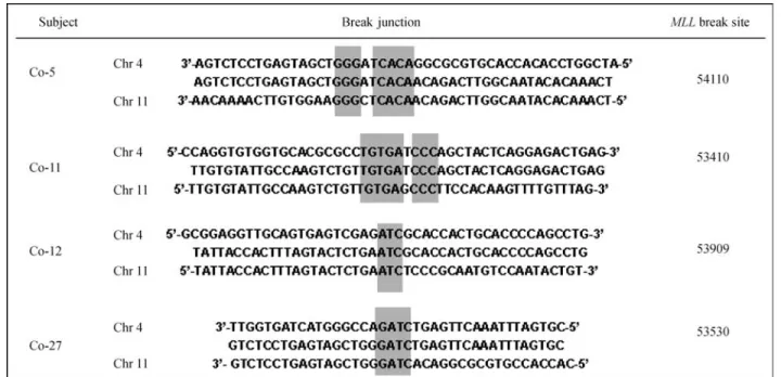
AF4and occurs mainly in acute lymphoblastic leukemia. The remaining translocations fusedMLLto sequences lo-cated on chromosomes 1, 2, 9 (7 cases), 12 (2 cases) and 19 (Table 1). Although these chromosomes contain known MLL partner genes, such as EPS15 (1q32), MLLT11 (1q21),AF9(9q34),CIP29(12q13),ELL(19q13) andEEN (19q13) (Atlas of Genetics and Cytogenetics in Oncology and Haematology), the partner sequences did not match with specific chromosome bands. Interestingly, sequence analysis of the breakpoint junctions revealed short microhomologies (1-8 bp) suggestive of non-homologous end joining repair (NHEJ) (Figure 2).
Chromosomal preparations from 49 individuals were also analyzed using the LSI MLL (Vysis) commercial probe, which allows the detection of different rearrange-ments at 11q23. At least 1000 nuclei were analyzed per in-dividual and the translocation frequencies were found to vary from zero to 0.3 events/100 cells (mean ± SD = 0.04±0.06). The specific probes also allowed the detection of extra signals, with frequencies ranging from zero to 0.79 signals/100 cells (mean±SD = 0.18±0.19) (Table 2).
Together, these results raised the question of whether MLLfusion genes were expressed at a transcriptional level. Since t(4;11) was the most frequent translocation, RNA samples from 22 donors were screened forMLL/AF4 tran-scripts using different primer sets that allowed the detection of all known fusion transcripts between exon 8 ofMLLand exon 7 ofAF4. No t(4;11)(q21;q23) transcripts were de-tected by RT-PCR in peripheral lymphocytes from healthy individuals (data not shown).
Discussion
The potential of the MLL gene for recombination makes it difficult to detect aberrations by classic methods. Consequently, the detection of MLL translocations is a challenge because although they have a known 5’ sequence their 3’ end can be one of a wide variety of translocation
partners. The use of inverse PCR eliminates this problem by amplifying circularized fragments derived from any seg-ment flanking a known DNA sequence. As shown here, MLLfusion genes were detected in the peripheral lympho-cytes of 49 of the 100 normal individuals examined in this work. The presence of these rearrangements was confirmed by FISH on interphase nuclei, and showed that 28.5% of the samples showed signal separation (based on the use of a specific dual-color “split-signal” DNA probe). The dis-crepancy between the results obtained with these two
meth-ods probably reflects the difference in their sensitivities: whereas FISH can detect one positive cell in a thousand, PCR-based techniques can detect one cell in a million. In-terestingly, 21 individuals who were negative for rear-rangements by FISH had extra signals for theMLLgene by PCR; these extra signals probably represented transloca-tions with other gene partners.
Direct DNA sequencing of the inverse PCR am-plicons showed that most of the fusion sequences were t(4;11)(q21;q23); however, noAF4/MLL transcripts were detected by RT-PCR (standardized for the study of minimal residual disease) in 22 RNA samples.
The results of this study show thatMLL rearrange-ments are not restricted to malignant cells but may also oc-cur in normal hematopoietic cells. As indicated above, several studies have reported the presence of leukemia-lymphoma-associated fusion genes (e.g., BCR/ABL1, IGH/BCL2, TCRb/g) in normal individuals.In tandem par-tial duplications ofMLLhave been detected in almost all bone marrow and peripheral blood samples from healthy
Table 1-MLLfusions detected by inverse PCR in peripheral blood lym-phocytes from healthy individuals.
Control Clone Translocation detected e-value*
Co 5 46 t(4;11) 3e-99; 8e-50 Co 8 47 t(4;11)(q21;q23) 2e-14; 6e-42
Co 11 48 t(9;11) 9e-60; 2e-17 49 t(9;11) 1e-74; 1e-15 Co 18 51 t(4;11)(q21;q23) 0.008
Co 21 52 t(2;11) 8e-04; 8e-04
Co 28 53 t(9;11) 2e-172
Co 30 54 t(4;11)(q21;q23) 3e-14; 8e-98 Co 31 55 t(4;11)(q21;q23) 3e-67;4e-131 55-2 t(9;11) 0.003; 7e-146
Co 33 56 t(4;11)(q21;q23) 3e-58 Co 37 57 t(4;11)(q21;q23) 6e-13; 1e-161
Co 39 59 t(1;11) 5e-37; 1e-114
Co 41 60 t(11;19)(q23;p13) 6e-46; 9e-42 60-2 t(4;11)(q21;q23) 2e-21; 5e-41
Co 43 61 t(4;11)(q21;q23) 6e-23; 4e-58 Co 44 62 t(4;11)(q21;q23) 0.046
Co 49 63 t(4;11)(q21;q23) 5e-38; 2e-145
Co 50 64 t(4;11)(q21;q23) 3e-57 Co 53 66 t(4;11)(q21;q23) 1e-09
Co 54 67 t(4;11)(q21;q23) 1e-12; 2e-14 68 t(4;11)(q21;q23) 2e-16; 7e-35
Co 58 70 t(4;11)(q21;q23) 6e-15; 5e-77
Co 61 72 t(4;11)(q21;q23) 2e-58
73 t(9;11) 2e-04
Co 63 74 t(4;11)(q21;q23) 7e-48 Co 70 75 t(11;12) 5e-37;2e-54 Co 71 115 t(4;11)(q21;q23) 4e-52;1e-24
Co 72 116 t(4;11)(q21;q23) 8e-18 Co 84 117 t(4;11)(q21;q23) 5e-63
Co 97 118 t(9;11) 1e-137; 8e-31 Co 98 119 t(4;11)(q21;q23) 0.057
Co 99 120 t(11;12) 3e-08; 7e-99
Co 113 121 t(9;11) 2e-13
Co 114 122 t(4;11)(q21;q23) 2e-57; 2e-36
(*) value obtained by BLASTn analysis.
Table 2- Frequencies ofMLLrearrangements and extra signals in periph-eral blood lymphocytes from healthy individuals analyzed by FISH.
Controls Rearrange-ments per
100 cells
Extra signals per
100 cells
Controls Rearrange-ments per
100 cells
Extra signals per
100 cells
Co-1 0.00 0.30 Co-33 0.00 0.20
Co-2 0.10 0.20 Co-34 0.10 0.30
Co-3 0.10 0.30 Co-35 0.10 0.30
Co-4 0.00 0.20 Co-36 0.00 0.00
Co-5 0.10 0.30 Co-37 0.10 0.00
Co-9 0.09 0.59 Co-38 0.00 0.20
Co-10 0.00 0.79 Co-39 0.20 0.10
Co-11 0.00 0.09 Co-40 0.10 0.10
Co-12 0.00 0.09 Co-41 0.00 0.00
Co-13 0.00 0.49 Co-42 0.10 0.20
Co-14 0.00 0.00 Co-43 0.00 0.00
Co-15 0.00 0.00 Co-44 0.00 0.10
Co-16 0.00 0.49 Co-45 0.00 0.00
Co-17 0.00 0.09 Co-46 0.00 0.40
Co-18 0.30 0.30 Co-48 0.00 0.00
Co-20 0.00 0.39 Co-50 0.00 0.00
Co-21 0.00 0.00 Co-51 0.00 0.00
Co-22 0.00 0.00 Co-52 0.00 0.00
Co-24 0.00 0.00 Co-55 0.00 0.02
Co-26 0.00 0.09 Co-57 0.00 0.10
Co-27 0.00 0.00 Co-59 0.00 0.20
Co-28 0.09 0.19 Co-63 0.10 0.00
Co-30 0.20 0.60 Co-64 0.00 0.20
Co-31 0.00 0.20 Co-65 0.00 0.00
donors (Schnittgeret al., 1998; Bäseckeet al., 2002, 2006), but there is only one report of translocations involvingMLL in normal individuals. Uckum et al. (1998) used nested PCR to show that rearrangements involvingMLLand the transcription factor AF4, resulted in the translocation t(4;11)(q21;q23) in bone marrow samples from fetuses and normal children, as well as in fetal liver samples.
These findings indicate that such translocationsper se do not define clinically apparent diseases, but rather that malignant progression appears to depend on additional fac-tors such as the occurrence of oncogenic secondary alter-ations. Leukemia-associated gene fusions are generally believed to occurin utero, before birth. For twins with con-cordant leukemia and MLL aberrations, the concordance rate reaches almost 100% (Greaves, 2002) and retrospec-tive studies have shown the clonality of the rearrangements (Gale et al., 1997). According to Greaves and Wiemels (2003), the Knudson model, in addition to the twin concor-dance data, indicates that for every child with a particular translocation-positive leukemia, there has to be a greater number of healthy individuals that harbor the same trans-location in a silent pre-leukemic clone. Similar studies of umbilical cord blood samples have shown that the frequen-cies ofETV6/RUNX1andRUNX1/ETO, for example, are 100 times higher in neonates than in pediatric leukemia pa-tients (Moriet al., 2002). These rearrangements may occur in a high proportion of developing fetuses, but without the production of functional chimeric proteins; alternatively, they could originate through inappropriate cellular condi-tions (Kim-Rouilleet al., 1999).
Specific breaks involving theMLL gene can be in-duced by a variety of stimuli associated with cellular stress
or apoptosis, such as serum starvation or treatment with cy-tosine arabinoside (Stanullaet al., 1997; Bettiet al., 2001; Vaughanet al., 2005). The activation of some components of the apoptotic process under these conditions has been demonstrated (Alamet al., 1999), and cells can recover the normal phenotype in the absence of phagocytic signals (Reddienet al., 2001). Based on these considerations, it seems plausible thatMLLrearrangements in normal indi-viduals could result from exposure to genotoxic agents. The involvement of epipodophylotoxins in anomalies of this gene in therapy-related leukemias, and the evidence that neonatal leukemia originatesin utero, have led to the hy-pothesis that maternal exposure to topoisomerase II inhibi-tors during pregnancy could be associated with an increased risk of leukemia (Ross, 2000). Synthetic and nat-ural flavonoids bind to topoisomerase II to form a cleavable complex, despite the paradoxical finding that in some cases these compounds are anticarcinogenic (Greaves, 1997). Strick et al. (2000) demonstrated that natural flavonols such as quercetin and fisetin induced the same level of breaks at 11q23 as did etoposide, whereas luteolin and genistein were two-fold less effective than this drug and, in some cases, their combination had a cumulative effect in in-ducingMLLcleavage.
Epidemiological studies have shown a significant as-sociation between infant leukemias and maternal exposure to various chemicals (Shuet al., 1996, 1999; Schuzet al., 2000; Maet al., 2002; Mucciet al., 2004). In the specific case of infant leukemia withMLL gene fusions, a case-control study identified significant variations in the inges-tion of herbal medicines, drugs (e.g., Dipyrone), and insec-ticides (Alexander et al., 2001). A similar study that
focused on maternal diet concluded that the ingestion of fruits and vegetables during pregnancy usually diminished the general risk of leukemia, although in the case of AML MLL(+) exposure to certain natural topoisomerase II inhib-itors appeared to increase the risk of disease (Spectoret al., 2005).
According to Wiemelset al.(1999), the exposure of mothers and fetuses to dietary, medicinal and environmen-tal substances that interact with topoisomerase II can be or-ders of magnitude lower in terms of dose level than for drugs used in chemotherapy. However, in some cases, these compounds are as biologically active as the topoisomerase II inhibitors used to treat cancer. The most abundant natural sources of topoisomerase inhibitors in a normal diet are fruits, vegetables and grains, which are rich in isoflavo-noids. The antioxidant effect of these substances has been widely demonstrated (Prior, 2003), although epidemiologi-cal studies have shown that a high ingestion of isofla-vonoids does not mean a reduced risk for all types of cancer (Hertoget al., 1994). In Asian countries, for example, the ingestion of isoflavonoids can reach 28 mg/day (Fukutake et al., 1996; Nakamuraet al., 2000). The plasma concentra-tion of these substances after ingesconcentra-tion is relatively high (Frankeet al., 1998; Watanabeet al., 1998) and can persist for two days. This finding suggests that the repeated inclu-sion of certain foods in the diet may ensure elevated plasma levels of these compounds (Hollmanet al., 1997; de Vries et al., 1998).
Wiemels et al. (1999) also suggested that inter-individual variation in drug metabolism by phase I and phase II detoxifying enzymes could play an important role in modulating the response to low doses of topoisomerase II inhibitors. Thus, for example, the frequency of NQ01 (NAD(P)H: quinone oxido-reductase) low-activity alleles is 2.5 times lower in patients withAF4/MLLfusions than in the normal population. Similarly, polymorphisms in CYP3A4(which converts epipodophylotoxins into catechol metabolites) have been associated with an increased risk of leukemia (Felixet al., 1999). Uncontrolled exposure to cer-tain substances and their metabolites can also contribute to gene fusions. Thus, hybrid genes that are present at low fre-quencies in peripheral blood of normal individuals tend to be more common in exposed populations, as in the case of theTCRb/ghybrid gene in agricultural workers exposed to pesticides (Lipkowitz et al.,1992) and the translocation t(14;18) in smokers (Bellet al., 1995).
Our study group consisted of healthy non-smokers with no previous history of drug treatment or chronic use of medicines. However, the presence ofMLLfusions in pe-ripheral blood lymphocytes of these individuals may have been related to previous exposures to substances from a va-riety of sources. Since lymphocytes circulate continuously they are considered to be more vulnerable to chemical or physical agents than other cell types (Tucker and Preston, 1996).
Tumor-associated translocations in peripheral lym-phocytes may be transitory since sequential blood samples were not always positive for gene fusions, as shown for the BCR/ABLhybrid gene (Biernauxet al., 1995). Other au-thors have suggested that such rearrangements may be ex-pressed in hematopoietic cells that have entered the apoptotic pathway and have already lost their relevance (Boseet al., 1998). On the other hand, whereas the genetic regulation mediated by MLL is important during hema-topoietic differentiation, the expression of this gene (or of the fusion products) may be irrelevant in mature cells.
This study is the first to report the presence ofMLL fusion genes at a genomic level in peripheral blood lym-phocytes of healthy adults. To date, all screenings of leuke-mia-associated rearrangements have been based on RT-PCR. TheAF4/MLL fusion transcripts were initially described in normal individuals (Uckumet al., 1998) but subsequent studies failed to detect any transcription of this rearrangement (Kim-Rouilleet al., 1999; Trkaet al., 1999). In agreement with the latter studies, our results show that blood cells do not express detectable levels ofAF4/MLL transcripts and there is noa fortiorisynthesis of the chime-ric protein. In addition, sequencing of the PCR products, which provides breakpoint information, showed that in most cases the translocations were not in frame, thus strengthening the hypothesis that they may be tolerated for years without adverse consequences.
The biological significance of fusion genes and their respective chimeras in differentiated cells is still uncertain, and an important question remains about their oncogenic potential in healthy individuals. Nevertheless, the high pro-portion ofMLLrearrangements in normal individuals sug-gests that 11q23 anomalies may possibly result from exposure to endogenous or exogenous DNA-damaging agents. In practical terms, the high susceptibility of the MLL gene to chemically-induced damage suggests that monitoring the aberrations associated with this gene in pe-ripheral lymphocytes may be a sensitive assay for assessing genomic instability in individuals exposed to genotoxic stress.
Acknowledgments
We thank all of the donors who generously cooper-ated in this study, and Sueli A. Neves, Luiz A. da Costa Jr. and Mendelson Mazucato for technical assistance. This work was supported by FAPESP (grant n. 02/13317-8 and 03/01915-0), CAPES and CNPq.
References
Adler HT, Chinery R, Wu DY, Kussick SJ, Payne JM, Fornace JR
and Tkachuk DC (1999) LeukemicHRXfusion proteins
tion of caspases during T lymphocyte stimulation results in selective substrate cleavage in nonapoptotic cells. J Exp Med 12:1879-1890.
Alexander F, Patheal S, Biondi A, Brandalise S, Cabrera ME,
Chan LC, Chen Z, Cimino G, Cordoba JC, Gu LJ et al.
(2001) Transplacental chemical exposure and risk of infant
leukemia withMLLgene fusion. Cancer Res 61:2542-2546.
Bäsecke J, Griesinger F, Trümper L and Brittinger G (2002) Leu-kemia- and lymphoma-associated genetic alterations in healthy individuals. Ann Hematol 81:64-75.
Bäsecke J, Podleschny M, Clemens R, Schnittger S, Viereck V, Trümper L and Griesinger F (2006) Lifelong persistence of
AML associated MLL partial tandem duplications (MLL
-PTD) in healthy adults. Leuk Res 30:1091-1096.
Bell DA, Liu Y and Cortopassi GA (1995) Occurrence ofBCL-2
oncogene translocation with increased frequency in the pe-ripheral blood of heavy smokers. J Natl Cancer Inst 87:223-224.
Betti CJ, Villalobos MJ, Diaz MO and Vaughan TM (2001)
Apoptotic triggers initiate translocations within the MLL
gene involving the nonhomologous end joining repair sys-tem. Cancer Res 61:4550-4555.
Biernaux C, Loos M, Sels A, Huez G and Stryckmans P (1995)
Detection of majorBCR-ABLgene expression at a very low
level in blood cells of some healthy individuals. Blood 86:3118-3122.
Bose S, Deininger M, Gora-Tybor J, Goldman JM and Melo JV
(1998) The presence of typical and atypicalBCR/ABLfusion
enes in leukocytes from normal individuals: Biologic sig -nificance and implications for the assessment of minimal re-sidual disease. Blood 92:3362-3367.
Brassesco MS, Camparoto ML, Tone LG and Sakamoto-Hojo ET
(2004) Analysis ofETV6/RUNX1fusions for evaluating the
late effects of cancer therapy in all (acute lymphoblastic leu-kemia) cured patients. Cytogenet Genome Res 104:346-351. de Vries JHM, Hiollman PCH, Meyboom S, Buysman MNCP, Zosk Pl, Van Staveren WA and Katan MB (1998) Plasma concentrations and urinary excretion of the antioxidant fla-vonols quercetin and kaempferol as biomarkers for dietary intake. Am J Clin Nutr 68:60-65.
Echlin-Bell DR, Smith LL, Li L, Strissel PL, Strick R, Gupta V,
Banerjee J, Larson R, Relling MV, Raimondi SC et al.
(2003) Polymorphisms in theMLLbreakpoint cluster region
(BCR). Hum Genet 113:80-91.
Eguchi-Ishimae M, Eguchi M, Ishii E, Miyasaki S, Ueda K, Kamada N and Mizutani S (2001) Breakage and fusion of
the TEL(ETV6) gene in immature B lymphocytes. Blood
97:737-743.
Ewing B and Green P (1998) Base-calling of automated sequencer traces using Phred. II. Error probabilities. Genome Res 8:186-194.
Ewing B, Hillier L, Wendl MC and Green P (1998) Base-calling of automated sequencer traces using Phred I. Accuracy as-sessment. Genome Res. 8:175-185.
Felix CA, Walker AH, Lange BJ, Williams TM, Winick NJ, Cheung NKV, Lovett BD, Nowell PC, Blair IA and Rebbeck
TR (1999) Association of CYP3A4with treatment-related
leukemia. Proc Natl Acad Sci USA 95:13176-13181. Franke AA, Custer LJ and Tanaka Y (1998) Isoflavones in human
breast milk and other biological fluids. Am J Clin Nutr 68:1466s-1473s.
and Wakayashi K (1996) Quantification of genistein and genistin in soybeans and soybean products. Food Chem Toxicol 34:457-461.
Gale KB, Ford AM, Repp R, Borkhardt A, Keller C, Eden OB and Greaves MF (1997) Backtracking leukemia to birth: Identi-fication of clonotypic gene fusion sequences in neonatal blood spots. Proc Natl Acad Sci USA 94:13950-13954. Greaves M (1997) Aetiology of acute leukaemia. Lancet
349:344-349.
Greaves M (2002) Childhood leukemia. Science, medicine, and the future. Br Med J 324:283-287.
Greaves M and Wiemels J (2003) Origins of chromosome translo-cations in childhood leukaemia. Nat Rev Cancer 3:1-7. Hertog MG, Feskens EJ, Hollman PC, Katan MB and Kromhout
D (1994) Dietary flavonoids and cancer risk in the Zutphen elderly study. Nutr Cancer 22:175-184.
Hollman PC, Van Tripj JM, De Vries MMJ and Katan MB (1997) Bioavailability of the dietary antioxidant flavonol quercetin in man. Cancer Lett 114:139-140.
Hunger SP and Cleary ML (1998) What significance should we attribute to the detection of MLL fusion transcripts? Blood 92:709-711.
Kim-Rouille MH, Macgregor A, Wiedermann LM, Greaves M
and Navarrete C (1999)MLL/AF4gene fusions in normal
newborns. Blood 93:1107-1108.
Lipkowitz S, Garry VF and Kirsh IR (1992) Interlocus V-J recom-bination measures genomic instability in agriculture work-ers at risk for lymphoid malignancies. Proc Natl Acad Sci USA 89:5301-5305.
Liu Y, Hernandez AM, Shibata D and Cortopassi GA (1994)
BCL2 translocation frequency rises with age in humans.
Proc Natl Acad Sci USA 91:8910-8914.
Ma X, Buffler PA, Gunier RB, Dahl G, Smith MT, Reinier K and Reynold P (2002) Critical windows of exposure to house-hold pesticides and risk of childhood leukemia. Environ Health Perspect 110:955-960.
Marculescu R, Le T, Simon P, Jaeger U and Nadel U (2002) V(D)J-mediated translocations in lymphoid neoplasms: A functional assessment of genomic instability by cryptic sites. J Exp Med 195:85-98.
Mori H, Colman SM, Xiao Z, Ford AM, Healy LE, Donaldson C, Howa JM, Navarrete C and Greaves M (2002) Chromosome translocations and covert leukemic clones are generated dur-ing normal fetal development. Proc Natl Acad Sci USA 99:8242-8247.
Mucci LA, Granath F and Cnattingius S (2004) Maternal smoking and childhood leukemia and lymphoma risk among 1.440.542 Swedish children. Cancer Epidemiol Biomarkers Prev 13:1528-1532.
Nakamura Y, Tsuji S and Tonogai Y (2000) Determination of the levels of isoflavonoids in soybean and soy-derived foods and estimation of the isoflavonoids in the Japanese daily in-take. J Aoac Int 83:635-650.
Nilson I, Lochner K, Siegler G, Greil J, Beck JD, Fey GH and Marschalek R (1996) Exon/intron structure of the human ALL-1 (MLL) gene involved in translocations to chromo-somal region 11q23 and acute leukaemias. Br J Haematol 93:966-972.
Popovic R and Zeleznik-Le NJ (2005)MLLHow complex does it
Prior RL (2003) Fruits and vegetables in the prevention of cellular oxidative damage. Am J Clin Nutr 78:570s-578s.
Quina AS, Gameiro P, Sá da Costa M, Telhada M and Parreira L
(2000)PML-RARAfusion transcripts in irradiated and
nor-mal hematopoietic cells. Genes Chrom Cancer 29:266-275. Reddien PW, Cameron S and Horvitz HR (2001) Phagocyte
pro-motes programmed cell death in C. elegans. Nature
412:198-202.
Ross JA (2000) Dietary flavonoids and theMLLgene: A pathway
to infant leukemia? Proc Natl Acad Sci USA 97:4411-4413. Ruazy O, Galoin S, Chale JJ, Adoue D, Albarede G, Delsol G and
Al Saati T (1998) Detection of t(14;18) carrying cells in bone marrow and peripheral blood from patients affected by non-lymphoid diseases. Mol Pathol 51:333-338.
Sai-Peng S and Liu LF (2001) Nucleolytic cleavage of the mixed leukemia breakpoint region during apoptosis. J Biol Chem 276:31590-315995.
Schnittger S, Wormann B, Hiddermann W and Griesinger F
(1998) Partial tandem duplications of theMLLgene
detect-able in peripheral blood and bone marrow of nearly all healthy donors. Blood 92:1728-1734.
Schuz J, Kaletsch U, Meinert R, Kaatsch P and Michaelis J (2000) Risk of childhood leukemia and parental self-reported occu-pational exposure to chemicals, dusts, and fumes: Results from pooled analyses of German population-based case-control studies. Cancer Epidemiol Biomarkers Prev 9:835-838.
Shu XO, Ross JA, Pendergrass TW, Reaman GH, Lampkin B and Robison LL (1996) Parental alcohol consumption, cigarrette smoking, and risk of infant leukemia: A children’s cancer group study. J Natl Cancer Inst 3:24-31.
Shu XO, Stewart P, Wen WQ, Han D, Potter JD, Buckley JD, Heineman E and Robison L (1999) Parental occupation ex-posure to hydrocarbons and risk of acute lymphoblastic leu-kemia in offpring. Cancer Epidemiol Biomarkers Prev 8:783-791.
Slany RK (2005) When epigenetics kills:MLLfusion proteins in
leukemia. Hematol Oncol 23:1-9.
Spector LG, Xie Y, Robison LL, Heerema NA, Hilden JM, Lange
B, Felix CA, Davies SM, Slavin J, Potter JDet al.(2005)
Maternal diet and infant leukemia: The DNA topoisomerase II inhibitor hypothesis: A report from the children’s oncol-ogy group. Cancer Epidemiol Biomarkers Prev 14:651-655. Stanulla M, Wang J, Chervinsky DS, Thandla S and Aplan PD
(1997) DNA cleavage within theMLLbreak cluster region is
a specific event which occurs as part of higher-order chro-matin fragmentation during the initial stages of apoptosis. Mol Cell Biol 17:4070-4079.
Strick R, Strissel P, Borgers S, Smith SL and Rowley JD (2000)
Dietary flavonoids induce cleavage in the MLLgene and
may contribute to infant leukemia. Proc Natl Acad Sci USA 97:4790-4795.
Strout MP, Marcucci G, Bloomfield CD and Caligiuri MA (1998)
The partial tandem duplication ofALL1 (MLL) is
consis-tently generated byAlu-mediated homologous
recombina-tion in acute myeloid leukemia. Proc Natl Acad Sci USA 95:2390-2395.
Summers KE, Goff LK, Wilson AG, Gupta RK, Lister TA and
Fitzgibbon J (2001) Frequency of theBCL2/IGH
rearrange-ment in normal individuals: Implications for the monitoring of disease in patients with follicular lymphoma. J Clin Oncol 19:420-424.
Trka J, Zuna J, Hrusak O, Michalova K, Muzikova K, Kalinova
M, Horak J and Stary J (1999) No evidence forMLL/AF4
ex-pression in normal cord blood samples. Blood 93:1106-1107.
Tucker JD and Preston RJ (1996) Chromosome aberrations, micronuclei, aneuploidy, sister chromatid exchanges and cancer risk assessment. Mutat Res 365:147-159.
Uckum FM, Herman-Haten K, Crotty ML, Sensel MG, Sather HN, Tuel-Ahlgren L, Sarquis MB, Bostrom B, Nachman JB,
Steinherz PGet al.(1998) Clinical significance ofMLL-AF4
fusion transcript expression in the absence of a cytogene-tically detectable t(4;11)(q21;q23) chromosomal translo-cation. Blood 92:810-821.
Van Dongen JIM, Macintyre EA, Delabesse E, Rossi V, Saglio G,
Gottardi E, Rambaldi A, Dotti G, Griesinger F, Parreira Aet
al. (1999) Standardized RT-PCR analysis of fusion
tran-scripts from chromosome aberrations in acute leukemia for detection of minimal residual disease. Leukemia 13:1901-1928.
Vaughan AT, Betti CJ, Villalobos MJ, Prenkumar K, Cline E, Jiang Q and Diaz MO (2005) Surviving apoptosis: A possi-ble mechanism of benzene-induced leukemia. Chem Biol Interact 153-154:179-185.
Watanabe S, Yamaguchi M, Sobue T, Takahashi T, Miura T, Arai Y, Mazur W, Wahala K and Adlercrutz H (1998) Pharma-cokinetics of the soybean isoflavones in plasma, urine and feces of men after ingestion of 60 g baked soybean powder (kinako). J Nutr 128:1710-1715.
Whitman SP, Strout MP, Marcucci G, Freud AG, Culley LL, Zeleznik-Le NJ, Mrozek K, Theil KS, Kees UR, Bloomfield CDet al.(2001) The partial nontandem duplication of the
MLL(ALL1) gene is a novel rearrangement that generates
three distinct fusion transcripts in B-cell acute lympho-blastic leukemia. Cancer Res 61:59-63.
Wiemels JL, Pagnamenta A, Taylor MG, Eden OB, Alexander FE, Greaves MF and United Kingdom Childhood Cancer Study Investigators (1999) A lack of a functional NAD(P)H:quinone oxidoreductaseallele is selectively
asso-ciated with pediatric leukemias that haveMLLfusions.
Can-cer Res 59:4095-4099.
Yasukawa M, Banso S, Dolken G, Sada E, Yakushjin Y, Fujita S
and Makino H (2001) Low frequency ofBCL2/JH
translo-cation in peripheral blood lymphocytes of healthy Japanese individuals. Blood 98:486-488.
Internet Resources
Atlas of Genetics and Cytogenetics in Oncology and Haematol-ogy, http://atlasgeneticsoncology.org/Genes/MLL.html, ac-cessed September 8th, 2007).
Basic Local Alignment Search Tool (BLAST),
http://www.ncbi.nlm.nih.gov/BLAST.
Associate Editor: Emmanuel Dias Neto