Acute leukemia in early childhood
1Divisão de Medicina Experimental, Centro de Pesquisa,
Instituto Nacional de Câncer, Rio de Janeiro, RJ, Brasil
2Escola Nacional de Saúde Pública, FIOCRUZ, Rio de Janeiro, RJ, Brasil
M. Emerenciano1,
S. Koifman2 and
M.S. Pombo-de-Oliveira1
Abstract
Acute leukemia in early childhood is biologically and clinically distinct. The particular characteristics of this malignancy diagnosed during the first months of life have provided remarkable insights into the etiology of the disease. The pro-B, CD10 negative immunopheno-type is typically found in infant acute leukemia, and the most common genetic alterations are the rearrangements of the MLL gene. In addi-tion, the TEL /AML1 fusion gene is most frequently found in children older than 24 months. A molecular study on a Brazilian cohort (age range 0-23 months) has detected TEL /AML1+ve (N = 9), E2A/PBX1+ve
(N = 4), PML /RARA+ve (N = 4), and AML1/ETO+ve (N = 2) cases.
Undoubtedly, the great majority of genetic events occurring in these patients arise prenatally. The environmental exposure to damaging agents that give rise to genetic changes prenatally may be accurately determined in infants since the window of exposure is limited and known. Several studies have shown maternal exposures that may give rise to leukemogenic changes. The Brazilian Collaborative Study Group of Infant Acute Leukemia has found that mothers exposed to dipyrone, pesticides and hormones had an increased chance to give birth to babies with infant acute leukemia [OR = 1.48 (95%CI = 1.05-2.07), OR = 2.27 (95%CI = 1.56-3.31) and OR = 9.08 (95%CI = 2.95-27.96)], respectively. This review aims to summarize recent clues that have facilitated the elucidation of the biology of early childhood leukemias, with emphasis on infant acute leukemia in the Brazilian population.
Correspondence
Correspondence M.S. Pombo-de-Oliveira Coordenação de Pesquisa, CPq Instituto Nacional de Câncer Rua André Cavalcanti, 37 20231-050 Rio de Janeiro, RJ Brasil
Fax: +55-21-3233-1470 E-mail: [email protected]
M.S. Pombo-de-Oliveira and S. Koifman are recipients of CNPq scholarships (Nos. 308532/2003-1 and 400813/2005-0, respectively), and M. Emerenciano is supported by the Fundação Ary Frauzino/Swiss Bridge Foundation (No. 2301504).
Received August 10, 2006 Accepted March 27, 2007
Key words
•Infant acute leukemia
•MLL
•Acute lymphoblastic leukemia
•Acute myeloid leukemia •Maternal exposures
•Molecular and exploratory epidemiology
Clinical characterization
Acute leukemia in early infancy com-prises a group of leukemias characterized by the diagnosis of acute lymphoblastic leuke-mia (ALL) or acute myeloid leukeleuke-mia (AML) during the first years of life. The term infant acute leukemia (IAL) is usually applied when the diagnosis is made within the first twelve months after birth. The medical services of some countries, however, extend this time frame to 18 months of age. Leukemia is the second most common malignancy that
prob-lems appearing later (4). Today it is possible to correlate these differences with the vari-able biological background of each indi-vidual case, such as cases with Down syn-drome that represent a distinct leukemia with spontaneous clinical remission (5).
Clinically, IAL is characterized by a high leukocyte count, hepatosplenomegaly, and central nervous system involvement. The pro-B immunophenotype, CD10 negative, and sometimes with aberrant expression of monocytoid differentiation is the one most frequently observed. These features indicate that the most common form of infant ALL originates in a stem cell still not fully com-mitted to lymphoid and/or myeloid differen-tiation. This is corroborated by the observa-tion that the multilineage gene expression profile precedes commitment of stem and progenitor cells to unilineage differentiation in the hematopoietic system, in which uni-lineage commitment is prefaced by a “pro-miscuous” phase of multilineage locus acti-vation (6,7). On the other hand, the presence of leukemia-specific translocations in primi-tive lymphoid-restricted CD34+CD19- cells
purified from t(4;11)-positive ALL samples has been recently reported (8), indicating a primitive lymphoid-restricted progenitor/ stem cell origin. In early infancy cases of myeloid origin (AML), the distribution of subtypes seems to be mostly M4 and M5, followed by the M7 subtype (characteristic of those with Down syndrome) (1). Surpris-ingly, however, in Brazil an apparent excess of acute promyelocytic leukemia has been reported in children younger than 18 months (9).
The most common genetic events occur-ring in children younger than 12 months, both in ALL and AML, are the rearrange-ments of the MLL gene on chromosome 11q23, which may be as high as 85% de-pending on the techniques applied (10,11). Infants diagnosed with acute leukemia har-boring an 11q23 rearrangement have a par-ticularly poor prognosis when compared to other children with acute leukemia (12). These singular characteristics seen in IALs -lack of CD10 expression, presence of my-eloid markers, and MLL gene rearrange-ments - are inter-correlated, and their pres-ence is inversely associated with age. For example, MLL rearrangement is associated with 90% of CD10- cases but only with 20%
of CD10+ cases. Also, the younger the child
the higher is the frequency of MLL translo-cations (10,13). In series of IAL cases from different regions of Brazil, the comparison between age, immunophenotyping, and MLL
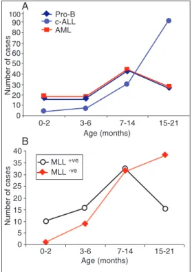
rearrangements demonstrated that up to 14 months of age the pro-B subtype and the
MLL-positive cases predominated (P < 0.0001), and among older cases, most of them were c-ALL and MLL-negative cases, as shown in Figure 1. Regarding the MLL
gene status, it is important to note that even in cases older than 14 months, some were
MLL positive (Figure 1B).
Regarding outcome, unfortunately, the prognosis in this age group still remains poor. At present, the overall 5-year survival for infant ALL patients remains
approxi-Number of cases
40 35 30 25 20 15 10 5 0
0-2 3-6 7-14 15-21
0-2 3-6 7-14 15-21
Age (months)
Age (months) Pro-B
c-ALL AML
MLL +ve MLL -ve A
B Figure 1. Distribution of infant
acute leukemia cases according to immuno-molecular character-ization and age. A, Immunophe-notyping profile. B, MLL status. Pro-B = CD19+/CD10- B precur-sor acute lymphoblastic leuke-mia; c-ALL = CD19+/CD10+ common acute lymphoblastic leukemia; AML = acute myeloid leukemia.
Number of cases
mately 40-50% (12,14). The above features and the short latency periods define IAL as a biologically and clinically distinct disease from childhood acute leukemia in general. Therefore, it seems probable that different genetic and environmental factors may be involved in the mechanisms of pathogenesis of IAL, childhood ALL and AML (1,13), and this knowledge is of great importance when dealing with this aggressive type of leukemia.
Mechanisms of pathogenesis
MLL gene rearrangements and topoisomerase
II inhibitors
The mixed-lineage leukemia gene (MLL /
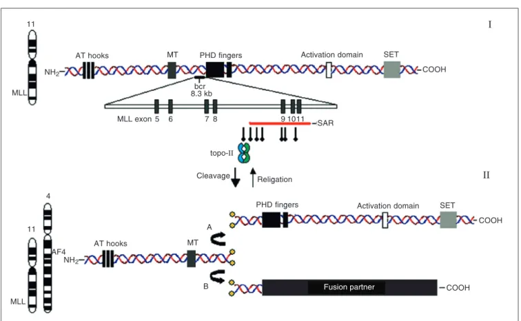
HRX/ALL1), located at cytogenetic band 11q23, is often altered in IAL, being rear-ranged in more than 80% of cases (12,13). This gene consists of at least 36 exons, en-coding a 3969-amino acid nuclear protein with a molecular weight of nearly 430 kDa that functions as a positive regulator of gene expression in early embryonic development and hematopoiesis. MLL translocation break-points cluster within an 8.3-kb region span-ning exons 5-11 (Figure 2I). In its germ-line form, MLL protein, a human homologue of the transcriptional regulator Trithorax of Dro-sophila, is an upstream transcriptional effec-tor of HOX genes, which play a key role in the regulation of hematopoietic development (15). To distinguish whether the MLL gene acts as an oncogene or whether the fusion
Figure 2.I, Structure of the MLL gene, with protein motifs indicated in the diagram. Three AT hooks are located near the N-terminal region. MT = DNA methyltransferase homology region; PHD = plant homeodomain zinc fingers; SET = su(var)3-9, enhancer of zeste, trithorax domain. Solid black bar just behind the PHD zinc fingers indicates the location of the breakpoint cluster region (bcr), and its exon/intron structure is highlighted. Solid red bar below exons 9, 10, and 11 indicates the high-affinity scaffold attachment region (SAR). Vertical arrows indicate the topo-II consensus sites. II, Consequences of topo-II cleavage. A, MLL double strand is correctly re-ligated, generating wild-type MLL. B, MLL is fused with frequent partner AF4, generating a fusion oncogene. The NCBI website (http://www.ncbi.nlm.nih.gov/) was extensively consulted for elaboration of this figure.
11
11 4
AT hooks AF4
NH2 NH2
AT hooks MT
MLL
MLL
MLL exon 5 6 7 8
PHD fingers Activation domain SET
COOH
PHD fingers Activation domain SET
COOH SAR
Religation topo-II
Cleavage
9 1011
COOH MT
A
B
I
II
Fusion partner bcr
proteins resulting from the translocations have dominant negative effects, in vitro ex-periments using hematopoietic progenitors from embryos of homozygous MLL knock-out mice were performed. The ability of wild-type AB2.1 embryonic stem (ES) cells and of single- or double-ALL-1 gene knock-out cells derived from them to differentiate along hematopoietic lineages was tested us-ing in vitro colony formation assays. The results showed that ALL-1 double-knockout ES cells formed a significantly greater num-ber of colonies with faster kinetics than wild-type and ALL-1 single-knockout ES cells. Furthermore, parental ES cells formed lin-eage-restricted colonies, whereas single- and double-knockout ES cells developed, at high frequency, immature and/or biphenotypic colonies, mimicking the aberrant hemato-poiesis typical of leukemia patients (16).
To date, over 87 chromosome partners of
MLL have been described with diverse func-tions and variable function domains, and 51 of the presumptive gene partners of MLL
have been cloned and analyzed at the molec-ular level (11). This marked promiscuity raises the question of whether the diverse partners contribute with common functions or have different effects in leukemogenesis.
MLL partners can be divided into two groups. First, the nuclear fusion partners, including AF4, AF9, AF10, ENL, ELL, AF17, and others, which are associated with different aspects of transcriptional regulation. The other group is mainly cytoplasmic and fre-quently associated with cytoskeleton-de-pendent signal transduction, including AF6,
Septin 6, ABI-1, EEN, and so on (17). Al-though the fusion partners display many dif-ferent features, all of them delete a large 3' portion of the MLL gene and connect the remaining part with the corresponding part-ner and, regardless of whether the fusion partner itself is nuclear or cytoplasmic, the MLL chimeric proteins consistently form punctuated patterns in the nucleus (18), indi-cating that the localization of the fusion
protein and its potential targets is mainly determined by the DNA-binding activity of the MLL portion and not by the fusion part-ner. Remarkable is the fact that the fre-quency and diversity of MLL-associated abnormalities vary according to de novo acute leukemia and therapy-related leukemias (19). Considering the role of the partner genes, these reports indicate that the transforming capacity of the truncated MLL alone is inef-fective, suggesting that a gain of MLL func-tion via a partner gene is crucial.
Although the mechanisms by which these rearrangements result in leukemia remain largely unknown, clues for understanding the mechanism of main translocations come from genomic details of the MLL gene. Sev-eral DNA motifs implicated in recombina-tion of DNA have been identified and local-ized within the MLL breakpoint cluster re-gion (bcr). These include topoisomerase-II (topo-II)-binding sites (20) and Alu se-quences (21). Also, a high-affinity scaffold attachment region (SAR) was identified within the telomeric (3') region of the bcr (Figure 2II). SARs are sites for binding of DNA to nuclear matrix proteins, functioning to maintain the structure of chromosomal loops and to allow regulation of transcrip-tion, DNA replicatranscrip-tion, and recombination (20).
Suggestively, most of the topo-II-con-sensus sites overlap the SAR, clustering in the telomeric part of the MLL bcr. The obser-vation of these recombination-prone se-quences in the MLL bcr region indicates that the rearrangement might result from DNA breakage and recombination events (20). Several in vitro studies support the link be-tween MLL cleavage and topo-II inhibitors in human hematopoietic cells (22,23). Nev-ertheless, these studies still do not prove the occurrence of recombination. The same bi-ased distribution of gene breakpoints within
the mechanisms of cleavage of the MLL
gene in IALs are similar to those of topo-II inhibitor-induced secondary leukemias (20). It was then suggested that the critical leuke-mogenic event(s) occurring in uterus might similarly involve prenatal exposure to topo-II inhibitors as represented by several natu-ral and medicinal substances, further dis-cussed in molecular and exploratory epide-miology.
Other molecular markers
It is clear that chromosome transloca-tions, especially those involving the MLL
gene, are the genetic alterations most fre-quently found in early IAL. However, not only MLL rearrangements but also other chro-mosome translocations may be found within this group, especially when the age limit is 18 months of age.
For instance, another hallmark in pediat-ric acute leukemias is the chromosomal trans-location t(12;21)(p13;q22), which fuses the
TEL and AML1 genes, resulting in the most common chromosomal alteration in child-hood cancer. The TEL /AML1 fusion gene occurs in approximately 25% of B-cell pre-cursor ALL in children 2-12 years old. There is evidence that TEL /AML1 usually arises prenatally as an early or initiating mutation. However, the low-twin concordance rate (~10%), the protracted and variable latency of the disease, and the transgenic modeling indicate that secondary postnatal genetic al-terations are also required (25,26). While
MLL-positive cases present a poor progno-sis and are often associated with CD10
-immunophenotypes, ALL cases expressing the TEL/AML1 fusion protein are associ-ated with CD10+ immunophenotypes and
excellent prognosis (27).
A molecular study conducted on a Bra-zilian cohort (age range 0 to 23 months) has detected TEL /AML1+ve (N = 9), E2A/PBX1+ve
(N = 4), PML /RARA+ve (N = 4), and AML1/ ETO+ve (N = 2) cases (10). It is unlikely,
however, that a single-chromosome translo-cation itself would be enough to cause overt leukemia. According to Greaves’ hypothesis (28), genetic alterations that impair differen-tiation probably cooperate with a second class of mutations that alter the proliferation and survival of a malignant clone. The short latency and the high-concordance rate be-tween twins with infant leukemia could sug-gest that the MLL-fusion gene should be sufficient to cause leukemia. Perhaps more likely, however, and accepting the same TEL / AML1 minimal two-step model for child-hood leukemia, the disruptive effect of the
MLL-gene fusion on, for example, DNA repair or cell-cycle regulation could facili-tate the rapid acquisition of additional, nec-essary genetic changes, particularly with continued exposure to a genotoxic chemical
in utero (29,30).
The gathering of multiple, independent and complementary genetic lesions as a re-quirement for abnormal development of a hematopoietic progenitor supports studies that aim to investigate other genes in the pathogenesis of early acute leukemia, bear-ing in mind the cell type and the correspond-ing chromosomal lesions. Accordcorrespond-ingly, sec-ondary chromosomal or genetic alterations are detected at diagnosis in a significant number of MLL-positive cases. In children
≤3 years old, among whom it is believed that the first genetic alteration required to fully develop childhood ALL occurs in utero, the leukemic cells prevailingly exhibit fetal-type
DJH junctions of the complementarily
deter-mining region 3 of the immunoglobulin H chain that lack the so-called N regions, which are added during DJH recombination events
considering the MLL translocation to be the first genetic hit undoubtedly arising in utero, but yet displaying N region-positive DJH
junctions, it is very likely that an exquisitely short latency period for only limited mu-tagenic events is further required (14,32). Furthermore, it was recently found that the receptor tyrosine kinase FLT3 is highly ex-pressed in MLL-rearranged ALL as com-pared with other leukemias (6). Moreover, it was found that approximately 20% of MLL -rearranged ALL cases possess activating mutations in the activation loop region (33). These data provide evidence that leuke-mogenic fusion proteins such as MLL fu-sions cooperate with activated kinases to promote leukemogenesis. Thus, the conun-drum of whether one or more secondary mutations are necessary for leukemia devel-opment in MLL-positive acute leukemias is a challenge that still needs to be fully eluci-dated. The combination of molecular and exploratory epidemiology methods is a good strategic model to test this hypothesis.
Molecular and exploratory epidemiology
What makes these leukemias so unique?
There is no doubt that the risks of devel-oping early acute leukemia are modulated by complex interactions between inherited predispositions, environmental exposure to damaging agents and chance events (28,34). Despite the fact that such leukemias are very rare, their investigation is absolutely neces-sary for the study of leukemogenesis be-cause they have a short latency period to-gether with the known immune molecular markers. The molecular epidemiological approach to genetic studies has suggested the concept that most, if not all, childhood acute leukemia cases originate in utero. The evidence for this is based on the following considerations: first, studies of identical twins with MLL-rearranged leukemias, presenting
the same fusion gene sequences, indicated that MLL rearrangements are acquired in utero. Such cases may be plausibly explained by the metastasis of a clonal event originat-ing in one twin to the other twin via placental anastomoses (30); second, the fusion gene sequences have been retrospectively identi-fied in archived neonatal heel blood spot cards of children in whom leukemia had subsequently developed (35). Finally, the same fetal origin has been established in older children through monozygotic twin studies or detection of other specific fusion gene sequences (e.g., TEL /AML1 and AML1/ ETO) in neonatal blood spots (25). These findings have led to the interpretation that many childhood leukemias originate prena-tally during fetal hematopoiesis. The mechan-isms of leukemogenesis in early infancy are related to the fact that the growing fetus and/ or child is more sensitive to the effects of potential DNA damage insults during the early stage of pregnancy and/or first semes-ter of life. Because reciprocal rearrangement of the MLL gene is the most common genetic feature, it is important to understand how these fusions could possibly result from the transplacental exposure to DNA-damaging substances. It has been well demonstrated that substances that target topo-II, inhibiting the re-sealing of a previously cleaved double-strand end by the same enzyme, trigger the most common mechanism in the formation of MLL rearrangement (model for therapy-related secondary AML (s-AML)) (22,36).
treat-ment-related leukemias has also been shown in IALs (24,38). Topo-II inhibitors include chemotherapeutic agents, benzene metabo-lites (such as benzoquinone), isoflavones, flavonoids, lignans, podophyllin resin, quino-lone antibiotics, and some pesticides. Spe-cific fruits and vegetables that contain quer-cetin, soybeans (genistein), tea, cocoa, wine (catechins), and caffeine have all been re-lated to an increased risk of infant AML (36,38-40). Thus, to better understand the environmental exposures to damaging agents that give rise to these genetic changes in utero, infants represent a more accurate group than older children, since the window of
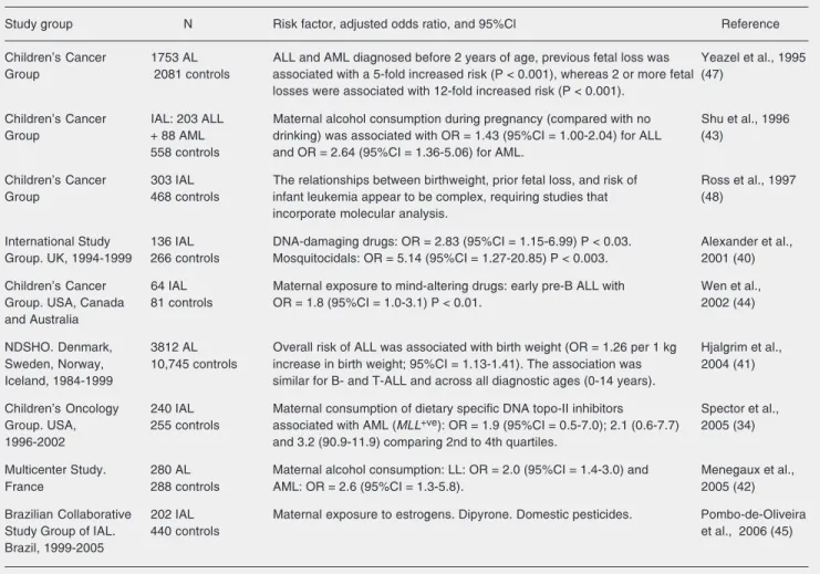
exposure is limited and known (shortly be-fore or during pregnancy). Although the epi-demiological data are still limited to support correlations, several studies have shown maternal exposures that may give rise to these leukemogenic genetic changes (34,40-44). In a literature review consulting the MEDLINE databank, we retrieved published papers focusing on IAL (1990-2006), sum-marized them in Table 1, and briefly dis-cussed their valuable results. These studies were searched through an intensive combi-nation of both MeSH terms and part of the text words and titles. The following terms were used: infant acute leukemia, childhood
Table 1. Data concerning leukemias reported in epidemiological literature.
Study group N Risk factor, adjusted odds ratio, and 95%Cl Reference
Children’s Cancer 1753 AL ALL and AML diagnosed before 2 years of age, previous fetal loss was Yeazel et al., 1995 Group 2081 controls associated with a 5-fold increased risk (P < 0.001), whereas 2 or more fetal (47)
losses were associated with 12-fold increased risk (P < 0.001).
Children’s Cancer IAL: 203 ALL Maternal alcohol consumption during pregnancy (compared with no Shu et al., 1996 Group + 88 AML drinking) was associated with OR = 1.43 (95%CI = 1.00-2.04) for ALL (43)
558 controls and OR = 2.64 (95%CI = 1.36-5.06) for AML.
Children’s Cancer 303 IAL The relationships between birthweight, prior fetal loss, and risk of Ross et al., 1997 Group 468 controls infant leukemia appear to be complex, requiring studies that (48)
incorporate molecular analysis.
International Study 136 IAL DNA-damaging drugs: OR = 2.83 (95%CI = 1.15-6.99) P < 0.03. Alexander et al., Group. UK, 1994-1999 266 controls Mosquitocidals: OR = 5.14 (95%CI = 1.27-20.85) P < 0.003. 2001 (40)
Children’s Cancer 64 IAL Maternal exposure to mind-altering drugs: early pre-B ALL with Wen et al.,
Group. USA, Canada 81 controls OR = 1.8 (95%CI = 1.0-3.1) P < 0.01. 2002 (44)
and Australia
NDSHO. Denmark, 3812 AL Overall risk of ALL was associated with birth weight (OR = 1.26 per 1 kg Hjalgrim et al., Sweden, Norway, 10,745 controls increase in birth weight; 95%CI = 1.13-1.41). The association was 2004 (41) Iceland, 1984-1999 similar for B- and T-ALL and across all diagnostic ages (0-14 years).
Children’s Oncology 240 IAL Maternal consumption of dietary specific DNA topo-II inhibitors Spector et al., Group. USA, 255 controls associated with AML (MLL+ve): OR = 1.9 (95%CI = 0.5-7.0); 2.1 (0.6-7.7) 2005 (34)
1996-2002 and 3.2 (90.9-11.9) comparing 2nd to 4th quartiles.
Multicenter Study. 280 AL Maternal alcohol consumption: LL: OR = 2.0 (95%CI = 1.4-3.0) and Menegaux et al.,
France 288 controls AML: OR = 2.6 (95%CI = 1.3-5.8). 2005 (42)
Brazilian Collaborative 202 IAL Maternal exposure to estrogens. Dipyrone. Domestic pesticides. Pombo-de-Oliveira
Study Group of IAL. 440 controls et al., 2006 (45)
Brazil, 1999-2005
“leuk(a)emia”, risk factor, and leukemia with
MLL gene rearrangements.
Ross et al. (43) suggested that maternal alcohol consumption during pregnancy re-sults in an increased risk for infant ALL (OR = 1.43; 95%CI = 1.00-2.04) and for infant AML (OR = 2.64; 95%CI = 1.36-5.06). The mechanism is explained by the ethanol in-duction of microsomal enzymes, such as cytochrome P450, which subsequently acti-vate pre-carcinogens (43). The same study showed that paternal smoking one month prior to pregnancy was associated with an increased risk (OR = 1.56; 95%CI = 1.03-2.36) of infant ALL. High-birth weights were also shown to be correlated with higher rates of infant ALL and AML. Concordant results were observed within the Brazilian Collabo-rative Study Group of Infant Acute Leuke-mia (BCSGIAL), using birth weight infor-mation obtained from the questionnaires of 202 IAL cases and 440 controls (45). Since insulin-like growth factor-1 is important in blood formation and regulation and has been shown to stimulate the growth of both my-eloid and lymphoid cells in culture, it was postulated that high levels of insulin-like growth factor-1 might produce large babies and contribute to the development of leuke-mia (46). A maternal history of fetal loss has also been associated with a five- to 12-fold increased risk of developing ALL or AML (47). However, Ross et al. (48) later showed that the relationships between birth weight, prior fetal loss, and risk of infant leukemia appear to be complex, and that only high-birth weight was in fact a significant risk factor of developing childhood leukemia.
An international collaborative study in-volving patients in Japan, Italy, China and Hong Kong, Greece, Egypt, Chile, and Bra-zil found the selective link between preg-nancy exposures to pesticides, in particular propoxur (a mosquitocidal compound, OR = 9.68; P = 0.003), or consumption during pregnancy of substances such as dipyrone (OR = 5.84; P = 0.001), and IALs with MLL
gene rearrangements (40). However, in this international study about transplacental chemical exposure and risk of IAL with
MLL gene fusion, there were a small number of cases molecularly classified and possible confounding factors related to selection of controls in the Brazilian settings. Hence, further analysis was required to confirm these apparent associations. More recently, the BCSGIAL conducted a molecular study com-bining epidemiological data obtained from interviews with mothers in order to re-evalu-ate the hypothesis that exposure to pharma-ceutical drugs is associated with IAL. The results of that study confirmed reports that mothers exposed to dipyrone and pesticides had an increased chance to give birth to babies with IAL (OR = 1.48; 95%CI = 1.05-2.07 and OR = 2.27; 95%CI = 1.56-3.31, respectively) (45). Furthermore, significant associations with risks for IAL were found, a strong positive association with maternal hormone intake during pregnancy (OR = 9.08; 95%CI = 2.95-27.96). The strong and significant association between IAL and es-trogen exposure observed in the BCSGIAL study deserves further investigation. Since a potential role of exogenous estrogens in other cancer was demonstrated in experimental models, and since the metabolite products in estrogen biosynthesis are semiquinones and quinines (49), we speculate that this high association found in the Brazilian series of early infancy acute leukemia could be ex-plained by the same pathway as that of topo-II inhibition caused by metabolite products in estrogen metabolism. On a hopeful note, a recently reported study demonstrated that maternal consumption of fresh vegetables and fruits during pregnancy was associated with a decreased risk of IAL (34).
Gene-environment interaction
Epidemiological and genetic studies sup-port the contention that the in utero origin of
transplacental chemical exposures (quinone-based chemicals) (38,40). The probability of the secondary event arising within a short time would be enhanced if the genetic back-ground conferred greater susceptibility of the individual to chemical damage. In this context, constitutive genetic vulnerability may not only act as a predisposing factor for the induction of MLL gene fusion but may also increase the risk of the occurrence of further mutations. For most leukemias, and also IALs, multiple genetic polymorphisms of xenobiotic metabolizing enzymes may interact with environmental, dietary, mater-nal, and other factors to modulate the devel-opment of acute leukemia. For example, quinones, which have been shown to cleave both the MLL gene and its frequent fusion partner AF4 at topo-II cleavage sites (50), may be poorly detoxified depending on the activity of NAD(P)H:quinone oxidoreduc-tase 1 (NQO1), an enzyme that detoxifies chemicals with quinone rings including ben-zene metabolites and flavonoids. The NQO1
gene is subject to polymorphisms that gener-ate an NQO1 protein with a significantly decreased enzymatic activity. Studies of Caucasian and Japanese patients have shown that the occurrence of alleles conferring low-activity variants of NQO1 was associated with an increased risk of IAL, especially with MLL /AF4 fusion genes (51,52). Sirma et al. (53) demonstrated that in pediatric ALL without MLL rearrangements, the null genotype of the NQO1 gene is not associated with the etiology of the disease. Recently, in a series of Italian IAL cases, contradictory results were obtained, with this polymor-phism appearing to be associated with infant ALL without MLL rearrangements, but not with MLL-positive infants (54). In the Bra-zilian series, preliminary results also did not show a significant increased risk (OR = 1.10; 95%CI = 0.62-1.95) of developing leukemia with rearrangements of the MLL gene in those individuals with low-activity variants of the NQO1 enzyme (Amorim MR, Silva
FA, Emerenciano M, Pombo-de-Oliveira MS, unpublished data).
Polymorphisms of folate-metabolizing enzymes have also been associated with the development of ALL. First, methylenetetra-hydrofolate reductase (MTHFR) 677C>T gene polymorphism has been linked to a decreased risk of pediatric ALL (55,56). This protective effect may be due to the greater availability of 5,10-methylenetetra-hydrofolate and thymidine pools and to an increased fidelity of DNA synthesis. We recently demonstrated interesting effects of MTHFR 677C>T and 1298A>C polymor-phisms in a Brazilian childhood series of cases including 62 infants. The results dem-onstrated a protective role of MTHFR 677C>T polymorphism, linked to a signifi-cant 2.18-fold decreased risk of developing ALL, whereas the 1298A>C polymorphism demonstrated a significant 2.01-fold in-creased risk for ALL in non-white children. It is possible that the opposite roles of 677C>T and 1298A>C polymorphisms in non-white children found in our study result from the different binding sites of MTHFR affected by the polymorphisms (57).
Another example is the inactivating poly-morphisms of detoxifying enzymes involved in carcinogen metabolism, such as glutathi-one S-transferases (GST) that have been associated with the development of ALL (58). A similar study showed the possible role of GST gene polymorphisms in suscep-tibility to IAL, but the authors analyzed the genotypes of the diseased infants’ parents. Surprisingly, they found that the deletion of both the GSST1 and GSTM1 genes in either parent might affect the risk of infant leuke-mia through a pathway independent of the
(OR = 2.7; 95%CI = 1.1-6.8; P = 0.04) (60). All of these findings need to be extended by larger studies with careful attention to ethnic and geographic diversity in the fre-quency of polymorphisms, and to socioeco-nomic status that directly influence the ex-posures and supplementation features.
Conclusions
Early childhood ALLs and AMLs con-sistently present MLL gene rearrangements, which, besides conferring an adverse out-come, have represented an important molec-ular diagnosis and monitoring marker, and have been of great importance to identify the unique molecular biological history of this disease. As reviewed here, many molecular features and environmental risk factors have been suggested to cooperate in the develop-ment of this malignancy. However, a
combi-nation of chances and interactions in the molecular-cellular differentiation pathways is necessary to producethe malignant phe-notype. Therefore, continued molecular epi-demiological studies are needed to better elucidate the biology of IAL, ultimately lead-ing to an improvement in the present out-come situation.
Acknowledgments
Many of the results reviewed here were made available by the physicians of the Bra-zilian Collaborative Study Group of Infant Acute Leukemia (BCSGIAL, see Appen-dix). The success of the BCSGIAL has de-pended on the help of many mothers who have given their time to contribute to the investigation. Therefore, we are very grate-ful to all involved.
References
1. Biondi A, Cimino G, Pieters R, Pui CH. Biological and therapeutic aspects of infant leukemia. Blood 2000; 96: 24-33.
2. Chessells JM, Eden OB, Bailey CC, Lilleyman JS, Richards SM. Acute lymphoblastic leukaemia in infancy: experience in MRC UKALL trials. Report from the Medical Research Council Working Party on Childhood Leukaemia. Leukemia 1994; 8: 1275-1279. 3. Ross JA, Davies SM, Potter JD, Robison LL. Epidemiology of
child-hood leukemia, with a focus on infants. Epidemiol Rev 1994; 16: 243-272.
4. Isaacs H Jr. Fetal and neonatal leukemia. J Pediatr Hematol Oncol 2003; 25: 348-361.
5. Magalhaes IQ, Splendore A, Emerenciano M, Figueiredo A, Ferrari I, Pombo-de-Oliveira MS. GATA1 mutations in acute leukemia in children with Down syndrome. Cancer Genet Cytogenet 2006; 166: 112-116.
6. Armstrong SA, Staunton JE, Silverman LB, Pieters R, den Boer ML, Minden MD, et al. MLL translocations specify a distinct gene expres-sion profile that distinguishes a unique leukemia. Nat Genet 2002; 30: 41-47.
7. Hu M, Krause D, Greaves M, Sharkis S, Dexter M, Heyworth C, et al. Multilineage gene expression precedes commitment in the hemopoi-etic system. Genes Dev 1997; 11: 774-785.
8. Hotfilder M, Rottgers S, Rosemann A, Schrauder A, Schrappe M, Pieters R, et al. Leukemic stem cells in childhood high-risk ALL/ t(9;22) and t(4;11) are present in primitive lymphoid-restricted CD34+. Cancer Res 2005; 65: 1442-1449.
9. Mendes WL, Coser VM, Ramos G, Pereira W, Lopes LF, de Oliveira
MS. The apparent excess of acute promyelocytic leukemia in infant acute leukemias in Brazil. Haematologica 2004; 89: ELT16. 10. Emerenciano M, Agudelo Arias DP, Coser VM, de Brito GD, Macedo
Silva ML, Pombo-de-Oliveira MS. Molecular cytogenetic findings of acute leukemia included in the Brazilian Collaborative Study Group of Infant acute leukemia. Pediatr Blood Cancer 2006; 47: 549-554. 11. Meyer C, Schneider B, Jakob S, Strehl S, Attarbaschi A, Schnittger
S, et al. The MLL recombinome of acute leukemias. Leukemia 2006; 20: 777-784.
12. Pui CH, Kane JR, Crist WM. Biology and treatment of infant leuke-mias. Leukemia 1995; 9: 762-769.
13. Greaves MF. Infant leukaemia biology, aetiology and treatment. Leukemia 1996; 10: 372-377.
14. Stam RW, den Boer ML, Pieters R. Towards targeted therapy for infant acute lymphoblastic leukaemia. Br J Haematol 2006; 132: 539-551.
15. Yu BD, Hanson RD, Hess JL, Horning SE, Korsmeyer SJ. MLL, a mammalian trithorax-group gene, functions as a transcriptional main-tenance factor in morphogenesis. Proc Natl Acad Sci U S A 1998; 95: 10632-10636.
16. Fidanza V, Melotti P, Yano T, Nakamura T, Bradley A, Canaani E, et al. Double knockout of the ALL-1 gene blocks hematopoietic differ-entiation in vitro. Cancer Res 1996; 56: 1179-1183.
17. Li ZY, Liu DP, Liang CC. New insight into the molecular mechanisms of MLL-associated leukemia. Leukemia 2005; 19: 183-190. 18. Yano T, Nakamura T, Blechman J, Sorio C, Dang CV, Geiger B, et
elements at the N terminus of the protein. Proc Natl Acad Sci U S A 1997; 94: 7286-7291.
19. Daser A, Rabbitts TH. Extending the repertoire of the mixed-lineage leukemia gene MLL in leukemogenesis. Genes Dev 2004; 18: 965-974.
20. Broeker PL, Super HG, Thirman MJ, Pomykala H, Yonebayashi Y, Tanabe S, et al. Distribution of 11q23 breakpoints within the MLL breakpoint cluster region in de novo acute leukemia and in trement-related acute myeloid leukemia: correlation with scaffold at-tachment regions and topoisomerase II consensus binding sites. Blood 1996; 87: 1912-1922.
21. Gu Y, Alder H, Nakamura T, Schichman SA, Prasad R, Canaani O, et al. Sequence analysis of the breakpoint cluster region in the ALL-1 gene involved in acute leukemia. Cancer Res 1994; 54: 2326-2330.
22. Ishii E, Eguchi M, Eguchi-Ishimae M, Yoshida N, Oda M, Zaitsu M, et al. In vitro cleavage of the MLL gene by topoisomerase II inhibitor (etoposide) in normal cord and peripheral blood mononuclear cells. Int J Hematol 2002; 76: 74-79.
23. Strick R, Strissel PL, Borgers S, Smith SL, Rowley JD. Dietary bioflavonoids induce cleavage in the MLL gene and may contribute to infant leukemia. Proc Natl Acad Sci U S A 2000; 97: 4790-4795. 24. Cimino G, Rapanotti MC, Biondi A, Elia L, Lo Coco F, Price C, et al.
Infant acute leukemias show the same biased distribution of ALL1 gene breaks as topoisomerase II related secondary acute leuke-mias. Cancer Res 1997; 57: 2879-2883.
25. Wiemels JL, Cazzaniga G, Daniotti M, Eden OB, Addison GM, Masera G, et al. Prenatal origin of acute lymphoblastic leukaemia in children. Lancet 1999; 354: 1499-1503.
26. Wiemels JL, Ford AM, Van Wering ER, Postma A, Greaves M. Protracted and variable latency of acute lymphoblastic leukemia after TEL-AML1 gene fusion in utero. Blood 1999; 94: 1057-1062. 27. Pui CH, Relling MV, Downing JR. Acute lymphoblastic leukemia. N
Engl J Med 2004; 350: 1535-1548.
28. Greaves MF. Molecular genetics, natural history and the demise of childhood leukemia. Eur J Cancer 1999; 35: 1941-1953.
29. Eguchi M, Eguchi-Ishimae M, Greaves M. Molecular pathogenesis of MLL-associated leukemias. Int J Hematol 2005; 82: 9-20. 30. Greaves MF, Maia AT, Wiemels JL, Ford AM. Leukemia in twins:
lessons in natural history. Blood 2003; 102: 2321-2333.
31. Wasserman R, Galili N, Ito Y, Reichard BA, Shane S, Rovera G. Predominance of fetal type DJH joining in young children with B precursor lymphoblastic leukemia as evidence for an in utero trans-forming event. J Exp Med 1992; 176: 1577-1581.
32. Fasching K, Panzer S, Haas OA, Borkhardt A, Marschalek R, Griesinger F, et al. Presence of N regions in the clonotypic DJ rearrangements of the immunoglobulin heavy-chain genes indicates an exquisitely short latency in t(4;11)-positive infant acute lympho-blastic leukemia. Blood 2001; 98: 2272-2274.
33. Gilliland DG, Griffin JD. The roles of FLT3 in hematopoiesis and leukemia. Blood 2002; 100: 1532-1542.
34. Spector LG, Xie Y, Robison LL, Heerema NA, Hilden JM, Lange B, et al. Maternal diet and infant leukemia: the DNA topoisomerase II inhibitor hypothesis: a report from the children’s oncology group. Cancer Epidemiol Biomarkers Prev 2005; 14: 651-655.
35. Gale KB, Ford AM, Repp R, Borkhardt A, Keller C, Eden OB, et al. Backtracking leukemia to birth: identification of clonotypic gene fusion sequences in neonatal blood spots. Proc Natl Acad Sci U S A 1997; 94: 13950-13954.
36. Hande KR. Etoposide: four decades of development of a topoisom-erase II inhibitor. Eur J Cancer 1998; 34: 1514-1521.
37. Andersen MK, Christiansen DH, Jensen BA, Ernst P, Hauge G, Pedersen-Bjergaard J. Therapy-related acute lymphoblastic leu-kaemia with MLL rearrangements following DNA topoisomerase II inhibitors, an increasing problem: report on two new cases and review of the literature since 1992. Br J Haematol 2001; 114: 539-543.
38. Ross JA. Maternal diet and infant leukemia: a role for DNA topo-isomerase II inhibitors? Int J Cancer Suppl 1998; 11: 26-28. 39. Frantz CE, Chen H, Eastmond DA. Inhibition of human
topoisomer-ase II in vitro by bioactive benzene metabolites. Environ Health Perspect 1996; 104 (Suppl 6): 1319-1323.
40. Alexander FE, Patheal SL, Biondi A, Brandalise S, Cabrera ME, Chan LC, et al. Transplacental chemical exposure and risk of infant leukemia with MLL gene fusion. Cancer Res 2001; 61: 2542-2546. 41. Hjalgrim LL, Rostgaard K, Hjalgrim H, Westergaard T, Thomassen
H, Forestier E, et al. Birth weight and risk for childhood leukemia in Denmark, Sweden, Norway, and Iceland. J Natl Cancer Inst 2004; 96: 1549-1556.
42. Menegaux F, Steffen C, Bellec S, Baruchel A, Lescoeur B, Leverger G, et al. Maternal coffee and alcohol consumption during preg-nancy, parental smoking and risk of childhood acute leukaemia. Cancer Detect Prev 2005; 29: 487-493.
43. Shu XO, Ross JA, Pendergrass TW, Reaman GH, Lampkin B, Robison LL. Parental alcohol consumption, cigarette smoking, and risk of infant leukemia: a Children’s Cancer Group Study. J Natl Cancer Inst 1996; 88: 24-31.
44. Wen W, Shu XO, Potter JD, Severson RK, Buckley JD, Reaman GH, et al. Parental medication use and risk of childhood acute lymphoblastic leukemia. Cancer 2002; 95: 1786-1794.
45. Pombo-de-Oliveira MS, Koifman S, Brazilian Collaborative Study Group of Infant Acute Leukemia. Infant acute leukemia and maternal exposures during pregnancy. Cancer Epidemiol Biomarkers Prev 2006; 15: 2336-2341.
46. Ross JA, Perentesis JP, Robison LL, Davies SM. Big babies and infant leukemia: a role for insulin-like growth factor-1? Cancer Causes Control 1996; 7: 553-559.
47. Yeazel MW, Buckley JD, Woods WG, Ruccione K, Robison LL. History of maternal fetal loss and increased risk of childhood acute leukemia at an early age. A report from the Children’s Cancer Group. Cancer 1995; 75: 1718-1727.
48. Ross JA, Potter JD, Shu XO, Reaman GH, Lampkin B, Robison LL. Evaluating the relationships among maternal reproductive history, birth characteristics, and infant leukemia: a report from the Children’s Cancer Group. Ann Epidemiol 1997; 7: 172-179.
49. Cavalieri EL, Stack DE, Devanesan PD, Todorovic R, Dwivedy I, Higginbotham S, et al. Molecular origin of cancer: catechol estro-gen-3,4-quinones as endogenous tumor initiators. Proc Natl Acad Sci U S A 1997; 94: 10937-10942.
50. Lovett BD, Lo Nigro L, Rappaport EF, Blair IA, Osheroff N, Zheng N, et al. Near-precise interchromosomal recombination and functional DNA topoisomerase II cleavage sites at MLL and AF-4 genomic breakpoints in treatment-related acute lymphoblastic leukemia with t(4;11) translocation. Proc Natl Acad Sci U S A 2001; 98: 9802-9807. 51. Eguchi-Ishimae M, Eguchi M, Ishii E, Knight D, Sadakane Y, Isoya-ma K, et al. The association of a distinctive allele of NAD(P)H: quinone oxidoreductase with pediatric acute lymphoblastic leuke-mias with MLL fusion genes in Japan. Haematologica 2005; 90: 1511-1515.
have MLL fusions. United Kingdom Childhood Cancer Study Investi-gators. Cancer Res 1999; 59: 4095-4099.
53. Sirma S, Agaoglu L, Yildiz I, Cayli D, Horgusluoglu E, Anak S, et al. NAD(P)H:quinone oxidoreductase 1 null genotype is not associated with pediatric de novo acute leukemia. Pediatr Blood Cancer 2004; 43: 568-570.
54. Lanciotti M, Dufour C, Corral L, Di Michele P, Pigullo S, De Rossi G, et al. Genetic polymorphism of NAD(P)H:quinone oxidoreductase is associated with an increased risk of infant acute lymphoblastic leukemia without MLL gene rearrangements. Leukemia 2005; 19: 214-216.
55. Franco RF, Simoes BP, Tone LG, Gabellini SM, Zago MA, Falcao RP. The methylenetetrahydrofolate reductase C677T gene poly-morphism decreases the risk of childhood acute lymphocytic leu-kaemia. Br J Haematol 2001; 115: 616-618.
56. Wiemels JL, Smith RN, Taylor GM, Eden OB, Alexander FE, Greaves MF. Methylenetetrahydrofolate reductase (MTHFR)
poly-morphisms and risk of molecularly defined subtypes of childhood acute leukemia. Proc Natl Acad Sci U S A 2001; 98: 4004-4009. 57. Zanrosso CW, Hatagima A, Emerenciano M, Ramos F, Figueiredo
A, Felix TM, et al. The role of methylenetetrahydrofolate reductase in acute lymphoblastic leukemia in a Brazilian mixed population. Leuk Res 2006; 30: 477-481.
58. Krajinovic M, Labuda D, Richer C, Karimi S, Sinnett D. Susceptibility to childhood acute lymphoblastic leukemia: influence of CYP1A1, CYP2D6, GSTM1, and GSTT1 genetic polymorphisms. Blood 1999; 93: 1496-1501.
59. Garte S, Taioli E, Crosti F, Sainati L, Barisone E, Luciani M, et al. Deletion of parental GST genes as a possible susceptibility factor in the etiology of infant leukemia. Leuk Res 2000; 24: 971-974. 60. Canalle R, Burim RV, Tone LG, Takahashi CS. Genetic
polymor-phisms and susceptibility to childhood acute lymphoblastic leuke-mia. Environ Mol Mutagen 2004; 43: 100-109.
Appendix
Membersof the Brazilian Collaborative Study Group of Infant Acute Leukemia (BCSGIAL)
Paulo Ivo C. Araújo3, Dora Márcia Alencar4, Silvia R. Brandalise5, Eni Guimarães Carvalho6, Virginia M. Coser7, Imaruí Costa7, José Carlos Córdoba8, Mariana Emerenciano1, Jane Dobbin1, Maria Célia Moraes Guerra3, Venâncio Gumes Lopes4, Isis Q. Magalhães8, Núbia Mendonça4, Andrea Gadelha9, Gilson Guedes9, Sérgio Koifman2, Flávia Pimenta9, Vitória P. Pinheiro5, Waldir Pereira7, Maria S. Pombo-de-Oliveira1, Gilberto Ramos10, Terezinha J.M. Salles11, Denise Bousfield da Silva12, Marcelo P. Land3, Elaine Sobral3, Fernando Werneck13, Carlos Scridelli14, Luis Gonzaga Tone14, Lincoln Vermondi12, Luis Fernando Lopes15, Wellington Mendes15.
1Centro de Pesquisa, Instituto Nacional de Câncer, Rio de Janeiro, RJ, Brazil 2Escola Nacional de Saúde Pública, FIOCRUZ, Rio de Janeiro, RJ, Brazil
3Instituto de Pediatria e Puericultura Martagão Gesteira, UFRJ, Rio de Janeiro, RJ, Brazil 4Sociedade de Oncologia da Bahia, Salvador, BA, Brazil
5Centro Infantil de Investigações Hematológicas Dr. Boldrini, Campinas, SP, Brazil 6Hospital Martagão Gesteira, Salvador, BA, Brazil
7Departamento de Hematologia, Universidade de Santa Maria, Santa Maria, RS, Brazil 8Hospital de Apoio Brasília, Unidade de Onco-Hematologia Pediátrica, Brasília, DF, Brazil 9Hospital Napoleão Laureano, João Pessoa, PB, Brazil
10Departamento de Pediatria, Faculdade de Medicina, UFMG, Belo Horizonte, MG, Brazil 11Hospital Oswaldo Cruz, CEON, Recife, PE, Brazil
12Serviço de Oncologia, Hospital Joana de Gusmão, Florianópolis, SC, Brazil
13Departamento de Pediatria, Hospital dos Servidores do Estado do Rio de Janeiro, Rio de Janeiro, RJ, Brazil