Variation in extragenic repetitive DNA sequences in
Pseudomonas syringae
and potential use of modified REP primers in the identification of closely
related isolates
Elif Çepni
1and Filiz Gürel
1,21
Department of Molecular Biology and Genetics, Faculty of Sciences, Istanbul University, Istanbul, Turkey.
2Center for Biotechnology and Genetic Engineering Research and Application, Istanbul University, Istanbul,
Turkey.
Abstract
In this study, Pseudomonas syringe pathovars isolated from olive, tomato and bean were identified by
spe-cies-specific PCR and their genetic diversity was assessed by repetitive extragenic palindromic (REP)-PCR. Re-verse universal primers for REP-PCR were designed by using the bases of A, T, G or C at the positions of 1, 4 and 11 to identify additional polymorphism in the banding patterns. Binding of the primers to different annealing sites in the genome revealed additional fingerprint patterns in eight isolates ofP. savastanoi pv. savastanoi and two isolates of P. syringae pv. tomato. The use of four different bases in the primer sequences did not affect the PCR reproducibility and was very efficient in revealing intra-pathovar diversity, particularly inP. savastanoi pv. savastanoi. At the pathovar level, the primer BOX1AR yielded shared fragments, in addition to five bands that discriminated among the pathovarsP. syringae pv. phaseolicola, P. savastanoi pv. savastanoi and P. syringae pv. tomato. REP-PCR with a modified primer containing C produced identical bands among the isolates in a pathovar but separated three pathovars more distinctly than four other primers. Although REP- and BOX-PCRs have been successfully used in the molecular identification ofPseudomonas isolates from Turkish flora, a PCR based on inter-enterobacterial repetitive intergenic concensus (ERIC) sequences failed to produce clear banding patterns in this study.
Key words:bacterial identification, biodiversity, PCR.
Received: October 11, 2011; Accepted: April 9, 2012.
Introduction
ThePseudomonas syringaegroup belongs to the fam-ily Pseudomonaceae and causes diseases in nearly all spe-cies of cultivated plants, including horticultural crops and ornamental and fruit trees (Younget al., 1996). Many of these pathovars, which are widely distributed on different plant parts (shoots, leaves, buds, pots, etc.), are highly spe-cific to one or a few related plant species and cause a variety of symptoms such as watersoaking, hypertrophic growth, cankers, chlorosis and necrosis (Murillo and Sesma, 2001). The Pseudomonas syringae group also has the best de-scribed epiphytic growth among phytopathogenic bacteria (Hirano and Upper, 2000). One member of this group,
Pseudomonas savastanoi, has been classified as a species rather than a pathovar based on DNA hybridization and ribotyping studies (Gardanet al., 1999). Pathogenic strains ofP. savastanoipv.savastanoiinfect olive trees and cause olive knot disease.
Pseudomonas syringae has previously been identi-fied based on fatty acid profiling (Stead, 1992), protein analyses (Van Zyl and Steyn, 1990) and plasmid profiles (King, 1989). More recently, DNA fingerprinting methods such as randomly amplified polymorphic DNA (RAPD), amplified fragment length polymorphism (AFLP) (Cirvil-leriet al., 2006; Sistoet al., 2007), restriction fragment length polymorphism (RFLP) (Manceau and Horvais, 1997) and insertion sequence-based methods (Weingart and Völksch, 1997; Oguiza et al., 2004; Quesada et al., 2008) have been used to diagnose and genotypeP. syringae
strains and pathovars of different origins. However, none of these methods is ideal for such identifications since each of them has technical disadvantages. PCR-based methods are still the most preferred approach for bacterial genotyping because their low-cost and high output make them efficient in providing new genomic sequence information. Bacterial extragenic non-coding regions have been widely used to obtain specific fingerprints that are more informative and reproducible than RAPD markers.
Repetitive extragenic palindromic (REP) sequences or elements were first described in Escherichia coli and
www.sbg.org.br
Send correspondence to Filiz Gürel. Department of Molecular Biol-ogy and Genetics, Faculty of Sciences, Istanbul University, Vezneciler 34134, Istanbul, Turkey. E-mail: [email protected].
Salmonella typhimurium operons (Higgins et al., 1982). The palindromic nature of these elements and their ability to form stable stem-loop structures in transcribed RNA suggest that they may have regulatory roles associated with transcriptional termination, mRNA stability and chromo-somal organization in bacteria (Versalovic et al., 1991). Amplifications based on REP elements in conjunction with other related families of repetitive elements, such as entero-bacterial repetitive intergenic concensus (ERIC) and BOX sequences, have been exploited in the molecular identifica-tion of bacteria pathogenic to plants. Three types of PCR (known as Rep-PCR) based on these elements have been used together and specific reproducible fingerprints were obtained more quickly and more cost-effectively than with other methods such as AFLP and RFLP (Louws et al., 1999).
Rep-PCR genotyping has been applied to many spe-cies of plant pathogenic bacteria but has limited ability in discriminating among pathovars and closely related iso-lates (de Bruijn, 1992; Judd et al., 1993; Woods et al., 1993). One reason for this poor discriminatory power is that REP-PCR may produce monomorphic bands in bacterial samples collected from distant geographic locations. An-other reason is the variability in the PCR amplification products obtained for the three types of repetitive elements in bacterial genomes. The universal primers of REP-PCR amplify the intervening DNA between two adjacent repeti-tive elements. We have modified reverse REP-PCR prim-ers produced by substituting A, T, G or C at three different positions to yield four primer combinations. By changing the annealing sites in the genome we obtained highly poly-morphic fingerprint patterns among P. savastanoi pv.
savastanoiisolates collected from olive trees. In the present study, we used a set of these primers with high discrimina-tory power to distinguish isolates and pathovars of P. savastanoi pv. savastanoi, P. syringae pv. phaseolicola
andP. syringaepv.tomato.
Materials and Methods
Bacterial strains
Pseudomonas syringaepathovars of tomato, bean and olive (Pss-14) (Table 1) were kindly provided by Prof. Hatice Ozaktan (Ege University), Dr. Aynur Karahan (Ankara Plant Protection Central Research In-stitute) and Prof. Kemal Benlioglu (Adnan Menderes University), respectively. All of the pathovars originated in Turkey except for NCPPB-52, which originated in the United Kingdom (UK). Pseudomonas savastanoi pv.
savastanoiisolates other than Pss-14 were isolated from olive knots of branches obtained from nurseries in the Marmara and Aegean regions (western and northwestern Turkey).
Isolation and identification ofP. savastanoipv.
savastanoi
P. savastanoi pv. savastanoi was isolated as de-scribed by Saygili (1995). Briefly, knots were washed in tap water and cut into small fragments (2 x 2 mm) with a sterile scalpel. The fragments were placed in sterile tubes containing 3 mL of sterile distilled water and left at room temperature for 30 min. Subsequently, ~20 mL aliquots were streaked on King B (KB) medium (Kinget al., 1954) in petri dishes and incubated at 28 °C for 72 h. The sus-pected colonies of P. savastanoi pv. savastanoi, which were flat and 2-3 mm in diameter with irregular margins and a grayish-white color, were selected and spread again on selective PVF-1 medium (Kado and Heskett, 1970) fol-lowed by incubation at 28 °C for 72 h. This subculturing was repeated 2-3 times to purify the isolates. The isolates were also Gram stained. All of theP. syringaestrains iden-tified (Table 1) were cultured in appropriate medium and stored at -80 °C.
For species-specific PCR and repetitive-PCR, a sin-gle colony of each strain ofP. syringaewas used as a source of template DNA. Primer sequences (Pss1: 5’-TGGGTTGCTACTTGTACCGGA-3’ and Pss2: 5’-CCGTGTACTACGTTCAGCGAG-3’) corresponding to the ptz (Basim and Ersoy, 2001) and iaaL (IAALF: 5’-GGCACCAGCGGCAACATCAA-3’ and IAALR: 5’-CGCCCTCGGAACTGCCATAC-3’) genes (Penyalver
et al., 2000) were used forP. savastanoipv.savastanoi. We also used primers derived from the argK (Psp1: 5’-CCATGAAGATTACAAGCCTG-3’ and Psp2: 5’-GCTAGCTATCAGGGGACGAC-3’) (Mosqueda-Ca-no and Herrera-Esterella, 1997) and cfI (Pst1: 5’-GGCGCTCCCTCGCACTT-3’ and Pst2: 5’-GGTATTGGCGGGGGTGC-3’) (Bereswill et al.,
Table 1-Pseudomonas savastanoiandP. syringaeisolates used in this study.
Pathovar Strain Isolate Host Location
Pseudomonas savastanoi
pv.savastanoi Pss-14 Olive Antalya
pv.savastanoi Pss-7A Olive Orhangazi pv.savastanoi Pss-8A Olive Orhangazi pv.savastanoi Pss-9A Olive Orhangazi pv.savastanoi Pss-7D Olive Orhangazi pv.savastanoi Pss-4B Olive Orhangazi
pv.savastanoi Pss-M9 Olive Akhisar
pv.savastanoi Pss-M25 Olive Akhisar
Pseudomonas syringae
pv.tomato Pst Tomato Izmir
pv.tomato Pst-101 Tomato Ankara
pv.phaseolicola Psp-3 Bean Ankara
pv.phaseolicola Psp-18 Bean Ankara
pv.phaseolicola Psp-R52 Bean Izmir
1994) genes for P. syringae pv. phaseolicola and P. syringaepv.tomato, respectively.
The reaction mixture consisted of 1x Ex-Taq DNA polymerase buffer (Takara), 2.5 mM MgCl2, 200mM of
each dNTP, 0.5 units of Ex-TaqDNA polymerase (Takara) and 50 pmol of each primer in a final volume of 25mL. The PCR conditions included an initial denaturation at 95 °C for 5 min, followed by 35 cycles of 95 °C for 1 min, 55 °C for 1 min and 72 °C for 2 min, with a final extension at 72 °C for 10 min. Three independent amplifications were done for each sample in a Techne thermocycler and the PCR prod-ucts were separated on 1% agarose gels and photographed under UV illumination.
Repetitive-PCR
Universal primers based on REP sequences were used for REP-PCR (Versalovicet al., 1991). The REP primers were modified by inserting a base (A, T, G or C) at one of three N positions in the reverse Rep-2-Dt primer (Table 2). We examined five primer sets that included different com-binations of REP primers (Dt/Rep-2-Dt, Rep-1R-Dt/Rep-2A, Rep-1R-Dt/Rep-2T, Rep-1R-Dt/Rep-2G and Rep-1R-Dt/Rep-2C). The primer used for BOX-PCR was BOXA1R (5’-CTACGGCAAGGCGACGCTGACG-3’) (Versalovicet al., 1994) while those used for ERIC-PCR were ERIC1 (5’-ATGTAAGCTCCTGGGGATTCAC-3’) and ERIC2 (5’-AAGTAAGTGACTGGGGTGAGCG-3’) (Versalovicet al., 1991).
The reaction mixture consisted of 1x Ex-Taq DNA polymerase buffer (Takara), 3 mM MgCl2, 200mM of each
dNTP, 1 unit of Ex-Taq DNA polymerase (Takara) and 50 pmol of each primer in a final volume of 25mL. The PCR conditions included an initial denaturation at 94 °C for 5 min followed by 40 cycles of 94 °C for 1 min, 34 °C for 1 min and 72 °C for 2 min, with a final extension at 72 °C for 15 min. Two independent amplifications were done for each sample in a Creon T-cy thermocycler and the PCR products were separated on 1.5% agarose gels and photo-graphed under UV illumination.
Data analysis
The bands for each strain and primer were scored as absent (0) or present (1) and the resulting fingerprints were
compared using MVSP 3.2 software. Jaccard’s coefficient of similarity index (Jaccard, 1908) was used to calculate similarity distances. Cluster analysis was done using the unweighted pair-group method with arithmetic average (UPGMA).
Results
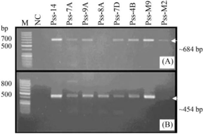
SevenP. savastanoipv.savastanoiisolates were ob-tained from 110 olive knots after elimination of suspected colonies. Isolates 7A and 7D (Table 1) were closely related samples obtained from two knots on the same branch. The bacteria were Gram-negative and grew well on selective PVF-1 medium. All of the isolates produced the expected bands of 684 bp and 454 bp with the primer pairs Pss1/Pss2 and IAALF/IAALR, respectively (Figure 1A,B). Bands of 650 bp and 1000 bp were amplified in allP. syringaepv. to-matoandP. syringaepv.phaseolicolastrains with primer pairs Pst1/Pst2 and 62a/63a, respectively (data not shown).
P. savastanoi pv. savastanoi genomic fingerprints were obtained with all of the five REP-PCR primer sets. Three primer sets (2-Dt, Rep-1R-Dt/Rep-2A and Rep-1R-Dt/Rep-2C) were more polymorphic among the isolates than the other two sets (Rep-1R-Dt/Rep-2T and Rep-1R-Dt/Rep-2G) (Figure 2). The corre-sponding PCR products ranged in size from 200 bp to 3000 bp (Figure 2). In twoP. syringaepv. tomatostrains from Ankara and Izmir, the REP primer sets produced very distinctive fingerprints, particularly with primer pairs Dt/Rep-2-Dt, Dt/Rep-2G and Rep-1R-Dt/Rep-2C (Figure 3). The corresponding PCR products ranged in size from ~200 bp to > 1500 bp.
At the pathovar level, each REP primer set was screened for well-defined, reproducible bands. The Rep1R-Dt and Rep-2C primer pairs distinguished among pathovars, as shown in Figure 4A. Amplification with the BOXA1R primer yielded PCR products of 270-2100 bp in each pathovar (Figure 4B). The arrows in Figure 4B indi-cate polymorphic bands for tomato, olive and bean
patho-Table 2- Universal and modified primers used for REP-PCR in this study.
Primer Primer sequence (5’-3’)
Rep-1R-Dt IIINCGNCGNCATCNGGC (forward) Rep-2-Dt NCGNCTTATCNGGCCTAC Rep-2A ACGACTTATCAGGCCTAC Rep-2T TCGTCTTATCTGGCCTAC Rep-2G GCGGCTTATCGGGCCTAC Rep-2C CCGCCTTATCCGGCCTAC
Letters in bold type indicate the bases that were altered in each primer at the positions indicated by N (underlined) in the first primer.
Figure 1 - Species-specific PCR fragments of P. savastanoi pv.
vars ofP. syringae (Psp-18, Pss-14 and Pst-101, respec-tively). REP-PCR yielded a higher number of bands and more complex patterns than BOX-PCR. P. syringae pv.
phaseolicola isolates were distinguishable from all the other isolates by the size of their PCR products (200-700 bp; lanes 1-4 in Figure 4B). Exceptions included the P. syringae pv. phaselicola pathovar from Ankara (Psp-3), which lacked the 700 bp band, and Psp-18, which
produced two additional bands of ~2100 bp and 1500 bp (lane 4 in Figure 4B).P. savastanoipv.savastanoicould be distinguished by a single specific fragment of 700 bp (lane 5 in Figure 4B).P. syringae pv. tomato strains differed completely from the other pathovars and from each other by producing two slightly different bands of ~250 bp (lanes 6 and 7 in Figure 4B). In contrast to REP-PCR which pro-duced several common bands in tomato pathovars, no com-mon bands were observed with BOX-PCR (lanes 6 and 7 in Figure 4B). The monomorphic and polymorphic bands ob-served with BOXA1R inP. syringaepv.phaselicola iso-lates of Turkish and UK origin demonstrated the conserved nature of BOX elements in theP. syringaegenome. No ob-servable inter-ERIC fingerprints were obtained in six local isolates or in isolate NCPPB-52 of UK origin.
The UPGMA dendrogram revealed two major divi-sions among the isolates (Figure 5) the P. syringae pv.
phaseolicola pathovars formed a group that includedP. savastanoiwhile theP. syringaepv. tomatoisolates formed a second group, with a genetic similarity of 0.25 between groups.The approximate genetic diversity among all iso-lates was 80%.
Discussion
The precise identification ofP. syringaepathovars, which are widespread pathogens, is important for basic studies related to genetic polymorphism and adequate agri-cultural management. Species-specific PCR is a powerful method for the rapid, convenient diagnosis of microbial pathogens in laboratory samples derived from plants with symptomatic phenotypes. The Pss1/Pss2 primer pair used by Basim and Ersoy (2001) for the molecular identification
Figure 2- Repetitive-PCR fragments ofP. savastanoipv.savastanoiisolates obtained with different REP primer combinations. (A) Rep-1R-Dt and Rep-2C, (B) Rep-1R-Dt and Rep-2-Dt, and Rep-1R-Dt and Rep-2A. M - DNA ladder.
ofP. savastanoipv.savastanoiwas particularly useful in the present study. Another set of primers (IAALF and IAALR for theiaaLgene) (Penyalveret al., 2000) was also useful for identifyingP. savastanoipv.savastanoistrains from naturally infected and asymptomatic olive trees. The use of combinations of modified primers in REP-PCR en-hanced the range of DNA fingerprints that could be de-tected. In this approach, it is the reverse primers rather than the forward primers that bind to the different annealing sites in the genome to produce the diversity of DNA amplicons.
The discriminatory potential of rep-PCR for different
P. syringaepathovars has been shown in previous reports (Louwset al., 1994; Weingart and Völksch, 1997; Steadet al., 2004; Vicente and Roberts, 2007; Kaluznaet al., 2010). A pathovar is defined as a subspecific group of strains that
can infect particular plants within a certain genus or spe-cies. Intra-pathovar diversity is generally of two types, namely, (a) isolates from the same pathovar that may have identical REP, BOX and ERIC fingerprints and (b) isolates that do not share common rep-PCR banding patterns (Louwset al., 1994). The first of these two groups was ini-tially identified in an analysis of samples that includedP. syringaepv. morsprunorumandXanthomonas campestris
pv. phaseoli; in this case, the number of monomorphic REP-PCR bands was greater than that of polymorphic bands in 12 isolates ofP. syringaepv. morsprunorum,P. syringaepv. syringaeandP. syringaepv. tomatofrom dis-tant locations in the United States (Louwset al., 1994). As shown here, the insertion of G and C in Rep2-Dt primers at positions 1, 4 and 11 yielded additional different sized bands in REP-PCR ofP. syringaepv.tomatoDNA (Figu-re 3). Despite the limited number of strains examined he(Figu-re, the use of modified REP-PCR primers enhanced the detec-tion of intra-pathovar diversity by expanding the number of annealing sites. There was a clear difference in the the abil-ity of the three types of rep-PCR to detect genetic diversabil-ity inP. syringae. Scortichiniet al.(2004) reported that REP primers were highly discriminatory in distinguishing amongP. savastanoiisolates from Italy. TheP. syringae
genome contains high copy numbers of REP elements that are related to insertion sequence elements (ISI) (Tobes and Pareja, 2006). This feature of REP elements may explain the abundance and diversity of fingerprints seen upon am-plification of the intervening regions.
Figure 4- REP-PCR fingerprints ofP. syringaepathovars obtained with Rep-1R-Dt and Rep-2C primers (A) and BOX-PCR fingerprints ofP. syringae
pathovars (B). M - 1 kb DNA ladder for (A) andlDNA/PstI DNA ladder for (B).
In our work, P. s. pv. savastanoi isolates showed more polymorphism than tomato and bean isolates by using REP primer combinations. One of the reasons for that is the bacterial population which was isolated from knots could be contained both epiphytic and pathogenic strains ofP. savastanoi. In addition, P. savastanoi pv. savastanoi
strains may have diverged extensively during their co-evolution with olive trees (Olea europaea) (Scortichiniet al., 2004), particularly since the latter are widely distrib-uted and well adapted in western Turkey.
The BOXA1R universal primer identified genetically distinctP. syringaepathovars and could therefore be poten-tially useful as a diagnostic tool, although the number of fingerprints it yielded was considerably lower than that tained with REP-PCR. No inter-ERIC fingerprints were ob-tained for any of the isolates. Although they have been used inP. syringaegenotyping (Louwset al., 1994; Scortichini
et al., 2004; Vicente and Roberts, 2007; Kaluznaet al., 2010), ERIC sequences have been found only in the Entero-bacteriaceae andVibriospecies (Wilson and Sharp, 2006). Theoretically, ERIC primers bind to sequences between ERIC copies and produce fragments of variable length, de-pending on the positions of these conserved elements. The amplifications reported for ERIC-PCR in several reports may have arisen from randomly binding primers and con-sist of homologous bands shared by isolates or pathovars. The random nature of ERIC-PCR can also be seen in the successful amplification of fungal DNA (Gürel et al., 2010), even though ERIC elements have not yet been re-ported in fungal DNA.
The availability of low-cost, fast and reliable methods for screening genetic diversity is a valuable tool in the diag-nosis of local bacterial populations. The work described here provides a procedure for the isolation ofP. savastanoi pv. savastoniisolates from olive knots and for reliable iden-tification using species-specific primers. The use of modi-fied primers allows the genetic variation among isolates in a particular environment to be monitored in a short time by colony PCR. The most important advantage in this ap-proach is that the primer combinations enhance the possi-bility of obtaining distinct and unique banding patterns among the genotypes that can extend our understanding of the genetic diversity of these organisms. As shown here, the banding patterns obtained with a cytosine-containing primer (Rep2-C) yielded information on genetic relation-ships and led to the identification of distinct pathovar groups (Figure 5). Variations based on extragenic repetitive DNA in local isolates ofP. savastanoimay provide clues to the nature of mutations associated with REP elements and the importance of the latter as regulators of bacterial gene expression.
Acknowledgments
We thank Prof. Kemal Benlioglu, Prof. Hatice Ozaktan and Dr. Aynur Karahan for providing the bacterial
strains used in this study. This work was supported by the Research Fund of Istanbul University (project no. T-719/30062005).
References
Basim H and Ersoy A (2001) Identification of Pseudomonas savastanoipv.savastanoi, olive knot pathogen, by polymer-ase chain reaction. In: Abstracts, Phytopathology Salt Lake 2001 APS/SON/MSA Joint Meeting, 25-29 August, 2001, Abstract S6.
Bereswill S, Bugert P, Völksch B, Ullrich M, Bender CL and Geider K (1994) Identification and relatedness of corona-tine-producing Pseudomonas syringaepathovars by PCR analysis and sequence determination of the amplification products. Appl Environ Microbiol 60:2924-2930.
Cirvilleri G, Scuderi G, Bonaccorsi A and Scortichini M (2006) Molecular characterization of Pseudomonas syringae pv.
syringaestrains from different host plants using fluorescent amplified fragment length polymorphism. J Plant Pathol 88:327-331.
de Bruijn FJ (1992) Use of repetitive (repetitive extragenic palin-dromic and enterobacterial repetitive intergenenic consen-sus) sequences and the polymerase chain reaction to finger-print the genomes ofRhizobium melilotiisolates and other soil bacteria. Appl Environ Microbiol 58:2180-2187. Gardan L, Shafik H, Belouin S, Broch R, Grimont F and Grimont
PAD (1999) DNA relatedness among the pathovars of Pseu-domonas syringaeand description ofPseudomonas tremae
sp. nov. andPseudomonas cannabinasp. nov. (ex Sutic and Dowson 1959). Int J Syst Bacteriol 49:469-478.
Gürel F, Albayrak G, Diken O, Çepni E and Tunali B (2010) Use of Rep-PCR for genetic diversity analyses in Fusarium culmorum.J Phytopathol 92:781-787.
Higgins CF, Ames GFL, Barnes WM, Clement JM and Hofnung M (1982) A novel intercistronic regulatory element of pro-karyotic operons. Nature 298:760-762.
Hirano SS and Upper CD (2000) Bacteria in the leaf ecosystem with emphasis onPseudomonas syringae- A pathogen, ice nucleus, and epiphyte. Microbiol Mol Biol Rev 64:624-653. Jaccard P (1908) Nouvelles recherches sur la distribution florale.
Bull Soc Vandoise Sci Nat 44:223-270.
Judd AK, Schneider M, Sadowsky MJ and de Bruijn FJ (1993) Use of repetitive sequences and the polymerase chain reac-tion technique to classify genetically related
Bradyrhizobium japonicum serocluster 123 strains. Appl Environ Microbiol 59:1702-1708.
Kado CI and Heskett MG (1970) Selective media isolation of
Agrobacterium, Corynebacterium, Erwinia,Pseudomonas
andXanthomonas.Phytopathology 60:969-976.
Kaluzna M, Ferrante P, Sobiczewski P and Scortichini M (2010) Characterization and genetic diversity of Pseudomonas syringae from stone fruits and hazelnut using repeti-tive-PCR and MLST. J Plant Pathol 92:781-787.
King GJ (1989) Plasmid analysis and variation inPseudomonas syringae. J Appl Bacteriol 67:489-496.
King EO, Ward NK and Raney DE (1954) Two simple media for the detection of pyocyanin and fluorescein. J Lab Clin Med 44:301-307.
XanthomonasandPseudomonaspathovars and strains gen-erated with repetitive sequences and PCR. Appl Environ Microbiol 60:2286-2295.
Louws FJ, Rademaker JLW and de Bruijn FJ (1999) The three Ds of PCR-based genomic analysis of phytobacteria: Diversity, detection, and disease diagnosis. Annu Rev Phytopathol 37:81-125.
Manceau C and Horvais A (1997) Assessment of genetic diversity among strains ofPseudomonas syringaeby PCR-restriction fragment length polymorphism analysis of rRNA operons with special emphasis onP. syringaepv.tomato.Appl Envi-ron Microbiol 63:498-505.
Mosqueda-Cano G and Herrera-Esterella L (1997) A simple and efficient PCR method for specific detection ofPseudomonas syringaepv.phaseolicolain bean seeds. World J Microbiol Biotechnol 13:463-467.
Murillo J and Sesma A (2001) The biochemistry and molecular genetics of host range definition:Pseudomonas syringae.J Phytopathol Medit 40:3-26.
Oguiza JA, Rico A, Rivas LA, Sutra L, Vivian A and Murillo J (2004)Pseudomonas syringaepv.phaseolicolacan be sepa-rated into two genetic lineages distinguished by the posses-sion of the phaseolotoxin biosynthetic cluster. Microbiology 150:473-482.
Penyalver R, Garcia A, Ferrer A, Bertolini E and Lopez MM (2000) Detection of Pseudomonas savastanoi pv.
savastanoiin olive plants by enrichment and PCR. Appl En-viron Microbiol 66:2673-2677.
Quesada JM, Perez-Martinez I, Ramos C, Lopez MM and Penyalver R (2008) IS53: An insertion element for molecu-lar typing ofPseudomonas savastanoipv.savastanoi. Res Microbiol 159:207-215.
Saygili H (1995) Fitobakteriyoloji. E.U. Ziraat Fakültesi Bitki Koruma Bölümü, Izmir, Turkey, 9944-5822-0-2.
Scortichini M, Rossi MP and Salerno M (2004) Relationship of genetic structure ofPseudomonas savastanoipv.savastanoi
populations from Italian olive trees and patterns of host ge-netic diversity. Plant Pathol 53:491-497.
Sisto A, Cipriani MG, Tegli S, Cerboneschi M, Stea G and Santilli E (2007) Genetic characterization by fluorescent AFLP of
Pseudomonas savastanoi pv. savastanoi strains isolated from different host species. Plant Pathol 56:366-372. Stead DE (1992) Grouping of plant pathogenic and some other
Pseudomonasspecies by using cellular fatty acid profiles. Int J Syst Bacteriol 42:281-295.
Stead DE, Simpkins SA, Weller SA, Hennesy J, Aspin A, Stan-ford H, Smith NC and Elphinstone JG (2004) Classification and identification of plant pathogenicPseudomonasspecies by REP-PCR derived genetic fingerprints. In: Iacobellis NS, Collmer A, Hutcheson SW, Mansfield JW, Morris CE, Mu-rillo J, Schaad NW, Stead DE, Surico G and Ullrich MS (eds)Pseudomonas syringaeand Related Pathogens. Biol-ogy and Genetics. Kluwer Academic Publishers, Dordrecht, pp 411-420.
Tobes R and Pareja E (2006) Bacterial repetitive extragenic palin-dromic sequences are DNA targets for insertion sequence el-ements.BMC Genomics 7:e62.
Van Zyl E and Steyn PL (1990) Differentiation of phytopa-thogenic Pseudomonas and Xanthomonas species and pathovars by numerical taxonomy and protein gel electrophoregrams. Syst Appl Microbiol 13:60-71. Versalovic J, Koeuth T and Lupski RJ (1991) Distribution of
re-petitive DNA sequences in eubacteria and application to fin-gerprinting of bacterial genomes. Nucleic Acids Res 19:6823-6831.
Versalovic J, Schneider M, de Bruijn FJ and Lupski JR (1994) Genomic fingerprinting of bacteria using repetitive se-quence-based PCR (rep-PCR). Methods Mol Cell Biol 5:25-40.
Vicente J and Roberts S (2007) Discrimination ofPseudomonas syringaeisolates from sweet and wild cherry using rep-PCR. Eur J Plant Pathol 117:383-392.
Weingart H and Völksch B (1997) Genetic fingerprinting of Pseu-domonas syringaepathovars using ERIC-, REP-, and IS50-PCR. J Phytopathol 145:339-345.
Wilson LA and Sharp PM (2006) Enterobacterial repetitive inter-genic concensus (ERIC) sequences inEscherichia coli: Evo-lution and implications for ERIC-PCR. Mol Biol Evol 23:1156-1168.
Woods CR, Versolovic J, Koeuth T and Lupski JR (1993) Whole-cell repetitive element sequence-based polymerase chain reaction allows rapid assessment of clonal relationship of bacterial isolates. J Clin Microbiol 31:1927-1931. Young JM, Saddler GS, Takikawa Y, De Boer SH, Vauterin L,
Gardan L, Gvozdyak R and Stead DE (1996) Names of plant pathogenic bacteria 1864-1995. Rev Plant Pathol 75:721-763.
Associate Editor: Celia Maria de Almeida Soares