Differences in the antimicrobial susceptibility profiles of
Moraxella bovis
,
M. bovoculi
and
M. ovis
Grazieli Maboni
1, Leticia T. Gressler
1, Julia P. Espindola
1, Marcelo Schwab
1,
Caiane Tasca
1, Luciana Potter
2, Agueda Castagna de Vargas
11Departamento de Medicina Veterinária Preventiva, Universidade Federal de Santa Maria, Santa Maria, RS, Brazil.
2Departamento de Zootecnia, Universidade Federal de Santa Maria, Santa Maria, RS, Brazil.
Submitted: January 22, 2014; Approved: August 15, 2014.
Abstract
The aim of this study was to determine the differences in the antimicrobial susceptibility profiles of
Moraxella bovis,M. bovoculiandM. ovis. Thirty-two strains ofMoraxellaspp. isolated from cattle
and sheep with infectious keratoconjunctivitis were tested via broth microdilution method to deter-mine their susceptibility to ampicillin, cefoperazone, ceftiofur, cloxacillin, enrofloxacin, florfenicol, gentamicin, neomycin, oxytetracycline and penicillin. The results demonstrated thatMoraxellaspp.
strains could be considered sensitive for most of the antimicrobials tested in this study, but differ-ences between the antimicrobial susceptibility profiles of these threeMoraxellaspecies were found. M. bovismight differ from other species due to the higher MIC and MBC values it presented.
Key words:bacterial resistance, broth microdilution, eye disease.
Introduction
Infectious keratoconjunctivitis (IK) affects cattle and sheep and is characterized by the development of conjunc-tivitis and corneal ulcers (Baptista, 1979). In cattle, the mi-croorganisms involved in IK are M. bovis (Henson and
Grumbles, 1960), M. ovis (Elad et al., 1988) and M. bovoculi(Angeloset al., 2007). In contrast, in sheep, the
main microorganisms isolated from IK lesions are
Mycoplasma conjunctivae,Chlamydophila psittaciandM. ovis(Eladet al., 1988; Dagnall, 1994).M. boviscan also be
isolated from sheep but it is found at a lower frequency (Libardoniet al., 2012). The treatment of this disease is
based on antimicrobial therapy, which should be adopted considering that it is necessary to combat two or more spe-cies of Moraxella present in the same lesion. However,
studies addressing the antimicrobial susceptibility of M. bovis,M. bovoculiandM. ovisare scarce in the literature.
To the best of our knowledge, this is the first study to report the antimicrobial susceptibility ofM. ovisisolated from IK
in sheep and the first study in Brazil to describe the antimicrobial susceptibility ofM. bovoculi.
It is important to determine the antimicrobial suscep-tibility ofMoraxellaspp., so that if the disease occurs the
best-possible treatment will be provided. In this context, the aim of this study was to determine the differences in the antimicrobial susceptibility profiles of M. bovis, M. bovoculiandM. ovis.
Materials and Methods
Samples characterization
Samples from clinical cases of infectious bovine and ovine IK occurring in southern Brazil were previously pro-cessed. These samples included different strains from the same herd, from different herds in similar locations and from different locations. A total of 32 samples were charac-terized asMoraxellaspp. based on Gram staining and
bio-chemical tests (Macfaddin, 2000). To perform molecular identification of species, DNA was extracted using the CTAB (cetyl-trimethylammonium bromide) method (Sam-brook and Russel, 2001). For DNA amplification by the polymerase chain reaction (PCR), the primers ISR fo for-ward (5’ ACCGACGCTTATCGCAGGTCACTA-3’) and DOI: http://dx.doi.org/10.1590/S1517-838246220140058
Send correspondence to A.C. Vargas. Laboratório de Bacteriologia, Universidade Federal de Santa Maria, Campus UFSM, prédio 44, sala 5137, 97105-900 Santa Maria, RS, Brazil. E-mail: [email protected].
ISR reverse (5’-GTGTCGAAGCA AAATCAGGGTCGT-3’) were used. Fragments with a length of 650 bp corresponding toM. bovisand those with a
length of 600 bp corresponding toM. bovoculiandM. ovis
were observed (Angelos and Ball, 2007). For the differenti-ation of Moraxella species, the enzyme RsaI was used,
which only cleaves the amplified DNA sequence fromM. bovoculi(600 bp) at a single restriction site, resulting in
two fragments of approximately 150 and 450 bp. This en-zyme does not cleave the DNA fragment of M. bovis
(650 bp) orM. ovis(600 bp) (Angelos and Ball, 2007). Ten
strains ofMoraxellaspp. were identified asM. bovis, 11 as M. bovoculiderived from cattle, and 11 asM. ovisstrains
isolated from sheep (Table 1).
Broth microdilution method
The MIC (minimal inhibitory concentration) and MBC (minimal bactericidal concentration) were investi-gated for ampicillin, gentamicin, neomicin, penicillin, cefoperazone, ceftiofur, cloxacillin, enrofloxacin, florfe-nicol and oxytetracycline. The MIC was determined in 96-well microplates, in which 100 mL of the bacterial
inoculum (105 cfu/mL) was added to 100 mL of Muller
Hinton broth (cation adjusted with 20 g/L of calcium and 10 g/L of magnesium), in this volume, 12 different concen-trations of each antimicrobial agent were diluted for 32 mg/mL to 0.015 mg/mL. All strains were analyzed in triplicate along with reference strains (M. bovis ATCC
10900,M. bovoculiATCC BAA1259 andM. ovisATCC
19575).Staphylococcus aureusATCC 25923 was used as
the standard quality control strain. Finally, the microplates were incubated under aerobic conditions at 35 °C for 24 h (CLSI, 2013).
To determine the MBC, 10mL of each antimicrobial
dilution corresponding to each strain, equal or higher than the MIC value, was transferred to Muller-Hinton agar plates and followed by incubation at 35 °C for 24 h.
As no standardized criteria for the interpretation of sensitivity exist forMoraxellaspp., breakpoints established
for Gram-negative pathogens related with bovine respira-tory disease were used. No established breakpoints were available for cloxacillin, cefoperazone and neomycin (CLSI, 2013). The MIC was defined as the lowest concen-tration of antibiotics that completely inhibited growth, and the MIC50and MIC90were defined as the lowest concentra-tions of antibiotics capable of inhibiting the growth of 50% and 90% of theMoraxellaspp. isolates, respectively. The
MBC50and MBC90have been defined as the lowest concen-trations of antimicrobial agents at which no bacterial growth is evident in 50% and 90% of isolates, respectively (CLSI, 2013).
Statistical analysis
The MIC and MBC values were evaluated via the Kruskal-Wallis test and subjected to the calculation of posi- Table
tion measurements. When differences between species of
Moraxellawere identified, the Bonferroni test was applied
to compare the modal MIC values.
Results and Discussion
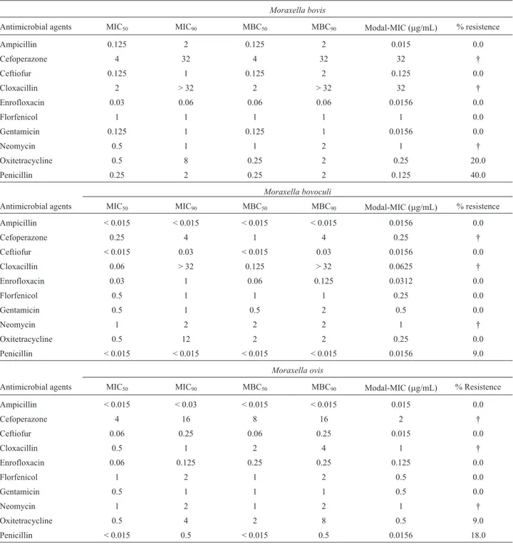
The results of the broth microdilution method for the 32 strains ofMoraxella spp. are shown in Table 2.
Gen-erally, the MIC and MBC values were similar, indicating that the same concentrations of the antimicrobial agents were able to both inhibit bacterial growth and kill the mi-croorganism (Table 2). Although there are no guidelines to define MICs forMoraxellaspp, interpretative criteria
de-rived from other pathogens have been proposed (Angeloset al., 2011). For instance, the critical breakpoints for
deter-mining ampicillin, ceftiofur, enrofloxacin, florfenicol,
gen-Table 2- Minimum inhibitory and bactericidal concentrations (MIC50, MIC90, MBC50and MBC90) modal/MIC and resistance ofMoraxella bovis,M.
bovoculiandM. ovis.
Moraxella bovis
Antimicrobial agents MIC50 MIC90 MBC50 MBC90 Modal-MIC (mg/mL) % resistence
Ampicillin 0.125 2 0.125 2 0.015 0.0
Cefoperazone 4 32 4 32 32 †
Ceftiofur 0.125 1 0.125 2 0.125 0.0
Cloxacillin 2 > 32 2 > 32 32 †
Enrofloxacin 0.03 0.06 0.06 0.06 0.0156 0.0
Florfenicol 1 1 1 1 1 0.0
Gentamicin 0.125 1 0.125 1 0.0156 0.0
Neomycin 0.5 1 1 2 1 †
Oxitetracycline 0.5 8 0.25 2 0.25 20.0
Penicillin 0.25 2 0.25 2 0.125 40.0
Moraxella bovoculi
Antimicrobial agents MIC50 MIC90 MBC50 MBC90 Modal-MIC (mg/mL) % resistence
Ampicillin < 0.015 < 0.015 < 0.015 < 0.015 0.0156 0.0
Cefoperazone 0.25 4 1 4 0.25 †
Ceftiofur < 0.015 0.03 < 0.015 0.03 0.0156 0.0
Cloxacillin 0.06 > 32 0.125 > 32 0.0625 †
Enrofloxacin 0.03 1 0.06 0.125 0.0312 0.0
Florfenicol 0.5 1 1 1 0.25 0.0
Gentamicin 0.5 1 0.5 2 0.5 0.0
Neomycin 1 2 2 2 1 †
Oxitetracycline 0.5 12 2 2 0.25 0.0
Penicillin < 0.015 < 0.015 < 0.015 < 0.015 0.0156 9.0
Moraxella ovis
Antimicrobial agents MIC50 MIC90 MBC50 MBC90 Modal-MIC (mg/mL) % Resistence
Ampicillin < 0.015 < 0.03 < 0.015 < 0.015 0.015 0.0
Cefoperazone 4 16 8 16 2 †
Ceftiofur 0.06 0.25 0.06 0.25 0.015 0.0
Cloxacillin 0.5 1 2 4 1 †
Enrofloxacin 0.06 0.125 0.25 0.25 0.125 0.0
Florfenicol 1 2 1 2 0.5 0.0
Gentamicin 0.5 1 1 1 0.5 0.0
Neomycin 1 2 1 2 1 †
Oxitetracycline 0.5 4 2 8 0.5 9.0
Penicillin < 0.015 0.5 < 0.015 0.5 0.0156 18.0
tamicin, penicillin and oxytetracycline efficacy against respiratory pathogens of cattle (Pasteurella multocida, Mannheimia haemolytica, and Haemophilus somnus)
could also represent interpretative data of M. bovis, M. bovoculiandM. ovissusceptibility to these antimicrobials.
According to these interpretative criteria, mostMoraxella
spp. strains were considered susceptible to most antimicro-bials, however some strains were considered resistant to penicillin and oxytetraciline, such asM. bovisstrains that
showed 40% (4/10) of resistance to penicillin and 20% (2/10) of resistance to oxytetracycline (> 2). Other studies foundM. bovisstrains, isolated in the United States,
resis-tant to gentamicin and oxytetracicline (Shryock et al.,
1998). Moreover,M. bovisstrains resistant to erythromycin
were also reported in South America (Conceição et al.,
2004).
According to the interpretative criteria used in this study, 9% (1/11) ofM. bovoculistrains could be considered
resistant to penicillin. A previous study reported similar re-sults forM. bovoculi, indicating higher resistance to
peni-cillin (12.3%) in comparison with other tested antimicro-bials (Angelos et al., 2011). The data reported in the
veterinary literature regarding the susceptibility of
Moraxellaspp. predate the description ofM. bovoculi,
con-sequently, it is mainly available from studies involvingM. bovis. (Webberet al., 1982; Shryocket al., 1998; Zielinski et al., 2002; Conceição et al., 2004). In this way, the
antimicrobial susceptibility ofM. bovoculiwas only
evalu-ated after the recent description of this species (Angeloset al., 2007), in a study in the United States (Angeloset al.,
2011). Thus, this is the second study evaluating the suscep-tibility profile ofM. bovoculi.
SomeM. ovisstrains examined in this study could be
considered resistant to oxytetracycline (9% - 1/11) and pen-icillin (18% - 2/11). The susceptibility data for M. ovis
available in the literature are scarce; the only study address-ing the antimicrobial susceptibility of M. ovis examined
isolates from cattle and reported resistance only for erythromycin (Catryet al., 2007). One reason for this lack
of information may be that this pathogen is not the primary agent involved in the etiology of the disease in sheep (Dagnall, 1994). It is important to note that the present re-port is the first to describe the susceptibility ofM. ovis
de-rived from sheep, and this etiological agent should also be controlled by antibiotics, especially to avoid exacerbation of lesions primarily caused by M. conjunctivae and C. pscittaci(Dagnall, 1994).
Oxytetracycline is usually the first choice for anti-microbial treatment of IK (Alexander, 2010). The MIC50 and MIC90 values obtained for oxytetracycline are pre-sented in Table 2. The previously reported MIC values for oxytetracycline for M. bovis (Shryocket al., 1998), M. bovoculi(Angeloset al., 2011), andM. ovis(Catryet al.,
2007) are lower than the values reported in this study. Al-though oxytetracycline is widely used in the treatment of
this disease, there are only two reports of resistance ofM. bovisto this drug in the literature (Shryocket al., 1998;
Senturket al., 2007). In the present study, 20% of theM. bovisstrains and 9% of theM. ovisstrains could be
consid-ered resistant to oxytetracycline. These results may suggest that the indiscriminate use of oxytetracycline over the years can be related with the selection ofMoraxellaspp. strains
resistant to this drug.
We observed significantly high MIC values (16mL/mL to > 32mL/mL) for cloxacillin. Susceptibility of M. bovisfor cloxacillin was reported (Webberet al., 1982),
and strains displaying high MIC values, similar to those found in the present study, were considered resistant. The MIC90 values for florfenicol obtained for M. bovis, M.
bovoculi and M. ovis were 1 mL/mL, 1 mL/mL and
2mL/mL, respectively. A previous study found 3.5% of
re-sistance for florfenicol amongM. bovoculistrains (Angelos et al., 2011). MIC90values for florfenicol, similar to those found in this study, were reported forM. bovisstrains
iso-lated in Argentina (Zielinski et al., 2002) and M. ovis
strains isolated in Belgium (Catryet al., 2007). Similar to
oxytetracycline, florfenicol has been reported to be an ef-fective treatment option for combating bovine IK (Gockeet al., 2002; Angeloset al., 2011), especially in cases where M. bovisis resistant to tetracycline antibiotic class
(An-geloset al., 2000).
The statistical analysis showed that the highest modal values of MIC occurred among theM. bovisstrains. In
con-trast,M. bovoculi displayed the lowest modal values of
MIC (Table 2). Based on the results obtained using the broth microdilution it can be suggested that there is differ-ence (p < 0.05) between the antimicrobial profile ofM. bovisand those ofM. bovoculiandM. ovis. Further studies
are necessary to determine the reason that higher concen-trations of antimicrobials are required to achieve inhibition ofM. bovis. According to the interpretative criteria used,
the in vitro results demonstrate that the three Moraxella
species showed the best susceptibility profile for ampi-cillin, ceftiofur, enrofloxacin, florfenicol and gentamicin.
References
Alexander D (2010) Infectious bovine keratoconjunctivitis: A re-view of cases in clinical practice. Vet Clin North Am Small Anim Pract 26:487-503.
Angelos JA, Ball LM (2007) Differentiation of Moraxella
bovoculisp. nov. from other coccoide moraxellae by the use
of polymerase chain reaction and restriction endonuclease analysis of amplified DNA. J Vet Diagn Invest 19:532-534. Angelos JA, Ball LM, Byrne BA (2011) Minimum inhibitory
con-centrations of selected antimicrobial agents forMoraxella
bovoculi associated with infectious bovine
keratoconjunc-tivitis. J Vet Diagn Invest 23:552-555.
Angelos JA, Spinks PQ, Ball LMet al.(2007)Moraxella bovoculi sp. nov., isolated from calves with infectious bovine kerato-conjunctivitis. Int J Syst Evol Microbiol 57:789-795. Baptista PJHP (1979) Infectious bovine keratoconjunctivitis: a
re-view. Br Vet J 135:225-242.
Catry BFB, Boyen F, Baele M et al. (2007) Recovery of
Moraxella ovisfrom the bovine respiratory tract and
differ-entiation ofMoraxellaspecies by tDNA-intergenic spacer PCR. Vet Microbiol 120:375-380.
Clinical and Laboratory Standards Institute (2013) Antimicrobial disk and dilution susceptibility tests for bacteria isolated from animals. Approved Standard - 4 edition. CLSI docu-ment VET01-A4, Wayne, PA.
Conceição FR, Bertoncelli DM, Storch BOet al.(2004) Antibi-otic susceptibility ofMoraxella bovisrecovered from out-breaks of infectious bovine keratoconjunctivitis in Argen-tina, Brazil and Uruguay between 1974 and 2001. Braz J Microbiol 35:364-366.
Dagnall GJ (1994) The role ofBranhamella ovis,Mycoplasma
conjunctivae and Chlamydia psittaci in conjunctivitis of
sheep. Br Vet J 150:65-71.
Elad D, Yeruham I, Bernstein M (1988)Moraxella ovisin cases of infectious bovine keratoconjunctivitis (IBK) in Israel. J Vet Med B 35:431-434.
Gokce HI, Citil M, Genc O (2002) Comparison of the efficacy of florfenicol and oxytetracycline in the treatment of naturally occurring infectious bovine keratitis. Ir Vet J 55:573-576.
Henson JB, Grumbles LC (1960) Infectious bovine kerato-conjunctivitis. I. Etiology. Am J Vet Res 21:761-766. Libardoni F, Scherer CFC, Farias L et al. (2012) Moraxella
bovoculiem casos de ceratoconjuntivite infecciosa bovina
no Rio Grande do Sul. Pesq Vet Br 32:743-746.
Macfaddin JF (2000) Biochemical testes for identification of medical bacteria, Lippincott Williams & Wilkins, Philadel-phia, P.A.
Sambrook J, Russel D (2001) Molecular cloning: a laboratory manual, Cold Spring Harbor Laboratory, New York, N.Y. Senturk S, Cetin C, Temizel Met al.(2007) Evaluation of the
clin-ical efficacy of subconjunctival injection of clindamycin in the treatment of naturally occurring infectious bovine keratoconjunctivitis. Vet Ophthalmol 10:186-189.
Shryock TR, White DW, Werner CS (1998) Antimicrobial sus-ceptibility ofMoraxella bovis. Vet Microbiol 61:305-309. Webber JJ, Fales WH, Selby LA (1982) Antimicrobial
suscepti-bility ofMoraxella bovisdetermined by agar disk diffusion and broth microdilution. AntimicrobAgentsChemother 21, 554-557.
Zielinski GC, Piscitelli HG, Perez-Monti Het al.(2000) Antibi-otic Sensitivity of an Argentine Strain Collection of
Moraxella bovis.Vet Ther 3:199-204.
Associate Editor: Odir Antonio Dellagostin