NIGELLA SATIVA ESSENTIAL OILS ON THE GROWTH OF ASPERGILLUS FUMIGATUS AND ASPERGILLUS FLAVUS
Khosravi, A.R.1*; Minooeianhaghighi, M.H.2; Shokri, H.3; Emami, S.A.4; Alavi, S.M.4; Asili, J.4
1
Mycology Research Center, Faculty of Veterinary Medicine, University of Tehran, Tehran, Iran; 2 Department of Basic Sciences, Faculty of Medicine, Gonabad University of Medical Sciences, Gonabad, Iran; 3 Department of Microbiology, Faculty of Veterinary Medicine, University of Mazandaran, Amol, Iran; 4 Department of Anatomy, Faculty of Medicine, Mashed University
of Medical Sciences, Meshed, Iran.
Submitted: June 02, 2009; Returned to authors for corrections: July 11, 2009; Approved: August 23, 2010.
ABSTRACT
The goals of this study were to evaluate the effectiveness of Cuminum cyminum, Ziziphora clinopodioides
and Nigella sativa essential oils to inhibit the growth of Aspergillus fumigatus and A.flavus and to evoke
ultrastructural changes. The fungi were cultured into RPMI 1640 media in the presence of oils at
concentrations of 8, 6, 5, 4, 3, 2, 1.5, 1.25, 1, 0.75 and 0.5 mg/ml in broth microdilution and 2, 1.5, 1 and 0.5
mg/ml in broth macrodilution methods with shaking for 48 h at 28oC. Conidial and mycelial samples exposed to 0.25, 0.5, 1, 1.5 and 2 mg essential oils/ml for 5 days in 2% yeast extract granulated plus 15%
Saccharose media were processed for transmission electron microscopy (TEM). Based on broth dilution
methods, C. cyminum and to a lesser extent Z. clinopodioides oils exhibited the strongest activity against A.
fumigatus and A.flavus with MIC90 ranging from 0.25 to 1.5 mg/ml, while the oil from N. sativa exhibited relatively moderate activity against two above fungi with MIC90 ranging from 1.5 to 2 mg/ml. The main changes observed by TEM were in the cell wall, plasma membrane and membranous organelles; in
particular, in the nuclei and mitochondria. These modifications in fungal structure were associated with the
interference of the essential oils with the enzymes responsible for cell wall synthesis, which disturbed
normal growth. Moreover, the essential oils caused high vacuolation of the cytoplasm, detachment of
fibrillar layer of cell wall, plasma membrane disruption and disorganization of the nuclear and mitochondrial
structures. Aspergillus fumigatus and A. flavus growth inhibition induced by these oils were found to be
well-correlated with subsequent morphological changes of the fungi exposed to different fungistatic
concentrations of the oils. Our results show the anti-Aspergillus activities of C. cyminum, Z. clinopodioides
and N. sativa essential oils, which strengthens the potential use of these substances as anti-mould in the
future.
Key words: Antifungal activity, Essential oil, Cuminum cyminum, Ziziphora clinopodioides, Nigella sativa ,
Aspergillus
INTRODUCTION
Moulds of the genus Aspergillus are among the most
common fungi in the environment, being found in the air, in
the soil, on plants and on decomposing organic matter (22).
Because of their powerful hydrolytic enzymes, fungi can cause
a high degree of deterioration when present in/on food- and
feed-stuffs (23). Thus, the presence and growth of these fungi
in food- and feed-stuffs threatens human and animals health. In
immunocompromised subjects, inhalation of spores gives rise
to aspergillosis, an invasive infection of the lungs or sinuses
and its dissemination to other organs (11).
Resistance of Aspergilli to some clinically used
antifungals brings a worrying clinical prognostic in people
attacked by aspergillosis (6, 9). The wide use and sometimes
misuse of antimicrobial agents in both human and animal
medicine has been responsible for rapid development of
resistant strains, toxicity and drug-drug interactions (16, 27).
Regarding the increasing clinical importance given to fungi
causing infections and the development of drug resistance,
many researchers focused on the antifungal properties of plant
products.
Plants from Iranian biomes, such as C. cyminum
(Apiaceae; known as Ziree), Z. clinopodioides (Labiatae;
known as Avishan) and N. sativa (Ranuculaceae; known as
black seed) have been used as natural medicines by local
populations in the treatment of several diseases (3, 30).
Previous studies revealed interesting antimicrobial effects from
their essential oils (1, 15, 20). To our knowledge, there is lack
of information about their effects on the kinetics of the
mycelial growth and germination of Aspergillus conidia. The
aims of this study were to evaluate the effect of C. cyminum, Z.
clinopodioides and N. sativa essential oils on the growth of two
important Aspergillus species, A. fumigatus and A. flavus,
recognized as potential air- and/or food-borne pathogens.
MATERIALS AND METHODS
Plant materials
The whole aerial parts of C. cyminum, Z. clinopodioides
and N. sativa plants, belonging to 3 plant families, were
collected from different regions of Khorasan province
(northeast of Iran) during 2008. The medicinal plants were
selected on the basis of traditional information regarding the
treatment of various diseases in Iran. Botanical identification
was performed at the Herbarium of Pharmacognosy
Department, School of Pharmacy, Mashhad University of
Medical sciences, Mashed, Iran.
Extraction of essential oils
Essential oils were isolated by water distillation for 3 h
from air-dried materials, using a Clevenger-type apparatus,
according to the procedure described in the European
Pharmacopoeia (8). The oils were stored at −4°C in sealed
brown vials until use. The essential oils were assayed at
concentrations of 8, 6, 5, 4, 3, 2, 1.5, 1.25, 1, 0.75 and 0.5 in
broth microdilution and 2, 1.5, 1 and 0.5 mg/ml in broth
macrodilution methods. The stock solutions of oils were
prepared according to Souza et al. (29). In this study, all
general chemical materials were purchased from Merck
Company (Darmstadt, Germany).
Fungal species and conidia preparation
Aspergillus fumigatus (ATCC 16913) and A. flavus
(ATCC 16013) strains were used as test microorganisms. In
addition, 6 isolates of A. fumigatus and A.flavus, obtained from
air samples, were included in this study. These strains were
taken from the Fungal Collection, Mycology Research Center,
Faculty of Veterinary Medicine, University of Tehran, Iran.
The fungal strains were precultured on Sabouraud glucose agar
using sterile distilled water containing 0.5% Tween 80. The
resulting mixture of conidia and hyphal fragments was
vortexed for 15 s and the heavy particles were allowed to settle
for 5 min. The resulting suspension was counted in a Neubauer
chamber and standardized to concentrations of 1×10⁷ conidia/ml. This suspension was further diluted 1:10 with
RPMI 1640 broth to final concentrations of 1×10⁶ conidia/ml.
Antifungal activity measurements
The MIC90 and MFC for 3 essential oils were determined by broth macro- and microdilution methods, according to the
protocol in M38-A for filamentous fungi with some
modifications (7). For the broth macrodilution method, 900 µl
of the final conidia suspensions were mixed with 100 µl of the
test essential oil in 12×75 mm test tubes and incubated at 28°C
for 48 h. The positive control tube contained 900 µl of conidial
suspension plus 100 µl of RPMI 1640, and the negative one
contained 1 ml of RPMI 1640 only. The lowest oil
concentration inhibiting fungal growth by 90% was identified
as the minimal inhibitory concentration (MIC90). In addition, flat-bottom microdilution plates containing 96 wells were
employed for the broth microdilution method. One hundred
microliters of final conidia suspension were added to each well
containing 100 µl of the oil. Positive control was the well
containing 100 µl of the inoculum suspension and 100 µl of the
RPMI only, and the negative control was a well containing 200
µl of RPMI 1640. The minimum fungicidal concentrations
(MFCs) were determined by subculturing 10 µl aliquot from all
MIC wells showing no visible growth on to Sabouraud glucose
agar plates. Each assay was performed 4 times and the results
were expressed as the average of the 4 repetitions.
Transmission electron microscopy (TEM)
Fungal materials obtained from 5-day-old cultures of 2%
yeast extracted granulated plus 15% Saccharose media and
treated with 0.25, 0.5, 1, 1.5 and 2 mg essential oils/ml were
processed for TEM (5). The samples were pre-fixed with 2.5%
glutaraldehyde in 0.1M sodium phosphate buffer (PBS), pH 7.2
for 3 h at room temperature, followed by thorough washing
with phosphate buffer (3 times, 30 min each). Specimens were
then post-fixed for 2 h in 1% aqueous osmium tetroxide (OsO4) at room temperature and washed in PBS buffer (pH=7.2, 3
times, 15 min each). Samples were dehydrated in a graded
acetone series (50%, 70%, 80% and 90%, one time at 20 min
for each dilution and 3 times at 30 min in 100% acetone) and
embedded in 25% spurr’s resin. Blocks were sectioned with a
diamond knife (ultramicrotome Richter OMU3). Sections
about 80 nm thick were collected on gold grids, stained
toluidine blue for 2 min and then examined under a Zeiss
transmission electron microscope at 80 kV (120-ILFORD
Delta 100 ASA, Zeiss, Germany).
Statistical analysis
Data were analyzed by the unpaired Student’s t test. A P
value of less than 0.05was considered statistically significant.
RESULTS
Inhibition of growth
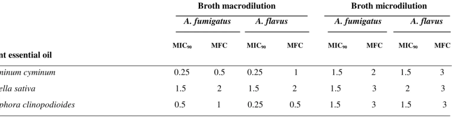
Results of the inhibitory activities of essential oils on the
growth of A. fumigatus and A. flavus are presented in Table 1.
Fungal growth inhibitions were found to be correlated with
dose dependent anti-mould activities. Based on broth
macrodilution method, C. cyminum oil exhibited the strongest
activity, with MIC90 value of 0.25 mg/ml against tested fungi. The MIC90values for Z. clinopodioides were 0.5 mg/ml for A. fumigatus and 0.25 mg/ml for A. flavus while the oil from N.
sativa exhibited relatively moderate activity with an MIC90 of 1.5 mg/ml for fungi. In broth microdilution method, C.
cyminum and Z. clinopodioides oils showed the MIC90 of 1.5 mg/ml for tested fungi while the MIC90 values for N. sativa oil were 1.5 and 2 mg/ml for A. fumigatus and A. flavus,
respectively. These results generally confirmed those obtained
Fungal development was completely inhibited during 48 h
of incubation at concentrations of 2 and 3 mg/ml of essential
oils in both macro- and microdilution methods, respectively.
Subcultures of these treated inoculums were negative,
confirming fungicidal effects against A. fumigatus and A. flavus
at these concentrations.
Table 1. Anti-Aspergillus susceptibility of Cuminum cyminum, Ziziphora clinopodioides and Nigella sativa in broth macro- and microdilution methods (mg/ml).
Broth macrodilution Broth microdilution A. fumigatus A. flavus A. fumigatus A. flavus
MIC90 MFC MIC90 MFC MIC90 MFC MIC90 MFC
Plant essential oil
Cuminum cyminum 0.25 0.5 0.25 1 1.5 2 1.5 3
Nigella sativa 1.5 2 1.5 2 1.5 3 2 3
Ziziphora clinopodioides 0.5 1 0.25 0.5 1.5 3 1.5 3
- The values in the table are an average of 4 experiments.
Transmission electron microscopy
In untreated fungi (controls), the cell wall was uniform
and thoroughly surrounded by an intact fibrillar layer. Plasma
membrane was unfolded with a uniform shape and all the
organelles, such as nuclei and mitochondria appeared normal
(Fig. 1a and 1b).
TEM observations of A. fumigatus and A. flavus hyphae
and conidia treated at sub-lethal essential oil concentrations
were illustrated in Figure 1. After treatment, the normal
morphologies of fungal hyphae and conidia were disturbed
when the concentration of essential oils increased in the culture
media.
The cell shapes were modified and lost their regularity in
comparison to the controls. The general alterations of A.
fumigatus and A. flavus hyphae and conidia treated with the
oils were similar. The major changes were found on the cell
wall, plasma membrane and membranous organelles specially
nuclei and mitochondria. The early changes in fungal
compartments in the presence of the lowest concentrations of
oils (0.25 and 0.5 mg/ml) were noticed in both hyphae and
conidia, showing abnormal shaped and swelled hyphae, high
vacuolation of the cytoplasm accompanied by vacuole fusion
(Fig. 1c and 1d). Subsequent events were loss of normal
conidia and hyphae shape, detachment of fibrillar layer of the
cell wall (Fig. 1e, 1f and 1g), destruction of hypha
memberanous organelles including nuclei and mitochondria,
and finally disorganization of cytoplasmic contents
accompanied by intensive degradation and lysis of the nucleus
and mitochondria (Fig. 1h). The most remarkable changes in
fungal compartments were observed in fungi treated with the
highest fungistatic concentrations of the oils (1.5-2 mg/ml).
Destruction and breaking down of plasma membrane at
different sites (Fig. 1i), disorganization of conidial and hyphal
cytoplasm and complete lysis of membranous organelles
- CW: Cell wall; PM: Plasma membrane; S: septum; W: Woronin body
Figure 1. Transmission electron micrographs of Aspergillus species: a) Control conidium of A. fumigatus (4400X); b) Control hyphae of A. flavus (50000X); c and d) Cross sections of A. fumigatus (c, 12000X) and A. flavus (d, 30000X) hyphae treated with
2 mg/ml N.sativa oil, showing clear separation of plasma membrane from the cell wall, detachment of fibrilar layer of the cell
wall, disruption of the cytoplasm and vacuolation of cytoplasm accompanied by vacuole fusion; e and f) Sections of A. flavus (e,
7000X) and A. fumigatus (f, 12000X) conidia treated with 0.5 mg/ml C. cyminum oil, showing increase in vacuolization
accompanied by complete detachment of plasma membrane from cell wall (e). Note strong cytoplasmic retraction of conidium (f);
g) A. fumigatus hyphae treated with 0.25 mg/ml Z. clinopodioides oil, showing degradation and discontinuity of plasma
membrane (7000X); h) A. flavus hyphae treated with 0.25 mg/ml Z. clinopodioides oil, showing destruction and lysis of hypha
memberanous organelles including nuclei and mitochondria and disorganization of cytoplasmic contents (12000X); i) Arrows
indicate destruction and breaking down of plasma membrane with massive formation of membrane-bounded vesicles (50000X); j
and k) Collapsed conidia treated with 0.25 mg/ml of C. Cyminum oil in A. fumigatus (j, 7000X) and A. flavus (k, 12000X), which
finally resulted in cells dead.
DISCUSSION
This study described the effectiveness of C. cyminum, Z.
clinopodioides and N. sativa essential oils against Aspergillus
species by both macro- and microdilution assays and
ultrastructural changes of the fungal species tested. In broth
macrodilution test, the oil concentrations of C. cyminum at 0.25
mg/ml, Z. clinopodioides at 0.5 and 0.25 mg/ml and N. sativa
at 1.5 mg/ml showed fungistatic activity against A. fumigatus
and A. flavus, respectively, while this effect was observed with
values of 1.5 mg/ml for C. cyminum and Z. clinopodioides, and
1.5 and 2 mg/ml for N. sativa oils in broth microdilution
method, with 90% growth inhibition after 48 h of incubation.
The MICs in broth macrodilution cultures were different from
the MICs for cultures in broth microdilution method, indicating
no significant difference between both tests. In several studies,
the microdilution MICs demonstrated interlaboratory
or similar to the macrodilution MICs and discrepancies
between the two tests were not statistically significant (13,14).
The different values for MIC90 obtained with the oils incorporated in broth macro- or microdilution tests showed that
the level of antifungal activity of essential oils was closely
dependent on the screening method used and fungi tested, as
previously reported (10).
This study demonstrated that C. cyminum oil had the
highest inhibitory effect against A. fumigatus and A. flavus
among the oils tested. The different activity of these oils may
be due to their different components, the structural
configuration of the constituent components and their
functional groups and possible synergistic interactions between
components (12). The main constituents of C. cyminum oil
were pinene, cineole and linalool, while the main components
found in Z. clinopodioides oil were pulegone,1,8-cineole and
limonene, and in N. sativa oil the components were trans
-anethole and p-cymene (4,21,24,25). Aligiannis et al. (2)
proposed a classification for plant materials, based on MIC
results in broth macrodilution test as follows: strong inhibitors
(MIC up to 0.5 mg/ml); moderate inhibitors (MIC between 0.6
and 1.5 mg/ml); weak inhibitors (MIC above 1.6 mg/ml).
According to Table 1, a strong activity against Aspergillus
species was indicated for the oils from C. cyminum and Z.
clinopodioides. The oil from N. sativa presented moderate to
weak activity. In respect to the oils activity, no significant
difference was observed between the Aspergillus species
tested. The MIC results for Aspergillus species in our study
were those found by Naeini et al. (20) for Candida albicans.
The TEM of essential oils-treated fungi in comparison
with untreated samples clearly showed dose-dependent changes
of fungal cells, especially on membranous structures.
Interestingly, growth inhibitions of A. fumigatus and A. flavus
were found to be well correlated with correspondence
morphological changes of the fungi exposed to different
fungistatic concentrations of the oils. As concerned the cell
wall, it was observed that the surfaces of the hyphae and
conidia treated by oils became rough in contrast to the control
group. As the concentration of oils increased, the cell gradually
became smaller, the cell wall was disrupted and became rough
and villiform. Subsequently, the cell wall became very thin and
even seemed to disappear in some old hyphae. In a study
conducted by Ghfir (18), a deformation of the apices of A.
fumigatus growing hyphae was observed in the presence of
Hyssopus officinalis essential oil. In another study, cell wall
degradation was also observed in C. albicans cells treated with
Carica papaya latex sap (17). Such modifications induced by
essential oils may be related to the interference of essential oil
components with enzymatic reactions involved in cell wall
synthesis, thus affecting fungal morphogenesis and growth;
however that remains to be proved.
The oils tested, besides the increase in vacuolization,
showed a strong alteration in the cytoplasmic membrane. The
plasma membrane of Aspergillus species was seen to be
irregular and dissociated from the cell wall and cut into small
fragments; these membrane segments were dispersed into the
cytoplasm. These changes were usually found in fungi treated
with imidazole components (26).
Finally, the membranous organelles were disrupted
following treatment with oils. The membrane-disruptive
activity of essential oil components may be closely associated
with the interference with enzymatic reactions of the
membrane, such as respiratory electron transport, proton
transport, and coupled phosphorylation steps (19). Moreover, a
marked depletion of cytoplasmic contents of hyphae
accompanied by lysis and disruption of membranes of major
organelles, such as nuclei and mitochondria indicated that in
high fungistatic concentrations the oils passed not only through
the cell wall but also through the plasma membrane and then
interacted with membranous structures of the cytoplasmic
organelles. The present results clearly showed that the oils
were able to inhibit fungal growth by changing the cell
uniformity via direct interaction with either cell wall or
significant morphological changes were found in the hyphae
and conidia exposed to the low concentration of the oils, but
the conidia exposed to relatively higher concentrations of oils
collapsed. There were minor differences in morphological
changes of different fungi based on the oils tested. It is
considered that this phenomenon is related to constituents of
oils that possess a polar functional group like hydroxyl group
or have a charged group such as anionic group or cationic
group (28).
From the results of this study, it might be concluded that
C. cyminum, Z. clinopodioides and N. sativa oils possess
antifungal activities to inhibit the growth of A. fumigatus and
A. flavus. The antifungal activity of the oils was evident at the
morphological level. Due to the antifungal activity of these oils
and their availability as natural volatile products, they might be
of use in future studies of antifungal agents.
ACKNOWLEDGMENT
This work was supported by the Research Council of
University of Tehran.
REFERENCES
1. Akgul, A.; Kivanc, M. (1988). Inhibitory effects of selected Turkish spices and oregano components on some foodborne fungi. Int. J. Food Microbiol. 6, 263-268.
2. Aligiannis, N.; Kalpotzakis, E.; Mitaku, S.; Chinou, I.B. (2001). Composition and antimicrobial activity of the essential oils of two
Origanum species. J. Agri. Food Chem. 40, 4168-4170.
3. Avicenna. (980-1037AD). Al-Qanun fi al Tibb, (The Canon of Medicine), Persian Edition by Sharaf-Kandi, A.R. (1985), Book II, 1st.edn. Soroush Press, Tehran, Iran.
4. Bakkali, F.; Averbeck, S. (2008).Biological effects of essential oils.
Food Chem. Toxicol. 46, 446-475.
5. Bozzola, J.J.; Russel, L.D. (1999). Electron Microscopy: Principles and Techniques for Biologist, Jones and Bartlet Publ., Sudbury.
6. Canuto, M.M.; Rodero, F.G. (2002). Antifungal drug resistance to azoles and polyenes. Lancet Infect. Dis. 2, 550-563.
7. CLSI/Clinical and Laboratory Standards Institute- Reference method for broth dilution antifungal susceptibility testing of conidium-forming filamentous fungi. Wayne, 2002. (Approved Standard M38-A).
8. Council of Europe. (1997). Methods of Pharmacognosy. In European Pharmacopoeia, 3rd edn, Strasbourg: European Department for the Quality of Medicines.
9. Curtis, L.; Conroy, L.; Coli, S.; Baker, K.; Our, C.H.; Hershow, R.; Norlock-Cruz, F.; Scheff, P. (2005). Aspergillus surveillance project at a large tertiary-care hospital. J. Hosp. Infect. 59, 188-196.
10. Delespaul, Q.; de Billerbeck, V.G.; Roques, C.G.; Michel, G.; Marquier-Viñuales, C.; Bessière, J.M. (2000). The antifungal activity of essential oils as determined by different screening methods. J. Essent..Oil Res. 12, 256-266.
11. Denning, D.W. (1998). Invasive aspergillosis. Clin. Infect. Dis. 26, 781-805.
12. Dorman, H.J.D.; Deans, S.G. (2000). Antimicrobial agents from plants: antibacterial activity of plant volatile oils. J. Appl. Microbiol. 88 (5), 308-316.
13. Espinel-Ingroff, A.; Kerkering, T.M.; Goldson, P.R.; Shadomy, S. (1991). Comparison study of broth macrodilution and microdilution antifungal susceptibility tests. J. Clin. Microbiol. 29, 1089-1094. 14. Espinel-Ingroff. A.; Kish, C.W.; Kerkering, T.M.; Fromtling, R.A.;
Bartizal, K.; Galgiani, J.N.; Villareal, K.; Pfaller, M.A.; Gerardens, T.; Rinaldi, S.M.G.; Fothergill, A. (1992). Collaborative comparison of broth macrodilution and microdilution antifungal susceptibility tests. J. Clin. Microbiol. 30(12), 3138-3145.
15. Fazly Bazzaz, B.S.;Haririzadeh, G. (2003). Screening of Iranian plants for antimicrobial activity. Pharmaceutical Biol. 41(8), 573-583. 16. Georgopapadakou, N.H. (2002). Infectious diseases 2001: drug
resistance, new drugs. Drug Res.5, 181-191.
17. Giordani, R.; Cardenas, M.L.; Moulin-Traffort, J.; Regli, P. (1996). Fungicidal activity of latex sap from Carica papaya and antifungal effect of D (+)-glucosamine on Candida albicans growth. Mycoses 39, 103-110.
18. Ghfir, B.; Fonvieille, J.L.; Dargent, R. (1997). Effect of essential oil of
Hyssopus officinalis on the chemical composition of the walls of
Aspergillus fumigatus. Mycopathologia 138, 7–12.
19. Knobloch, K.; Weigand, H.; Weiss, N.; Scharm, H.M.; Vigenschow, H. (1986). Action of terpenoids on energy metabolism. In Progress in essential oil research. Edited by Walter de Gruyter & Co., Berlin. 20. Naeini, A.; Khosravi, A.R.; Chitsaz, M.; Shokri, H.; Kamlnejad, M.
(2009). Anti-Candida albicans activity of some Iranian herbs used in traditional medicine. (In Press, Journal De Mycologie Medicale). 21. Nickavara, B.; Mojaba, F.; Javidniab, K.; Amoli, M.A. (2003). Chemical
composition of the fixed and volatile oils of Nigella sativa L. from Iran.
Z. Naturforsch 58, 629-631.
22. Nolard, N.; Detandt, M.; Beguin, H. (1988). Ecology of Aspergillus
Plenum Press.
23. Overy, D.P.; Seifert, K.A.; Savard, M.E.; Frisvad, J.C. (2003). Spoilage fungi and their mycotoxins in commercially marketed chestnuts. Int. J..Food Microbiol. 88, 69-77.
24. Ozturk, S.; Ercisli, S. (2007). Antibacterial activity and chemical constitutions of Ziziphora clinopodioides. Food Control 18(5), 535-540. 25. Salehi Surmaghi, H. (2006). Medicinal plants and phytotherapy.
Donyaee Taghazie, Tehran, Iran.
26. Scott, E.M.; Gorman, S.P.; Millership, J.S.; Wright, L.R. (1986). Effect of miconazole and clotrimazole on K+ release and inhibition of ergosterol biosynthesis in Trichophyton mentagrophytes and related
ultrastructural observations. J. Antimicrob. Chemother. 17, 423-432. 27. Shahi, S.K.; Shukla, A.C.; Bajaj, A.K.; Medgely, G.; Dikshit, A. (1999).
Broad spectrum antimycotic drug for the control of fungal infection in human beings. Curr. Sci.76, 836-839.
28. Sikkema, J.; de Bont, J.A.; Poolman, B. (1995). Mechanisms of membrane toxicity of hydrocarbons. Microbiol. Rev. 59(2), 201–222. 29. Souza, E.L.; Lima, E.O.; Freire, K.R.L.; Sousa, C.P. (2005). Inhibition
action of some essential oils and phytochemicals on the growth of moulds isolated from foods. Braz. Arch. Biol. Techno. 48, 245-250. 30. Tadjbakhsh, H. (2003). History of human and veterinary medicine in