A
rti
c
le
J. Braz. Chem. Soc., Vol. 20, No. 4, 644-651, 2009. Printed in Brazil - ©2009 Sociedade Brasileira de Química 0103 - 5053 $6.00+0.00
*e-mail: [email protected]
Antiparasitic Activities of Acridone Alkaloids from Swinglea glutinosa (Bl.) Merr.
Djalma A. P. dos Santos,a Paulo C. Vieira,*,a M. Fátima das G. F. da Silva,a João B. Fernandes,a Lauren Rattrayb and Simon L. Croftb
aDepartamento de Química,Universidade Federal de São Carlos, CP 676, 13565-905 São Carlos-SP, Brazil
bDepartment of Infectious and Tropical Diseases, London School of Hygiene & Tropical Medicine,
London, WC1E 7HT, UK
Onze alcalóides acridônicos isolados de Swinglea glutinosa (Bl.) Merr. foram avaliados para suas atividades in vitro contra linhagens de Plasmodium falciparum sensíveis a cloroquina 3D7,
Trypanosoma brucei rhodesiense STIB9000 e Leishmania donovani L82. Ensaios com células KB foram também executados com o objetivo de se determinar o grau de toxicidade das substâncias ativas contra os parasitas. Nove dos compostos apresentaram IC50 entre 0,3 e 11,6 µM contra P. falciparum. Em contraste, um pequeno número de compostos mostrou atividade significativa contra T. brucei rhodesiense e nenhum apresentou atividade contra L. donovani. Entre os alcalóides três tiveram IC50 < 1,0 µM contra P. falciparum, enquanto que contra T. b. rhodesiense cinco mostraram IC50 < 10 µM. A caracterização dos alcalóides,
1,3,5-triidróxi-4-metóxi-10-metil-2,8-bis(3-metilbut-2-enil)acridin-9(10H)-ona (1), 2,3-diidro-4,9-diidróxi-2-(2-hidróxipropan-2-il)-11-metóxi-10-metilfuro[3,2-b]acridin-5(10H)-ona (2) e 3,4-diidro-3,5,8-triidróxi-6-metóxi-2,2,7-trimetil-2H-pirano[2,3-a]acridin-12(7H)-ona (3), é aqui discutida. Discute-se também a relação estrutura-atividade para todos os compostos ensaiados. O isolamento e dados espectrais para os alcalóides 1-3 estão sendo aqui descritos pela primeira vez, embora em trabalho anterior tenham sido relatadas as suas atividades citotóxicas.
Eleven acridone alkaloids isolated from Swinglea glutinosa (Bl.) Merr. were examined for
in vitro activity against chloroquine-sensitive Plasmodium falciparum 3D7, Trypanosoma brucei rhodesiense STIB900 and Leishmania donovani L82. An assay with KB cells was developed in order to compare in vitro toxicity of alkaloids with the selective action on the parasites. Nine of the compounds had IC50 values ranging from 0.3 to 11.6 µM against P. falciparum. In contrast, a small number of compounds showed significant activity against T. brucei rhodesiense and none had activity against L. donovani. Among the alkaloids three had IC50 < 1.0 µM against P. falciparum, whereas against T. b. rhodesiense five had IC50 < 10 µM. The characterization of the acridone
alkaloids, 1,3,5-trihydroxy-4-methoxy-10-methyl-2,8-bis(3-methylbut-2-enyl)acridin-9(10H)-one (1), 2,3-dihydro-4,9-dihydroxy-2-(2-hydroxypropan-2-yl)-11-methoxy-10-methylfuro[3,2-b] acridin-5(10H)-one (2) and 3,4-dihydro-3,5,8-trihydroxy-6-methoxy-2,2,7-trimethyl-2H -pyrano[2,3-a]acridin-12(7H)-one (3), is discussed, as well as the structure-activity relationship of all compounds assayed. Isolation and spectral data of alkaloids 1-3 are described for the first time although their citotoxicities to cancer cells have been described before.
Keywords: antiparasitic acridone alkaloids, malaria, Swinglea glutinosa, Rutaceae
Introduction
Parasitic protozoa are the causative agents of human and livestock diseases infecting hundreds of millions of people every year and are collectively one of most important causes of human misery.1 Human African trypanosomiasis (HAT), or sleeping sickness, malaria, Chagas’ disease and
leishmaniasis are major health problems in many countries. HAT promoted by Trypanosoma brucei rhodesiense and
falciparum to chloroquine, a drug which formed the basis for cheap and effective treatment and for prophylaxis of this disease.4 Each year, approximately 300 to 500 million malaria infections lead to over one million deaths. In many endemic countries, malaria is responsible for economic stagnation, lowering the annual economic growth in some regions by up to 1.5%.5,6 Leishmaniasis is a disease caused by protozoa of the genus Leishmania. According to WHO 88 countries are affected, with 350 million people at risk. 90% of cases of visceral leishmaniasis occur in India, Sudan, Bangladesh and Brazil.7 Present chemotherapy for these diseases is inadequate or toxic, or becoming ineffective due to an increase in resistance.8
The family Rutaceae contains many secondary metabolites such as alkaloids, flavonoids, coumarins, limonoids and lignans with a large spectrum of biological activities.9 Studies showed that acridone alkaloids are compounds with promising activity against P. falciparum,10,11 and also have antiviral12 and antiproliferative effects on cancer cell lines.13,14The Asian genera Citrus and Swinglea are members of the Rutaceae and are included in the subfamily Aurantioideae. Citrus species
have been investigated and characterized by possessing acridone alkaloids. These data stimulated an investigation of Swinglea glutinosa (Bl.) Merr. in a search for lead acridones.
From the MeOH extract of the stem bark of S. glutinosa three new acridone alkaloids were identified: 1,3,5-trihydroxy-4-methoxy-10-methyl-2,8-bis (3-methylbut-2-enyl)acridin-9(10H)-one (1), 2,3-dihydro- 4,9-dihydroxy-2-(2-hydroxypropan-2-yl)-11-methoxy-10-methylfuro[3,2-b]acridin-5(10H)-one (2) and 3,4-dihydro-3,5,8-trihydroxy-6-methoxy-2,2,7-trimethyl-2H-pyrano[2,3-a]acridin-12(7H)-one (3). In addition, eight known alkaloids were characterized by the analysis of their NMR spectra and compared with reference data: 1,3,5-trihydroxy-2,8-bis (3-methylbut-2-enyl)-10-methyl-9-acridone (4),10 glycocitrine-IV (5),15 citrusinine-II (6),16 citrusinine-I (7),17 citibrasine (8),16 5-hydroxynoracronycine (9),18 pyranofoline (10)19 and bis-5-hydroxynoracronycine (11)20. These alkaloids were tested in vitro against chloroquine-sensitive P. falciparum 3D7, T. b. rhodesiense
Antiparasitic Activities of Acridone Alkaloids from Swinglea glutinosa (Bl.) Merr. J. Braz. Chem. Soc.
646
Results and Discussion
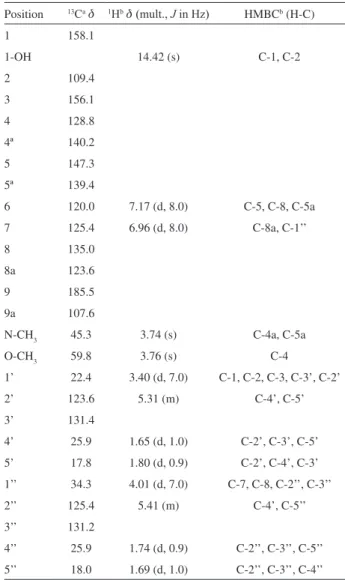
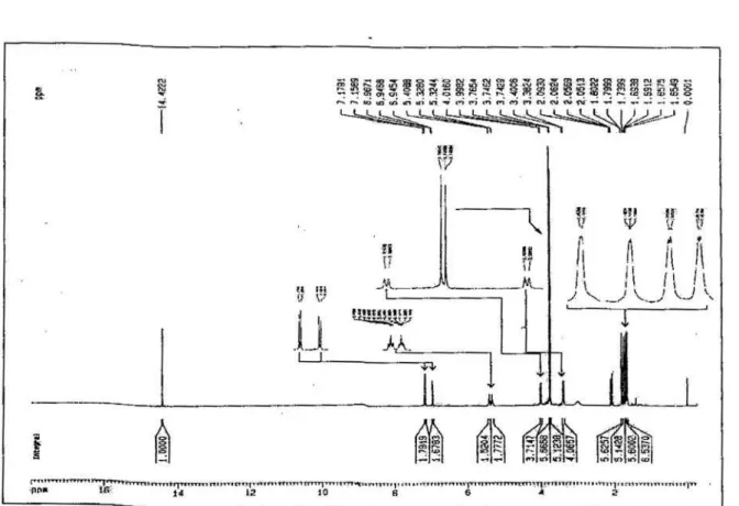
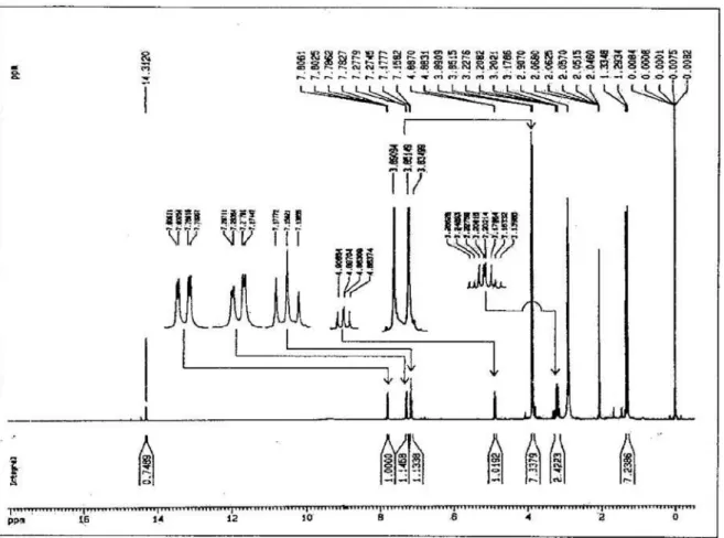
Compound 1 was isolated as an amorphous powder, the molecular formula was determined as C25H29NO5 by HRESIMS, showing an [M+H]+ peak at m/z 424.2124. The UV absorptions bands at λmax/nm: 222, 265, 285, 332 and 410 nm, IR absorptions at νmax/cm-1: 3500 and 1625 cm and the characteristic signal of a hydrogen-bonded hydroxyl proton at d14.42 in the 1H NMR spectra, exchangeable with D2O, suggested the presence of a hydroxyl group. These data together with analyses of the 13C NMR spectrum, suggested that compound 1 had a 1-hydroxy-9-acridone framework.16-22
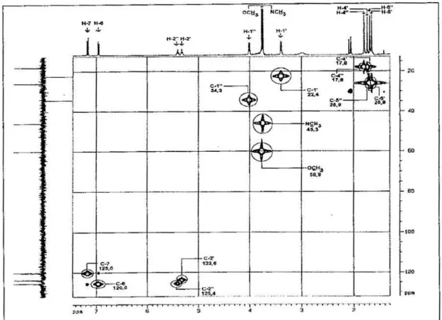
The 1H NMR spectrum of the compound 1 showed two AB type aromatic protons at d 7.17 (1 H, d, J 8.0Hz, H-6) and 6.96 (1H, d, J 8.0Hz, H-7), one N-methyl group at d 3.74 and one O-methyl group at d 3.76. Also signals of two trisubstituted double bonds at d 5.41(1H, m, H-2''), 5.31 (1H, m, H-2'), two methylene protons at d 4.01 (2H, d, J 7.0Hz, H-1’’), 3.40 (2H, d, J 7.0Hz, H-1’), the former protons being deshielded by the 9-carbonyl group, and four vinyl methyl groups [d 1.80 (3H,d, J 0.9Hz, H-4’ E), 1.65 (3H,d, J 1.0Hz, H-5’ Z), 1.74 (3H,d, J 0.9Hz, H-4’’ E), 1.69 (3H,d, J 1.0Hz, H-5’’ Z)] were observed, which suggested the presence of two prenyl groups (3-methylbut-2-enyl). Their positions were confirmed at C-2 and C-8 by HMBC.
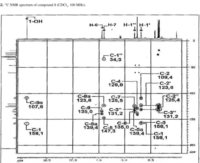
The position of a prenyl group at C-2 was confirmed by the correlation of the methylene protons at d 3.40 (H-1') with C-1 (d158.1), C-2 (d 109.4), C-3 (d 156.1), C-2' (d123.6) and C-3' (d 131.4) in the HMBC spectrum. In addition, the correlation of 1-OH at d14.42 with C-1 and C-9a (d 107.6) confirmed that the first prenyl group was attached to C-2. The position of the second prenyl group was confirmed by the correlation of the deshielded methylene protons at d 4.01 (H-1'') with C-7 (d 125.4), C-8 (d 135.0), C-2'' (d 125.4) and C-3'' (d 131.2), confirming that the second prenyl group was attached to C-8. The N-methyl protons showed correlation with C-4a and C-5a at d 140.2 and 139.4, respectively. The
O-methyl protons showed correlation with C-4 at d 128.8. This value is consistent with the acridone possessing an
O-methyl group at C-4.17,23 Based on these data, the structure of 1 was characterized as 1,3,5-trihydroxy-4-methoxy-10-methyl-2,8-bis(3-methylbut-2-enyl)acridin-9(10H)-one. Table 1 shows the data of 1H, 13C NMR and correlations observed in the HMBC experiment.
Compound 2 was isolated as an amorphous powder, the molecular formula C20H21NO6 was determined on the basis of HRESIMS, exhibiting an [M+H]+ peak at m/z 372.1447. The UV and IR spectra were identical to those of 1, however some differences were observed in the 1H and 13C NMR. In the 1H NMR spectrum, the characteristic signal of a
hydrogen-bonded hydroxy proton at d14.42, exchangeable with D2O suggested the presence of a hydroxyl group. These data suggested that compound 2 had a 4-hydroxy-5-acridone skeleton.16-22
The 1H NMR spectrum of 2 showed an ABX type aromatic spin system at d 7.15 (1 H, t, J 7.8Hz, H-7), 7.29 (1H, dd, J 7.8, 1.4Hz, H-8) and 7.80 (1H, dd, J 7.8, 1.4Hz, H-6), this last proton being deshielded by the 5-carbonyl group. The spectrum also showed one N-methyl group at d 3.85 and one O-methyl group at d 3.89. The presence of a hydroxyisopropyldihydrofuran moiety was suggested by an oxymethine proton at d 4.88 (1H, dd, J 9.4, 7.8Hz, H-2), methylene protons as twodd in an AB system at d 3.20 (1H, dd, J 15.5, 7.8Hz, H-3a) and 3.26 (1H, dd, J 15.5, 9.4Hz, H-3b), two methyl groups at d 1.33 (3H, s, H-2') and 1.29 (3H, s, H-3'). The signal at d 93.0 and 71.5 in the 13C NMR spectrum supported the presence of this substituent in the acridone nucleus.23
Table 1. 1H and 13C NMR and HMBC data for compound 1 (acetone-d
6)
Position 13Cad 1Hbd (mult., J in Hz) HMBCb (H-C)
1 158.1
1-OH 14.42 (s) C-1, C-2
2 109.4 3 156.1 4 128.8 4ª 140.2 5 147.3 5ª 139.4
6 120.0 7.17 (d, 8.0) C-5, C-8, C-5a
7 125.4 6.96 (d, 8.0) C-8a, C-1’’
8 135.0
8a 123.6
9 185.5
9a 107.6
N-CH3 45.3 3.74 (s) C-4a, C-5a
O-CH3 59.8 3.76 (s) C-4
1’ 22.4 3.40 (d, 7.0) C-1, C-2, C-3, C-3’, C-2’
2’ 123.6 5.31 (m) C-4’, C-5’
3’ 131.4
4’ 25.9 1.65 (d, 1.0) C-2’, C-3’, C-5’
5’ 17.8 1.80 (d, 0.9) C-2’, C-4’, C-3’
1’’ 34.3 4.01 (d, 7.0) C-7, C-8, C-2’’, C-3’’
2’’ 125.4 5.41 (m) C-4’, C-5’’
3’’ 131.2
4’’ 25.9 1.74 (d, 0.9) C-2’’, C-3’’, C-5’’
5’’ 18.0 1.69 (d, 1.0) C-2’’, C-3’’, C-4’’
The presence of a linear orientation of the hydroxyisopropyldihydrofuran moiety was confirmed by correlations of the methylene protons at d 3.20 (H-3) with C-3a (d109.4), C-2 (d 93.0) and C-1' d 71.5 and the correlations of the 4-OH at d 14.31 with C-4a (d 108.4), C-4 and C-3a in the HMBC spectrum. The spectrum also showed the correlation of the signals of the N-methyl protons at d 3.85 with d 143.5 and 138.0 assigned to C-11a and C-9a, respectively. The O-methyl proton at d 3.89 showed correlation with C-11 at d 126.6. The correlation at H-6, H-7 and H-8 in the HMBC spectrum allowed the assignments of the carbons C-9, C-9a, C-8, C-7, C-6, C-5a and C-5. Based on the above evidence, compound
2 could be defined as 2,3-dihydro-4,9-dihydroxy-2-(2-hydroxypropan-2-yl)-11-methoxy-10-methylfuro[3,2-b] acridin-5(10H)-one. Table 2 shows 1H, 13C NMR data for
2 and the correlations observed in the HMBC.
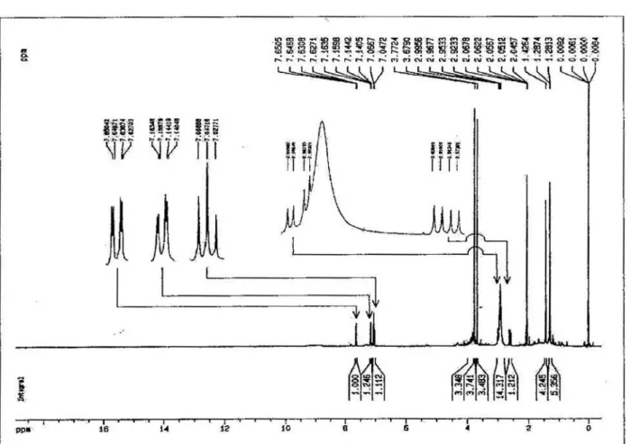
Compound 3 was isolated as an amorphous powder, the molecular formula was determined as C20H21NO6 by HRESIMS, showing an [M + H]+ peak at m/z 372.1447.
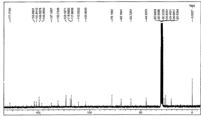
The UV absorption band at λmax 265, 285, 332 and 410 nm, IR absorptions at νmax 3509 and 1620 cm-1 and comparison with 1H and 13C NMR data characterized an acridone nucleus.16-22 The lack of a characteristic signal of hydrogen-bonded hydroxy proton in the 1H NMR spectrum, in acetone-d
6 and DMSO-d6, and the shielded
value of C-12 at d 177.7 in the 13C NMR spectrum suggested that there is no intramolecularly hydrogen-bonded hydroxyl group as observed for 1 and 2. The 1H NMR spectrum showed an ABX type aromatic spin system at d 7.04 (1H, t, J 7.8Hz, H-10), 7.15 (1H, dd, J
7.8, 1.5Hz, H-9) and 7.64 (1H, dd, J 7.8, 1.5 Hz, H-11). The spectrum also showed one N-methyl group at d 3.65 and one O-methyl group at d 3.77. The presence of a 3-hydroxy-2,2-dimethyldihydropyran moiety was suggested by an oxymethine signal at d 3.88 (1H, m, H-3), two methylene protons Ha at d 3.00 (1H, dd, J 16.8, 5.7Hz, Ha-4), Hb 2.61 (1H, dd, J 16.8, 7.8Hz, Hb-4) and two methyl groups at d 1.43 (3H, s, H-1') and 1.28 (3H, s, H-2'). The resonances at d 69.1 and 78.1 in the 13C NMR spectrum supported the presence of this substituent on the acridone nucleus.
In the HMBC spectrum of 3, the methylene protons at d 2.61 (H-4) showed correlations with C-4a (d 103.9), C-5 (d 153.3), C-3 (d 69.1) and C-2 (d 78.1), indicating that the substituent was located at an angular position. This spectrum also showed the correlation of the
N-methyl protons at d 3.65 with C-6a (d 143.2) and C-7a (d 137.1). The O-methyl protons at d 3.77 showed correlation with C-6 at d 130.4; this value is consistent with an acridone having an O-methyl group in this position.17,23 Correlations of H-9, H-10 and H-11 allowed the assignments of the carbons C-8, C-8a, C-9, C-10, C-11, C-11a and C-12. Therefore, compound 3 was identified as 3,4-dihydro-3,5,8-trihydroxy-6-methoxy-2,2,7-trimethyl-2H-pyrano[2,3-a]acridin-12(7H )-one. Table 3 shows the data of 1H, 13C NMR and the correlation observed in the HMBC.
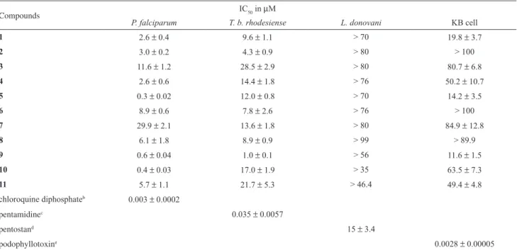
The eleven acridone alkaloids isolated from S. glutinosa
were tested for in vitro activity against P. falciparum, T. b. rhodesiense and L. donovani. An assay with KB cells indicated in vitro cytotoxicity. The results are summarized in Table 4. To facilitate the discussion, IC50 values were assigned as IC50T,P,K against T. b. rhodesiense, P. falciparum and KB cells, respectively.
Nine out of the eleven acridone alkaloids showed IC50P below 10 µM, four showed IC50T below 10 µM and none displayed significant activity against L. donovani.
Related acridone alkaloids from Thamnosma rodesica (Bak. F.), showed activity against promastigote and amastigote forms of Leishmania major. These Table 2. 1H and 13C NMR and HMBC data for compound 2 (acetone-d
6)
Position 13Cad 1Hbd (mult., J in Hz) HMBCb (H-C)
2 93.0 4.88 dd (9.4, 7.8) C-2’/C-3’, C-1’,C-3’
2ª 161.0
3 28.6 3.20 dd (15.5, 7.8, Ha) 3.26 dd (15.5, 9.4, Hb)
C-4, C-3a, C-2a, C-2
3ª 109.4
4 155.2
4-OH 14.31 s C-4a, C-4, C-3a
4a 108.4
5 182.6
5a 125.4
6 117.2 7.80 dd (7.8, 1.4) C-8, C-9a, C-5
7 120.4 7.15 t (7.8) C-9, C-5a
8 123.4 7.29 dd ( 7.8, 1.4) C-9a, C-6
9 148.8
9ª 138.0
11 126.6
11ª 143.5
1’ 71.5
2’ 26.1 1.33 s C-2, C-1’, C-3’
3’ 27.5 1.29 s C-2, C-1’, C-2’’
N-CH3 46.9 3.85 s C-11a, C-9a
O-CH3 60.9 3.89 s C-11
Antiparasitic Activities of Acridone Alkaloids from Swinglea glutinosa (Bl.) Merr. J. Braz. Chem. Soc.
648
alkaloids have a methyl 2,3-dihydroxypropanoate chain at C-3 and C-4,23 indicating that the substitution in the acridone skeleton is important for activity in this class of compounds.
According to the data, compound 5, with one prenyl group at C-2, was the most active against P. falciparum
with ICP
50 0.3 µM. Comparison of 5, 1 (IC P
50 2.6 µM) and 4 (ICP
50 2.6 µM) indicates that the second prenyl at C-8 was responsible for reducing the activity of this series. This fact was also observed by Weniger et al.,10 who performed the assay with four alkaloids from S. glutinosa on a Nigerian chloroquine-sensitive strain of P. falciparum. Analysis of the results for compounds 2, 3, 9, 10 and 11
suggests that the presence of a pyran ring is important for activity against P. falciparum and the position of this group, angular pyrano[2,3-c] (9) or linear pyrano[3,2-b] (10), did not alter the results of ICP
50.
The activity against P. falciparum observed for 6,
7 and 8,ICP
50 8.9, 29.9 and 6.1 µM, respectively, shows that presence of an O-methyl group at C-2 improves the activity.
From the 11 alkaloids tested against T. b. rhodesiense, compound 9 was the most active with an ICT
50 1.0 µM. Alkaloids 1-3 had their citotoxicity to cancer cells described in an earlier paper,30 however here we disclose for the first time their isolation, spectral data and structure elucidation.
Table 3. 1H and 13C NMR and HMBC data for compound 3 (acetone-d
6)
position 13Cad 1Hbd (mult.,
J in Hz)
HMBCb (H-C)
2ª 151,8*
2 78.1
3 69.1 3.88 m
4 27.3 3.00 dd (16.8, 5.7, Ha)
2.61 dd (16.8, 7.8, Hb)
C-3, C-2, C-1
4ª 103.9 5 153.3* 6 130.4 6ª 143.2 8 149.0 7ª 137.1
9 118.8 7.15 dd (7.8, 1.5) C-7a, C-11
10 123.1 7.04 t (7.8) C-8, C-11a
11 117.8 7.64 dd ( 7.8, 1.5) C-7a, C-9, C-12
11a 130.7
12 177.7
12a 110.9
1’ 20.5 1.43 s C-3, C-2, C-2’
2’ 26.0 1.28 s C-3, C-2, C-1’
N-CH3 44.9 3.65 s C-4a, C-5a
O-CH3 59.7 3.77 s C-4
a Recorded at 100 MHz; b Recorded at 400 MHz; * Assignments may
be exchanged.
Table 4. In vitro activity against P. falciparum 3D7, T. b. rhodesiense STIB 900, L. donovani L82 and KB cellsa
Compounds IC50 in µM
P. falciparum T. b. rhodesiense L. donovani KB cell
1 2.6 ± 0.4 9.6 ± 1.1 > 70 19.8 ± 3.7
2 3.0 ± 0.2 4.3 ± 0.9 > 80 > 100
3 11.6 ± 1.2 28.5 ± 2.9 > 80 80.7 ± 6.8
4 2.6 ± 0.6 14.4 ± 1.8 > 76 50.2 ± 10.7
5 0.3 ± 0.02 12.0 ± 0.8 > 70 14.2 ± 3.5
6 8.9 ± 0.6 7.8 ± 2.6 > 76 > 100
7 29.9 ± 2.1 13.6 ± 1.8 > 80 84.9 ± 12.8
8 6.1 ± 1.8 8.9 ± 0.9 > 99 > 89.9
9 0.6 ± 0.04 1.0 ± 0.1 > 56 11.6 ± 1.5
10 0.4 ± 0.03 17.0 ± 1.9 > 35 63.5 ± 7.3
11 5.7 ± 1.1 21.7 ± 5.3 > 46.4 49.4 ± 4.8
chloroquine diphosphateb 0.003 ± 0.0002
pentamidinec 0.035 ± 0.0057
pentostand 15 ± 3.4
podophyllotoxine 0.0028 ± 0.00005
a The mean IC
50 values of the test compounds and standard drug (n = 3, ±, σ, n = number of tests performed in three series). Drugs used as positive
Experimental
General
Optical rotations were measured using a Perkin Elmer polarimeter. IR spectra were recorded on a Bomem M-B Series spectrophotometer. UV absorptions were recorded using a Varian 500 SCAN UV-Vis-NIR spectrophotometer. 1H and 13C NMR data were recorded on Bruker ARX-200 and Bruker DRX-400 spectrometers. Spectra were recorded in acetone-d6 and DMSO-d6 with TMS as internal standard. All 2D NMR data were recorded at 400MHz (Bruker DRX-400), HSQC J 145 Hz; HMBC
J 8 Hz. HR-MS data were recorded on a Micromass Q-Tof (QqTOF) spectrometer; column chromatography was on silica gel 60 (Merck) and Sephadex LH-20 (Pharmacia). Preparative HPLC was performed on a Shodex Asahipak GS-310 2G column. TLC was carried out using Merck aluminum-backed silica gel 60 F254.
Plant material
Leaves, stem and root bark were collected in Campinas (SP) at the Instituto Agronômico de Campinas, and dried in the shade. The plant was identified by Prof. Dr. Maria Inês Salgado. A voucher specimen is deposited at the Herbarium of the Departamento de Botânica of the Universidade Federal de São Carlos (HUFSCar) as number 7110.
Extraction and isolation
The parts of the plant were extracted separately with n-hexane at room temperature for 3 days, filtered and evaporated under reduced pressure at 40 oC. This procedure was repeated 3 times to yield the crude hexane extract. The residue was extracted, as above, using MeOH to yield the crude MeOH extract. The crude MeOH extract (60 g) was fractioned by VCC over 1 kg of silica gel 60 (70-239 mesh, Merck) eluted with 1.5 L of the solvents: 100% hexane (fraction 1), 100% CH2Cl2 (fraction 2), 3:1 CH2Cl2:MeOH (fraction 3); 2:1 CH2Cl2:MeOH (fraction 4); 1:1 CH2Cl2:MeOH (fraction 5) and 100% MeOH (fraction 5). Fractions 2 and 3 were combined and chromatographed on silica gel using CH2Cl2 as mobile phase, the polarity was increased by addition of 5%, 10%, 13%, 18%, 20% and 25% of MeOH in a gradient system to yield 15 fractions. Lupeol was crystallized from fractions 1 to 3. Fraction 5 was rechromatographed on Si gel, being eluted with 1:3 n-hexane-acetone followed by Sephadex LH-20 with MeOH to give alkaloids 1 (16.3 mg), 4 (30.4 mg),
5 (3.5 mg), 8 (11.2 mg), 9 (13.4 mg), 10 (9.8 mg) and 11
(5.4 mg). Further purification of fraction 9 on Sephadex LH-20 with MeOH and HPLC using MeOH as mobile phase, UV detection at 254 and 365nm, and a flow rate of 3.0 mL min-1 afforded compounds 6 (13.5 mg), 7 (11.2 mg) and 8 (7.3 mg). Fraction 10 was dissolved in MeOH and submitted to Sephadex LH-20, being eluted with MeOH to give compounds 2 (5.2 mg) and 3 (4.3 mg).
1,3,5-Trihydroxy-4-methoxy-10-methyl-2,8-bis(3-methylbut-2-enyl)acridin-9(10H)-one,(1)
Amorphous powder, IR (liquid film) νmax/cm-1: 3500, 2964, 1625, 1566. UV (MeOH) λmax/nm (log ε) 265 (3.47), 285 (3.08), 332 (3.02) and 410 (2.57). 1H, 13C NMR and HMBC correlations see Table 1. HRESIMS m/z 424.2124 [M +H]+ (calc. for C
25H30NO5, 424.2124).
2,3-Dihydro-4,9-dihydroxy-2-(2-hydroxypropan-2-yl)-11-methoxy-10-methylfuro[3,2-b]acridin-5(10H)-one,(2)
Optically inactive, amorphous powder, IR (liquid film) νmax/cm
-1: 3475, 2964, 1630. UV (MeOH)λ
max/nm (log ε): 259 (4.21), 274 (4.62), 284 (4.65). 1H, 13C NMR and HMBC correlations see Table 2. m/z 372.1447 [M + H]+ (calc. for C20H22NO6, 372.1447).
3,4-Dihydro-3,5,8-trihydroxy-6-methoxy-2,2,7-trimethyl-2H-pyrano[2,3-a]acridin-12 (7H)-one,(3)
[α]25
D −21.4 (c, 0.0022 MeOH), amorphous powder, IR (liquid film) νmax/cm-1:3509, 2978, 1620, 1572. UV (MeOH) λmax/nm (log ε): 264 (3.39), 285 (3.13), 332 (2.99). 1H, 13C NMR and HMBC correlations see Table 3. HRESIMS m/z 372.1447 [M+ H]+ (calc. for C
20H22NO6, 372.1447).
Biological assays
Stock solutions of the compounds, plus control drugs, were prepared at a concentration of 20 mg mL-1 in DMSO (Sigma, UK), and diluted to appropriate concentrations prior to assays. IC50 values were calculated with MSXLFIT (IDBS, UK).
P. falciparum
Antiparasitic Activities of Acridone Alkaloids from Swinglea glutinosa (Bl.) Merr. J. Braz. Chem. Soc.
650
series and cultures incubated for a total of 48 h at 37 ºC in a 5% CO2-air mixture. After 24 h, {3H} hypoxanthine (0.2 mCi) was added to each well.25,26 At the end of the assay, plates were rapidly freeze-thawed, harvested using a Tomtec Mach III cell harvester (Tomtec, CT) onto a 96-well format filtermat and MeltilexTM solid scintillant (both Wallac, Finland) added prior to reading in a Microbeta 1450 scintillation counter (Wallac, Finland) at 1 min per well.
T. brucei rhodesiense
T. b. rhodesiense STIB900 bloodstream form trypomastigotes were maintained in HMI-18 medium,27 with 15% heat-inactivated fetal calf serum (Harlan Sera Lab, UK) at 37 °C in a 5% CO2-air mixture. Prior to drugging, trypomastigotes were washed and resuspended in fresh medium at a concentration 2 × 10⁵ trypanosoma/mL and 100 µL of this suspension was added to the drug dilutions. The top concentration for the test compounds was 30 mg mL-1. Pentamidine was included as the standard drug. Plates were incubated for 72 h at 37 ºC in a 5% CO2 -air mixture.28 At 72 h AlamarBlue was added to the plates. Plates were read after 4-5 h on a Gemini fluorescent plate reader (Sofimax Pro. 3.1.1, Molecular Devices, UK) at EX/EM 530/585 nm with a filter cut-off at 550 nm.
L. donovani
L. donovani L82 amastigotes were harvested from an infected hamster (Mesocricetus auratus) spleen and used to infect murine peritoneal exudate macrophages (PEM) at a ratio of 7:1. In brief, infected cells were exposed to drug for a total of 5 days.29 The percentage of infected cells was evaluated microscopically and the percentage inhibition in comparison with untreated controls was calculated.
Cytotoxicity assays
96-well plates were seeded with KB cells at 4 × 10⁴ mL-1 (100 µL per well). Drugs at 300, 30, 3 and 0.3 µg mL-1 were added in fresh overlay after 24 h, in triplicate at each concentration. Plates were incubated for 72 h at 37 °C in a 5% CO2-air mixture. At 72 h AlamarBlue was added to the plates. Plates were read after 4-5 h on a Gemini fluorescent plate reader (Sofimax Pro. 3.1.1, Molecular Devices, UK) at EX/EM 530/585 nm with a filter cut-off at 550 nm. IC50 values were calculated against the blanks and control samples.
Acknowledgments
The authors thank CNPq - Conselho Nacional de Desenvol vimento Científico e Tecnológico and Márcia
Ortiz Mayo Marques from the Instituto Agronômico de Campinas. Lauren Rattray and Simon Croft received support from UNDP/World Bank/WHO Research Programme for Tropical Diseases (TDR).
Supplementary Information
Available free of charge at http://jbcs.org.br, as PDF file.
References
1. Barrett, M. P.; Burchmore, R. J. S., Stich, A.; Lazzari J. O.; Frasch, A. C.; Cazzulo, J.; Krishnas, J.; Lancet2003, 362, 1469.
2. Hoet, S.; Opperdoes, F.; Brun R.; Quetin-Leclercq, J. Q.; Nat. Prod. Rep. 2004, 21, 353.
3. Legros, D.; Ollivier, G.; Gastellu-Etchegory, M.; Paquet, C.; Burri, C.; Jannin, J.; Buscher, P.; Lancet Infect. Dis. 2002, 2, 437.
4. Wernsdorfer, W. H.; Acta Tropica 1994, 56, 143. 5. Sachs, J.; Malaney, P.; Nature 2002, 415, 680.
6. WHO; www.rbm.who.int, accessed in February 18, 2004. 7. Sundar, S.; Trop. Med. Int. Health 2001, 6, 849.
8. Croft, S. L.; Parasitology 1997, 114, S3.
9. Da Silva, M. F. G. F.; Gottlieb, O. R.; Ehrendorfer, F.; Pl. Syst. Evol. 1988, 161, 97.
10. Weniger, B.; Um, B-H.; Valentin, A.; Estrada, A.; Lobstein, A.; Anton, R.; Maillé, M.; Sauvain, M.; J. Nat. Prod. 2001, 64, 1221. 11. Yamamoto, N.; Furukawa, H.; Ito, Y.; Yoshida, S.; Maeno, K.;
Nishiyama, Y.; Antiviral Res. 1989, 12, 21.
12. Kawaii, S.; Tomono, Y.; Katase, E.; Ogawa, K.; Yano, M.; Takemura, Y.; Motoharu, J.; Ito, C.; Furuhawa, H.; J. Nat. Prod. 1999, 62, 587.
13. Chaya, N.; Terauchi, K.; Yamagata, Y.; Kinjo, J.; Okabe, H.;
Biol. Pharm. Bull. 2004, 27, 1312.
14. Wu, T-S.; Kuoh, C-S.; Furukawa, H.; Phytochemistry 1983, 22,
1493.
15. Ito, C.; Kondo, Y.; Wu, T-S.; Furukawa, H.; Chem. Pharm. Bull. 2000, 48, 65.
16. Wu, T-S.; Furukawa, H.; Chem. Pharm. Bull. 1983, 31, 901. 17. Furukawa, H.; Yogo, M.; Wu, T-S.; Chem. Pharm. Bull. 1983,
31, 3084.
18. Wu, T-S.; Kuoh, C-S.; Furukawa, H.; Chem. Pharm. Bull. 1983,
31, 895.
19. Wu, T-S.; Furukawa, H.; Kuoh, C-S.; Hsu, K-S.; J. Chem. Soc., Perkin Trans. 1 1983, 1681.
20. Takemura, Y.; Wada, M.; Ju-ichi, M.; Ito, C., Furukawa, H.;
Chem. Pharm. Bull. 1998, 46, 693.
21. Pegel, K. H.; Wright, W.G.; J. Chem. Soc. (C) 1969, 2327. 22. Fraser, A. W.; Lewis, J.R.; J. Chem. Soc., Perkin Trans.1 1973,
23. Wu, T-S.; Chen, C-M.; Chem. Pharm. Bull. 2000, 48, 85. 24. Ahua, K. M.; Ioset. J-R.; Ransijn, A.; Mauël, J.; Mavi, S.;
Hostettmann, K.; Phytochemistry 2004, 65, 968.
25. Desjardins, R. E.; Canfield, C. J.; Haynes, J. D.; Chulay, J.D.;
Antimicrob. Agents Chemother. 1979, 16, 710.
26. O’Neill, M. J.; Bray, D. H.; Boardman, P.; Phillipson, J. D.; Warhurst, D.C.; Planta Med. 1985, 61, 394.
27. Hirumi, H.; Hirumi, K.; J. Parasitol. 1989, 75, 985.
28. Räz, B.; Iten, M.; Grether-Buler, Y.; Kamisky, R.; Brun, R.;
Acta Tropica 1997, 68, 139.
29. Neal, R.A.; Croft, S.L.; J. Antimicrob. Chemother. 1984, 14, 463.
30. Braga, P. A. C.; dos Santos, D. A. P.; da Silva, M. F. D. G. F.; Vieira, P. C.; Fernandes, J. B.; Houghton, P.J.; Fang, R.; Nat. Prod. Res. 2007, 21, 47.
Received: November 27, 2008
Web Release Date: April 3, 2009
S
u
p
p
le
m
e
nta
ry
Inf
o
rm
a
ti
o
n
J. Braz. Chem. Soc., Vol. 20, No. 4, S1-S8, 2009. Printed in Brazil - ©2009 Sociedade Brasileira de Química 0103 - 5053 $6.00+0.00
*e-mail: [email protected]
Antiparasitic Activities of Acridone Alkaloids from
Swinglea glutinosa
(Bl.) Merr.
Djalma A. P. dos Santos,a Paulo C. Vieira,*,a M. Fátima das G. F. da Silva,a
João B. Fernandes,a Lauren Rattrayb and Simon L. Croftb
aDepartamento de Química,Universidade Federal de São Carlos, CP 676, 13565-905 São Carlos-SP, Brazil
bDepartment of Infectious and Tropical Diseases, London School of Hygiene & Tropical Medicine,
London, WC1E 7HT, UK
Figure S1. 1H NMR spectrum of compound 1 (CDCl
Figure S2. 13C NMR spectrum of compound 1 (CDCl
3, 100 MHz).
dos Santos et al. S3 Vol. 20, No. 4, 2009
Figure S4. HMQC spectrum of compound 1 (CDCl3).
Figure S6. 1H NMR of compound 2 (CDCl
3, 400 MHz).
Figure S7. 13C NMR of compound 2 (CDCl
dos Santos et al. S5 Vol. 20, No. 4, 2009
Figure S8.HMBC spectrum of compound 2 (CDCl3).
Figure S10. High resolution mass spectrum of compound 2.
Figure S11. 1H NMR of compound 3 (CDCl
dos Santos et al. S7 Vol. 20, No. 4, 2009
Figure S12.13C NMR of compound 3 (CDCl
3, 100 MHz).
Figure S14. HMQC spectrum of compound 3 (CDCl3).