Artigo
Brazilian Journal of Pharmacognosy17(1): 01-07, Jan./Mar. 2007 Received 12/29/06. Accepted 02/26/07
Plants traditionally used against malaria: phytochemical and
pharmacological investigation of
Momordica foetida
Sonja Froelich
1, Bernadina Onegi
2, Adrian Kakooko
2, Karsten Siems
3, Carola Schubert
1,
Kristina Jenett-Siems
1*
1Institut für Pharmazie (Pharmazeutische Biologie), Freie Universität Berlin, Königin-Luise
Str. 2+4, D-14195, Berlin, Germany,
2Department of Pharmacy (Pharmacognosy Unit), Makerere University, Kampala, Uganda,
3Analyticon Discovery GmbH, Hermannswerder Haus 17, D-14473, Potsdam, Germany
RESUMO: “Plantas tradicionalmente utilizadas contra malária: investigação fi toquímica e farmacológica de Momordica foetida”.Folhas de Momordica foetida tradicionalmente usadas para tratar sintomas de malária em partes da África Oriental foram estudadas in vitro com respeito à sua atividade antimalárica. Utilizando-se o teste de incorporação de hipoxantina [3H] a atividade
antiplasmódica dos extratos hidrofílico e lipofílico contra a cepa sensíveis a cloroquina poW e o clone multiresistente Dd2 de Plasmodium falciparum foi determinada. O extrato éter de petróleo/ acetato de etila mostrou atividade signifi cativa com valores de CI50de 7,3 g/mL (poW) e 13,0
g/mL (Dd2). A citotoxicidade foi estimada em duas linhas de células humanas HepG2 e ECV-304 e o índice de seletividade (IS) foi calculado, variando entre 12 e 15. A análise fi toquímica levou ao isolamento de um número de glicosídeos fenólicos, por exemplo eriodictiol-, 5,7,4’-triidroxifl avanona, kaempferol e 5,7-diidroxicromona-7-O-ß-D-glicopiranosideo, descritos pela primeira vez de M. foetida. Na tentativa de provar um possível modo de ação foi avaliada a infl uência dos extratos brutos bem como dos compostos isolados sobre a degradação da heme usando-se cloroquina como composto de referência. O extrato acetato de etila mostrou inibição da degradação da heme em torno de 88%, bastante similar a cloroquina (84%) e também o eriodictiol-7-O-ß-D-glicopiranosideo (2) inibiu a degradação da heme em 86%.
Unitermos:Momordica foetida, Cucurbitaceae, glicosídeos de fl avonóides, teste de GSH-haemin, malária, medicina tradicional.
ABSTRACT: Leaves from Momordica foetida traditionally used to treat symptoms of malaria in parts of East Africa were studied for in vitro antimalarial activity. Using an [3H]
hypoxanthine-incorporation assay the antiplasmodial activity of hydrophilic and lipophilic extracts against the chloroquine-sensitive strain poW and the multiresistant clone Dd2 of Plasmodium falciparum was determined. The petrol ether/ethyl acetate extract showed signifi cant activity with IC50 values of 7.3g/mL (poW) and 13.0 g/mL (Dd2). Cytotoxicity was estimated on two human cell lines HepG2 and ECV-304 and a selectivity index (SI) was calculated, ranging between 12 and 15. Phytochemical analysis led to the isolation of a number of phenolic glycosides, e.g. eriodictyol-, 5,7,4’-trihydroxyfl avanone-, kaempferol- and 5,7-dihydroxychromone-7-O-ß-D-glucopyranoside, not previously known from M. foetida. In order to prove one possible mode of action the infl uence of the crude extracts as well as the isolated compounds on the heme degradation with chloroquine as reference compound was evaluated. The ethyl acetate extract showed about 88% inhibition of heme degradation, quite similar to chloroquine (84 %), and also eriodictyol-7-O-ß-D-glucopyranoside (2) inhibited heme degradation by 86 %.
Keywords: Momordica foetida, Cucurbitaceae, fl avonoid glycosides, GSH-haemin multiwell assay, malaria, traditional medicine.
INTRODUCTION
Momordica foetida Schumach. & Thonn. is a perennial climbing herb with tendrils, the fl owers are cream often with a reddish or orange centre. Male and female fl owers are found on the same plant. The characteristic fruit is bright orange with prickles. This species with its beautiful habit but a strong and unpleasant smell belongs to the Cucurbitaceae family.
The Cucurbitaceae consist of about 120 genera and 850 species that are widely distributed (Jeffrey, 1990). Many species are commercially grown for their nutritive value and in some cases they are used for medicinal purposes. In East and Central Africa, M. foetida is used to treat a number of ailments which include hypertension, diabetes mellitus, fever and especially symptoms of malaria (Hakizamungu et al., 1992; Rwangabo, 1993).
isolation of cucurbitane triterpenoids from a leaf extract (Mulholland, 1997), alkaloids and glycosides from the complete plant (Olaniyi; Marquis, 1975; Olaniyi, 1975) and the identifi cation of sitosteryl glycoside, 5,25-stigmastadien-3ß-yl-glucoside and 1ß-hydroxyfriedel-6-en-3-one (Olaniyi, 1980). In vivo studies with water extracts showed that M. foetida orally given in different doses prolonged the survival of Plasmodium berghei infected mice (Waako et al., 2005). In this study, we have examined the antiplasmodial activity, cytotoxicity and effects on the GSH-dependent heme degradation of crude plant extracts and isolated compounds from M. foetida.
MATERIAL AND METHODS
Spectrometric methods
1H NMR spectra were recorded on a Bruker
AVANCE DPX 400-spectrometer (400 MHz) using TMS as internal standard. Samples were dissolved in acetone-d6, chloroform-d1or methanol-d4. FAB-MS were obtained on a Varian MAT CH5DF instrument with DMSO/ Glycerin, Xenon; CHCl3/slm-Nitrobenzylalkohol, Xenon or MeOH/Glycerin, Xenon as solvents. EI-MS were recorded on a Varian MAT CH7A, (70 eV).
Plant material
M. foetida was collected in Kisaasi, a place 10 km north of Kampala. It was identifi ed by Mr. Adrian Kakooko, the botanist and plant taxonomist from the Makerere University, Kampala, Uganda. The general habitat range is in clearings and along forest edges. Voucher specimens are deposited in the herbarium of the Department of Pharmacy, Makerere University, Kampala, Uganda.
Extraction and isolation of compounds
Air dried leaves (345 g) were extracted with methanol (3 x 1.5 L). The extract was concentrated under reduced pressure yielding 52.2 g of the crude methanol extract (CME). The dry CME was acidifi ed with a 2 % (m/v) aqueous solution of tartaric acid. The aqueous layer was extracted with petrol ether (3 x 0.30 L), CH2Cl2 (3 x 0.30 L), EtOAc (3 x 0.30 L) and butanol (3 x 0.30 L). The evaporated EtOAc-extract (2.0 g) was dissolved in 5 mL methanol, and subjected to chromatography on a glass column (2.5 x 34 cm) packed with Sephadex LH 20 (40 g) and eluted with 0.25 L methanol to give 50 fractions. From fractions 13-17 compound 1 (4 mg) crystallized spontaneously. From fractions 11-12 compound 3 was isolated by preparative TLC (HCO2H-H2O-EtOAc 9:9:82, Rf 0.49, 7 mg).
Fractions 18-24 yielded three compounds 2 (Rt = 370 s, 2.6 mg), 4 (Rt = 723 s, 4.1 mg) and again 1 (Rt = 492 s, 4.2 mg) upon preparative HPLC (H2O-MeOH
70:30, after 20 min 30:70, after 30 min 1:99 MeOH). The CH2Cl2-extract (0.29 g) was subjected to column chromatography on LiChroprep® RP-18 material [Merck, 40-63 m] (H2O-MeOH 60:40 to MeOH) yielding 123 fractions. From fractions 5-14 crystals were obtained, which were further purifi ed by preparative TLC (CHCl3-EtOAc 60:40) to give compound 5 (Rf 0.6, 12.6 mg).
Fractions 15-64 were purifi ed by preparative TLC (cyclohexane-EtOAc 50:50) and compound 6 (Rf 0.06, 5 mg) was obtained.
Chromatographic system
Analyses were carried out on a Knauer HPLC Pump 64 apparatus, Knauer HPLC Programmer 50, barspec Chrom-A-Scope Detector (Knauer, Berlin, Germany).
A reversed phase mode column Eurospher 100 C18 (250 x 4.0 mm I.D., 5 m) was employed (Knauer, Berlin, Germany) at room temperature of 25°C, fl ow rate of 1.5 mL/min and wavelength of 225 nm. Solvents used were of HPLC grade (LGC Promochem, Wesel, Germany) and were degassed by sonication before use.
5 , 7 , 4 ’ Tr i h y d ro x y f l a v a n o n e 7 O ß D -glucopyranoside (Prunin) (1): (-) FAB-MS m/z433 [M-H]-;1H NMR (Acetone-d
6, 400 MHz): į 2.77 (1H, dd,J =
3.0 Hz, J = 17.0 Hz, H-3eq), 3.23 (1H, dd,J= 13.0 Hz, J = 17.0 Hz, H-3ax), 3.47 (2H, t,J = 8.8 Hz, H-2’’, H-3’’), 3.56 (1H, t,J = 8.8 Hz, H-4’’), 3.60 (1H, m, H-5’’), 3.70 (1H,dd,J= 5.3 Hz, J= 11.8 Hz, H-6’’a), 3.88 (1H, dd, J = 2.34 Hz, J = 11.8 Hz, H-6’’b ), 5.07 (1H, d,J = 7.4 Hz, H-1’’), 5.49 (1H, dd,J= 3.0 Hz, J = 13.0 Hz, H-2ax), 6.14 (1H, d,J = 2.2 Hz, H-6), 6.17 (1H, d,J = 2.2 Hz, H-8), 6.90 (2H, d,J = 8.6 Hz, H-3’,H-5’), 7.38 (2H, d,J = 8.6 Hz, H-2’, H-6’), 12.07 (1H, s, 5-OH).
5,7,3’,4’-Tetrahydroxyflavanone-7-O-ß-D-glucopyranoside (Eriodictyol-7-O-ß-D-5,7,3’,4’-Tetrahydroxyflavanone-7-O-ß-D-glucopyranoside) (2): (-) FAB-MS m/z449 [M-H]-;1H NMR (Acetone-d
6,
400 MHz): į 2.77 (1H, dd,J = 3.0 Hz, J = 17.1 Hz, H-3eq), 3.20 (1H, dd,J = 12.6 Hz, J = 17.1 Hz, H-3ax), 3.47 (2H,t,J = 9.3 Hz, H-2’’, H-3’’), 3.53 (1H, t,J= 8.9 Hz, H-4’’), 3.61 (1H, m, H-5’’), 3.70 (1H, dd,J = 5.4 Hz, J= 11.9 Hz, H-6’’a), 3.89 (1H, dd,J = 2.9 Hz, J = 11.9 Hz, H-6’’b), 5.08 (1H, d, 7.6 Hz, H-1’’), 5.44 (1H, dd,J = 3.0 Hz,J = 12.6 Hz, H-2ax), 6.13 (1H, d,J = 2.0 Hz, H-8), 6.16 (1H, d,J = 2.0 Hz, H-6), 6.88 (2H, s, H-2’, H-5’), 7.04 (1H, s, H-6’), 12.07 (1H, s, 5-OH).
5 , 7 D i h y d r o x y c h r o m o n e 7 O ß D -glucopyranoside (3): (+) FAB-MS m/z363 [M+Na]+;1H
K a e m p f e ro l - 7 - O - ß - D - g l u c o p y r a n o s i d e (Populnin) (4): (-) FAB-MS m/z447 [M-H]-; 1H NMR
(Acetone-d6, 400 MHz): į 3.28 (1H, m, H-5’’), 3.36 (1H, t,J = 8.5 Hz, H-2’’), 3.45 (2H, m, H-3’’, H-4’’), 3.51 (1H, dd,J = 5.5 Hz, J = 11.5 Hz, H-6’’a), 3.64 (1H, dd,J = 3.0 Hz,J = 11.5 Hz, H-6’’b), 5.26 (1H, d,J = 7.5 Hz, H-1’’), 6.29 (1H, d,J = 1.9 Hz, H-6), 6.53 (1H, d,J = 1.9 Hz, H-8), 6.98 (2H, d,J = 8.9 Hz, H-3’, H-5’), 8.15 (2H, d,J = 8.9 Hz, H-2’, H-6’), 12.44 (1H, s, 5-OH).
5,7-Dihydroxychromone (5): EI-MS m/z (rel. int. %) 178 [M]+ (100), 150 [M-CO] (35), 124 (26), 111 (14),
97 (22). 1H NMR (Methanol-d
4, 400 MHz): į 6.14 (1H, d,
J = 6.0 Hz, H-3), 6.15 (1H, d,J = 2.0 Hz, H-8), 6.27 (1H, d,J = 2.0 Hz, H-6), 7.93 (1 H, d,J= 6.0 Hz, H-2).
2-Hydroxybenzoic acid (Salicylic acid) (6): EI-MSm/z (rel. int. %) 138 [M]+ (54), 120 [M-H
2O] (100),
92 (53), 64 (19), 39 (16). 1H NMR (CDCl
3, 400 MHz): į
6.90 (1H, dt, 0.5 Hz, 8.0 Hz, H-4/H-5), 7.0 (1H, dd, 0.5 Hz, 8.0 Hz, H-3), 7.48 (1H, dt, 0.5 Hz, 8.0 Hz, H-4/H-5), 7.90 (1H, dd, 0.5 Hz, 8.0 Hz, H-6), 10.62 (1H, br s, -COOH).
Materials
RPMI 1640 medium was purchased from Gibco-BRL. GSH and diethylenetriamine-penta-acetic acid (DETEPAC) were obtained from Lancaster. Haemin, HEPES, Na phosphates, NaHCO3 and DMSO were obtained from Roth. [3H] Hypoxanthine was purchased
from Amersham. Flat-bottomed 96-well plates were obtained from Neolab.
In vitro antiplasmodial activity
The chloroquine-sensitive strain of P. falciparum poW (IC50 of chloroquine = 0.011 M) and a chloroquine-resistant clone Dd2 (IC50 of chloroquine = 0.12 M) were maintained in continuous culture in human red blood cells (A+) diluted to 5% haematocrit in RPMI 1640 medium
supplemented with 25 mM HEPES, 30 mM NaHCO3, and 10% human A+ serum (Trager and Jensen, 1976).
The extracts were prepared as follows: 100 g of powder were extracted twice with equal volumes of petrol ether /ethyl acetate (300 mL) at room temperature
O
O OH O
O
OH
O
O OH O
O
OH OH CH2OH
OH
R
HO
O
O OH O
O
OH
O
O OH O
O
OH OH CH2OH
HO
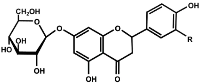
1 R = H = 5,7,4’-Trihydroxyfl avanone-7-O-ß -D-glucopyranoside = Prunin
2 R = OH = 5,7,3‘,4‘-Tetrahydroxyfl avanone-7-O-ß -D-glucopyranoside = Eriodictyol-7-O-ß-D-glucopyranoside
Figure 1. Structures of fl avanone glycosides isolated fromM. foetida.
for 24 hours. Afterwards the plant powder was air dried and treated three times with 300 mL methanol. In either case, the solvents were evaporated from the combined solutions of each batch affording the corresponding crude drug extracts. The latter were dissolved in DMSO (1 mg/50 L) and diluted in RPMI 1640 medium. For determination of the IC50 values the samples were diluted to give fi nal concentrations between 100 and 1.56 g/ mL.
Antiplasmodial activity tests were performed in 96-well culture plates (Corning™, Sigma Aldrich) as described by Desjardins et al. (1979). Briefl y, aliquots of 150 L of parasitized culture (2.5% haemocrit, 0.5% parasitaemia) were exposed to two-fold dilutions of test substances. After incubation in a candle jar for 24 h, 0.5 Ci of [3H] hypoxanthine (1mCi/mL) was added to each
well and the plates incubated for a further 18 h.
Cells were harvested onto glass fi bre fi lters (Wallac) with a cell harvester (Inotech) and incorporated radioactivity was determined by a liquid scintillation counter (1450 Microbeta plus). All tests were performed in triplicate. The percentage of growth inhibition was calculated as: [1-(cpm in drug treated cultures/cpm in untreated cultures)] x 100. The concentration at which growth was inhibited by 50% (IC50) was estimated by interpolation.
Multiwell plate GSH-haemin interaction assay
The GSH-haemin interaction assay was performed as described by Steele et al. (2002). In brief, three stock solutions were prepared: 1mM DETEPAC in
O
OH
O OH O
O
OH
O
OH
O OH O
O
OH OH
CH2OH
OH HO
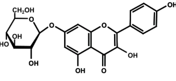
Figure 3. Kaempferol-7-O-ß-D-glucopyranoside = Populnin (4).
84
21
4 2
86
4 13
88
44 38
0 10 20 30 40 50 60 70 80 90 100
Chlo roqu
ine Quin
ine
Arte mis
inine [1
]
[2] [3] [4]
EtOAc -ext
ract
Dic hloro
meth an-e
xtra ct
Petrol ethe
r ext ract
%
I
n
hi
bi
ti
o
n
Plant species extract P. falciparum a
IC50 (µg/mL)
P. falciparum b
IC50 (µg/mL)
ECV-304 IC50 (µg/mL)
SI ECV/Pf a
HepG2 IC50 (µg/mL)
SI HepG2/Pf a
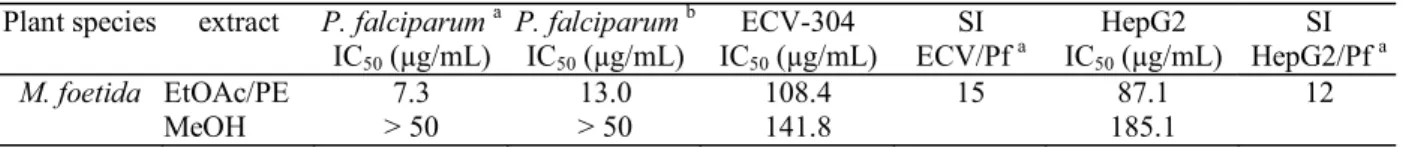
M. foetida EtOAc/PE 7.3 13.0 108.4 15 87.1 12
MeOH > 50 > 50 141.8 185.1
Table 1. In vitro antiplasmodial activity of Momordica foetida leaf extracts on Plasmodium falciparum (poW- and Dd2-strain) and cytotoxicity on human cell lines ECV 304 and HepG2.
10 mM Na phosphate pH 7.0; 2 mM haemin in DMSO (prepared fresh daily); 100 mM GSH, 1 mM DETEPAC, 10 mM Na phosphate pH 6.8. For the experiments, working solutions were as follows: ‘A’, 4 vol. of DETEPAC/phosphate stock + 1 vol. of ethanol; ‘B’, 5 L of haemin stock solution per mL of solution ‘A’; ‘C’, 0.15 mL of GSH stock solution per mL of solution ‘A’.
Assays were carried out in 96-well (400 L)fl at-bottomed plates. Solution A (100 L) was added, followed by drug (2 L of 2 mM drug stock in DMSO) or solvent control in eight parallel samples. Solution B (200 L) was then added to all wells followed by 50 L of solution C. Final concentrations of drug, haemin and crude extract were 11 M, 5.7 M and 100 g/mL, respectively. The absorbance at 360 nm (A360) was measured after 1 and 30 min with a plate reader Tecan Spectra Fluor to determine the∆A
360. The effect of the haemin-binding compounds
was evaluated as the percentage decrease compared with control absorbance. Mean and SD for eight parallel samples of at least three independent experiments were calculated.
Cytotoxicity
Cytotoxicity of the extracts was estimated against human hepatocellar carcinoma (HepG2) and human urinary bladder carcinoma (ECV-304) (derivative of T-24) cells. Cytotoxicity was estimated by a proliferation assay using the MTT-assay (Mosmann, 1983; Freshney, 1990). Crude extracts were dissolved in DMSO and diluted with medium to the desired concentrations.
Human urinary bladder carcinoma cells (ECV-304) were cultivated in Earle Medium 199 supplemented with 10% fetal calf serum in 96 well plates in an atmosphere of 5% CO2at 37°C in a humidifi ed environment. The ECV-304 cells were seeded at a density of approximately 1000 cells per well. After 24 h they were supplemented with 100 l extract or substance in medium and cultivated for further 4 days. The cell viability was measured by the MTT-assay using DMSO to dissolve the formed purple formazan. The absorbance was quantifi ed at 580 nm with a plate reader. Data are presented as the mean of 3 independent experiments with 8 parallel samples for each concentration. The IC50 values were calculated by linear regression.
The human hepatocellar carcinoma (HepG2) cells were cultivated in MEM Earle medium supplemented with 10% fetal calf serum, 5 mL sodium pyruvate solution and 5 mL non-essential amino acids solution in 96 well plates in an atmosphere of 5% CO2 at 37°C in a humidifi ed environment. The HepG2 cells were seeded at a density of approximately 2000 cells per well.
RESULTS AND DISCUSSION
Antiplasmodial activity and cytotoxicity of extracts
In this study, we evaluated the in vitro antiplasmodial activity of hydrophilic and lipophilic extracts of M. foetida leaves against the chloroquine-sensitive strain poW and the multiresistant clone Dd2 using an [3H] hypoxanthine-incorporation assay. The
petrol ether/ ethyl acetate extract showed signifi cant antimalarial activity with IC50 values of 7.3 (poW) and 13.0g/mL (Dd2).
Cytotoxicity of the extracts was estimated against human hepatocellar (HepG2) and human urinary bladder carcinoma (ECV-304, derivative of T-24) cells.
Both extracts did not show pronounced toxicity (IC50 values > 50 g/mL) and the selectivity indices (SI), defi ned as ratio of cytotoxic and antiplasmodial activity, which are presented in table 1, varied between 12 (HepG2) and 15 (ECV-304) (see table). These results might support the traditional use of M. foetida as antimalarial remedy.
Phytochemical analysis
The ethyl acetate soluble fraction was subjected to chromatography on Sephadex LH 20. Elution with methanol afforded compounds 1-4 (Fig. 1-3). Final purifi cation was achieved by reverse phase HPLC and TLC. The compounds were identifi ed as the known
flavanone derivatives 5,7,4’-trihydroxyfl avanone-7-O-ß-D-glucopyranoside (1), also known as prunin, which was isolated before from the bark of Salix acutifolia Willd. (Zapesochnaya et al., 2002), 5,7,3’,4’-tetrahydroxyflavanone-7-O-ß-D-glucopyranoside (Eriodictyol-7-O-ß-D-glucopyranoside) (2) (Mun’im et al., 2003), the fl avonole glycoside kaempferol-7-O-ß -D-glucopyranoside (Populnin) (4) (Harbourne, 1994) and 5,7-dihydroxychromone-7-O-ß-D-glucopyranoside (3)
apoW-strain = IC
50 value for chloroquine = 0.011 M bDd2-strain = IC
(Simon et al., 1994). None of these substances has been reported from M. foetida before.
Column chromatography of the dichloromethane extract of M. foetida leaves and further purifi cation by TLC afforded 5and6. Compound 5 turned out to be 5,7-dihydroxychromone, which is known, e. g. from Calluna vulgaris (Ericaceae) (Simon et al., 1994), but has never been detected before in the genus Momordica.
Compound6 was identifi ed as salicylic acid on the basis of its 1H NMR and EI-MS spectra, a compound
widespread in the plant kingdom (Hegnauer, 1964).
Infl uence on GSH-dependent haemin degradation
In order to check one possible antiplasmodial mode of action we evaluated the infl uence of the crude extracts of M. foetida as well as the isolated compounds on the glutathione-dependent heme degradation with chloroquine as reference compound.
WhenPlasmodium falciparum infects a human erythrocyte, it feeds on the degradation of hemoglobin. Hemoglobin is ingested from the host cell and is digested inside the parasites food vacuole. The by-product of this digestion is toxic heme or ferriprotoporphyrin, which is detoxifi ed by forming an insoluble polymer, malaria pigment or hemozoin. Recently, an alternative heme detoxifi cation mechanism has been described (Ginsburg et al., 1998).
Nonpolymerised heme exits the food vacuole into the parasites cytosol where it is degraded by glutathione. Quinoline antimalarials like chloroquine have been shown to interfere with both ways. Chloroquine accumulates to high levels inside the acid food vacuole where it complexes with heme and prevents its polymerisation. Free or complexed heme exits the food vacuole where its glutathione-dependent degradation is also inhibited. Heme thus accumulates in membranes and permeabilises them to cations, leading to parasite’s death.
The reaction of hemin with glutathion and the interference of antimalarials can be monitored spectrophotometrically. The broad absorption of hemin is immediately changed to a peak at 364 nm upon addition of glutathione. During 30 min this peak decreases to approximately half without further decrease. Addition of quinoline antimalarials to hemin results fi rst in the alteration of the hemin spectrum due to immediate formation of an alkaloid-hemin-complex and then leads to altered effects of glutathione. In the case of chloroquine the 364 nm peak is not formed and there is little change over 30 min. These fi ndings were used by Steele and co-workers to develop a microassay, which allows the identifi cation of compounds that are able to inhibit the degradation of heme by GSH.
Among the crude extracts of M. foetida, only the ethyl acetate extract was able to interfere with haemin degradation, inhibiting it by ~ 88 % at 100 g/
mL (Fig. 4). Compounds 1-4, which were obtained from this extract, were evaluated too. Eriodictyol-7-O-ß -D-glucopyranoside (2) showed an inhibition of 86 % similar to chloroquine with 84 %. Thus, a catechol sub-structure seems to be necessary for an interaction with haemin in this type of compounds. This is additionally stressed by the activity of quercetin in this assay (results not shown). Nevertheless, the antiplasmodial potency of 2 remains to be proven.
ACKNOWLEDGEMENTS
The authors are grateful to Mrs. U. Ostwald (Institut für Organische Chemie, FU Berlin) for providing the FAB-MS spectra. Thanks are due to Mrs. U. Friedrich for technical assistance in the cell laboratory.
REFERENCES
Desjardins RE, Canfi eld CJ, Haynes JD, Chulay JD 1979. Quantitative assessment of antimalarial activity by a semi-automated microdultion technique. Antimicrob Agents Chemother 16: 710-718.
Freshney RI 1990. Tierische Zellkulturen: Ein Methoden-Handbuch 2. Aufl age. Berlin, New York: De Gruyter p. 259-260.
Ginsburg H, Famin O, Zhang J, Krugliak M 1998. Inhibition of glutathione-dependent degradation of heme by chloroquine and amodiaquine as a possible basis for their antimalarial mode of action. Biochem Pharmacol 56: 1305-1313.
Hakizamungu E, Van Puyvelde L, Wery M 1992. Screening of Rwandese medicinal plants for anti-trichomonas activity. J Ethnopharmacol 36: 143-146.
Harbourne JB 1994. The Flavonoids. London, New York:
Chapmann & Hall.
Hegnauer R 1964. Chemotaxonomie der Pfl anzen III. Basel und Stuttgart: Birkhäuser Verlag Vol. 3, p. 34-39.
Jeffrey C 1990. Cucurbitaceae in Flora of Tropical East Africa. London: Crown Agents.
Mosmann T 1983. Rapid colorimetric assay for cellular growth and survival: application to proliferation and cytotoxicity assays. J Immunol Methods 65: 55-63. Mulholland DA, Sewram V, Osborne R, Pegel KH, Connolly
JD 1997. Cucurbitane triterpenoids from the leaves of
Momordica foetida.Phytochemistry 45: 391-395. Mun’im A, Osamu N, Ozawa T 2003. Antioxidative compounds
from Crotalaria sessilifl ora. Biosci Biotechnol Biochem 67: 410-414.
Olaniyi AA 1975. A neutral constituent of Momordica foetida. J Nat Prod 38: 361-362.
Olaniyi AA, Marquis VO 1975. Phytochemical and preliminary pharmacological investigation of the alkaloid obtained fromMomordica foetida.Journal of Pharmacy (Yaba Nigeria) 6: 117-119.
Olaniyi AA 1980. 1 ß-Hydroxyfriedel-6(7)-ene-3-one from
Momordica foetida.J Nat Prod 40: 303-305. Rwangabo PC 1993. La medicine traditionnelle au Rwanda.
Paris: Edition Karthala and ACCT.
Simon A, Chulia AJ, Kaouadji M, Delage C 1994.
chromones from Calluna vulgaris. Phytochemistry 36: 1043-1045.
Steele JCP, Phelps RJ, Simmonds MSJ, Warhurst DC, Meyer DJ 2002. Two novel assays for the detection of haemin-binding properties of antimalarials evaluated with compounds isolated from medicinal plants. J Antimicrob Chemoth 50: 25-31.
Trager W, Jensen JB 1976. Human malaria parasites in continuous culture. Science 193: 673-675.
Waako PJ, Gumede B, Smith P, Folb PI 2005. The in vitro
and in vivo antimalarial activity of Cardiospermum halicacabumL. and Momordica foetida Schumach. et Thonn.J Ethnopharmacol 99: 137-143.
Zapesochnaya GG, Kurkin VA, Braslavskii VB, Filatova NV 2002. Phenolic compounds of Salix acutifolia Bark.