REVIEW
Human lagochilascariasis—A rare helminthic
disease
Dulcinea Maria Barbosa Campos1*, Alverne Passos Barbosa2, Jayrson Arau´jo de Oliveira3, Giovana Galvão Tavares1, Pedro Vitor Lemos Cravo1,2,4, Alejandro
Luquetti Ostermayer2
1Programa de Po´s-Graduac¸ão em Sociedade Tecnologia e Meio Ambiente e Curso de Farma´cia, Centro Universita´rio de Ana´polis–UniEVANGE´ LICA, Ana´polis, Goia´s, Brazil,2Instituto de Patologia Tropical e Sau´de Pu´blica, Universidade Federal de Goia´s, Goiaˆnia, Goia´s, Brazil,3Instituto de Ciências Biolo´gicas, Universidade Federal de Goia´s, Goiaˆnia, Goia´s, Brazil,4Instituto de Higiene e Medicina Tropical, Universidade Nova de Lisboa, Lisboa, Portugal
Abstract
Lagochilascariasis is a parasitic disease caused by a helminth of the order Ascaroidea, genusLagochilascaristhat comprises 6 species, among which onlyLagochilascaris minor Leiper, 1909, is implicated in the human form of the disease. It is remarkable that the major-ity of cases of human lagochilascariasis in the Americas have been reported in Brazil. The natural definitive hosts of this parasite seem to be wild felines and canines. Lagochilascaria-sis is mostly a chronic human disease that can perLagochilascaria-sist for several years, in which the para-site burrows into the subcutaneous tissues of the neck, paranasal sinuses, and mastoid.L. minorexhibits remarkable ability to migrate through the tissues of its hosts, destroying even bone tissue. Fatal cases have been described in which the parasite was found in the lungs or central nervous system. Treatment is often palliative, with recurrence of lesions. This paper summarizes the main features of the disease and its etiologic agent, including preva-lence, life cycle, clinical course, and treatment.
Introduction
Robert T. Leiper, 1909, a helminthologist at the London School of Tropical Medicine and Hygiene who received specimens of a Nematoda recovered from subcutaneous abscesses of 2 patients from Trinidad (off the coast of South America), was the first to describe Lagochilas-caris minor. At that time, the digestive tract of a carnivore was suggested as the probable habitat forL.minor. He proposed that the finding of parasites in subcutaneous abscesses in humans from Trinidad was remarkable evidence that an animal, other than a human, could be the definitive host for this helminth [1]. Since then, more than 100 cases of purulent abscesses in humans have been reported in different countries in the Americas.L.minorhas been found in subcutaneous abscesses in the cervical region [1–5], mastoid [4–9], rhino-oro-pharynx [4,5,9,10], tonsils [4,5,11,12], auditory meatus [4,5,10,13], nasal sinuses [9,13–15], lungs [4,5,16], central nervous system [6,13,17],sacral region [4], eyes [10,18], and dental alve-oli [5,9,19] of humans. Different stages of the life cycle (eggs, larvae, and adult worm) have also
a1111111111 a1111111111 a1111111111 a1111111111 a1111111111 OPEN ACCESS
Citation:Campos DMB, Barbosa AP, Oliveira JAd, Tavares GG, Cravo PVL, Ostermayer AL (2017) Human lagochilascariasis—A rare helminthic disease. PLoS Negl Trop Dis 11(6): e0005510.
https://doi.org/10.1371/journal.pntd.0005510
Editor:Philip J. Cooper, Universidad San Francisco de Quito, ECUADOR
Published:June 22, 2017
Copyright:©2017 Campos et al. This is an open access article distributed under the terms of the
Creative Commons Attribution License, which permits unrestricted use, distribution, and reproduction in any medium, provided the original author and source are credited.
Funding:The authors received no specific funding for this work.
been found [5,14,20,21]. It is remarkable that all these organs differ from the digestive tract, the normal habitat of all other ascarides. Corroborating Leiper’s, 1909, assumption, a wild felid,Puma concolor, naturally infected byL.minorwas recently found in Mexico [22].
Methods
A literature review on the subject was conducted, using publications from this group and other scientific articles published in indexed journals in the Latin American and Caribbean Health Sciences database (LILACS), Scientific Electronic Library Online (SciELO), andIndex Medicus(MEDLINE). The authors of this manuscript did not participate in any clinical activ-ity involving humans. Human involvement was limited to the contribution of parasitological diagnosis in case reports, whose photographs were authorized by the patients. From the stand-point of experiments using animals, based on Brazil’s Federal Law No. 11,794 of 8 October 2008, the projects of this group were carried according to the protocol approved by the Ethics Committee on Animal and Human Medical Research of the Clinical Hospital at the Federal University of Goia´s and, subsequently, by the Ethics Committee on Animal Use (CEUA) of the Federal University of Goia´s, which is subordinate to the National Council for the Control of Animal Experimentation (CONCEA).
Geographical distribution and prevalence
Human lagochilascariasis has been recorded in Trinidad and Tobago [1,2,18,23], Surinam [14,20,24,25], Mexico [9,26], Costa Rica [27], Venezuela [6,15,28], Colombia [11,12], Bolivia [29], Ecuador [30], Paraguay [7], Brazil [3–5,10,13,16,21,31–35] and Peru [36] (Fig 1). As for its geographic distribution by country, the highest concentration is found in Brazil, represent-ing 78.1% (100/128) of the total number of cases reported in the literature [36]. Among the cases in Brazil, the majority (60%, 60/100) were recorded in the state of Para´, followed by Ron-doˆnia, Tocantins, Mato Grosso, Acre, Roraima, São Paulo, and Parana´ (Fig 1). It should be noted that only 1 case of human lagochilascariasis has been recorded in each of the following states: Maranhão, Paraı´ba, Mato Grosso do Sul, and Goia´s [36].
The disease in humans
Lagochilascariasis in humans is often misdiagnosed as an abscess of common causes, mainly of bacterial etiology. It presents more often as a subcutaneous abscess on the neck (Fig 2), mid-dle ear (Fig 3), mastoid, tonsils, and nasal sinuses, which physicians in countries in the Neo-tropical region should check.
Other common localizations of the parasite are the central nervous system, lungs, sacral region, eyeballs, and dental alveoli [21,34,35]. Usually, the patient seeks medical help after sev-eral days of discomfort and is examined by sevsev-eral physicians, showing poor response to anti-biotics [9]. The lesion usually evolves slowly over weeks or months. Some patients report the formation of a tumor in the neck, initially small, without fistula and with no pain [4,5,21,35]. As the disease progresses, the tumorous lesion becomes painful, with or without spontaneous fistula, with drainage of a serous purulent exudate, generally containing small whitish worms, eggs, and larvae [4,5,21,35]. The size of the tumorous lesion may vary from 5 to 12 cm and is usually found in the cervical region, with the aspect of a pseudocyst, nodule, or abscess [8,37]. It is usually a painful lesion with a hard consistency and undefined limits. The migration of the parasite through the host’s tissues originates secondary lesions that can be located close to or very far from the initial abscess [4,5].
be able to control the pathogenic processes, as well as limit the establishment of new lesions [5].
Some reports refer to chronic abscesses of the auditory meatus (with purulent exudate for 1 or 2 years) and painful tumor in the mastoid [7,9], which may progress to neurological involvement [13,17]. Otalgia and purulent otorrhea have been recorded. Otoscopy in the right ear shows retroauricular swelling, polyps in the external ear canal, and fistula with drainage of pus [38]. In cases of otitis and mastoiditis, the X-ray examination shows extensive areas of osteolysis in the mastoid region [13]. The osteolytic capacity of this parasite has also been reported in the destructive lesion of the sacral bone, as well as of the 4thand 5thlumbar verte-brae and soft tissues adjacent to the sacrum [4]. In other cases, lesions in the middle ear and mastoid extend to the base of the skull, evolving into extradural abscesses and instances of neck stiffness. The preceding phase may be characterized by a history of ear drumming, intense headache with the pain radiating to the hemiface, and, finally, elimination of worms through the oral cavity [13,32]. Other authors have reported finding only 1 nodule with fistula
Fig 1. Geographical distribution of human lagochilascariasis in the Americas.
in the neck in patients at the time of diagnosis and elimination of the adult worms through the oral cavity and auditory meatus a few months later [4,5,11].
Neurological involvement due toL.minorinfection may develop in the absence of lesions in the neck, with clinical manifestations such as seizures, headache, paresthesia, motor alter-ations, cerebellar ataxia, and mental confusion [17]. Vomiting, papilledema, and facial paraly-sis have been described as well [13]. A fatal case of encephalopathy due toL.minorwith a subacute, progressive disease characterized by headache, stupor, and coma has been reported [17]. In this patient, death occurred 3 months after the beginning of the illness. Neuropatho-logical examination revealed diffuse foci of necrosis of the cerebral hemispheres and cerebel-lum and presence of the nematode in the parenchyma and in the cisterns at the base of the brain [17].
Bilateral bronchopneumonia, with hundreds of abscesses measuring 2–5 mm scattered throughout the pulmonary parenchyma, has been reported [16]. Microscopically, in lungs, in each low-power field, at least 1 abscess was visible, the majority of them containing fourth stage larvae, young adults, or mature adult worms. In addition to the unusualness of its loca-tion in the lungs, all the evoluloca-tionary stages of the worm were found in the affected organ, i.e., eggs, larvae, and adult worms [16], a fact that characterizes the autoinfecting cycle of the para-site. There are reports of pneumonia occurring with fever and dyspnea, which progressed to
Fig 2. Cervical lesion draining pus in a patient infected withLagochilascaris minor.
cyanosis, respiratory insufficiency, and death less than 3 months after the onset of symptoms [16].
Chronic tonsillitis occurring with the sensation of worms moving through the throat, elimi-nation of worms through the mouth, sensation of ingesting worms, headache, hearing loss, and overall debility have been observed in infections of tonsils and the middle ear [11].
An important clinical issue is the distinction between otitis, mastoiditis, sinusitis, and ton-sillitis caused byL.minorinfection and other related diseases. Clinicians, particularly otorhi-nolaryngologists and neurologists, working in Neotropical regions should be attentive to information about the discharge of adult worms through nasal sinuses, mouth, or auditory meatus [5]. The proteolytic enzymes inL.minorcan facilitate its migration through the host’s tissues by hydrolyzing collagens of the extracellular matrix [39].
The life cycle
An experimental model involving mice and domestic cats was described in an attempt to unravel the life cycle ofL.minor[21,40]. Mice act as intermediate hosts and domestic cats as definitive hosts of this helminth [21,40]. In that study, eggs recovered from human lesions were stored in 1% formaldehyde at room temperature (20–33˚C) for a period of approximately 30 days in order to obtain the third stage (infecting stage) larvae [21,34,41,42].
In the intermediate host (mouse) orally inoculated with infecting eggs, larvae hatched in the later part of the small intestine and cecum 4 to 6 hours after infection [21,40–42] (Fig 4).
Approximately 6 h after inoculation, early third stage larvae were first observed passing through the mucosa in distal portions of the small intestine and cecal mucosa (Fig 5).
Fig 3. Adult worm ofLagochilascaris minormigrating from the mastoid to the external auditory meatus.
Following the hatching period, larvae were found inside lymphatic vessels and the hepatic portal vein, reaching the hepatic parenchyma (Fig 6) and lungs in 24–48 hours [21,40–42].
After migration, larvae encyst in skeletal muscles and subcutaneous tissues [21,40–42] (Fig 7). Nodules were distributed irregularly in the muscles of the cervical, thoracic, abdominal, lumbar, axillary, and paw regions of the mice [21,41]. Encysted larvae were also found in the liver, lungs, and heart. Adult worms may also sometimes be found inside nodules of experi-mentally inoculated mice [41].
When cats (definitive hosts) ingest infecting eggsper os, the parasites do not reach sexual maturity [21,34,40,41]. However, when cats are fed with mouse carcasses infected with third stage larvae, larval hatching from cysts occurs in the stomach [21,34,40]. After hatching, larvae migrate to upper regions of the digestive tract, reaching the adult stage in tissues of rhino and oropharynx (tonsils and soft palate, including unilateral or bilateral lesions), nasal sinuses, middle ear, mastoid, cervical lymph nodes, lungs, and brain [21,34,40]. After 3 hours of inocu-lation, third stage larvae are found almost exclusively in the stomach, although some have also
Fig 4.Lagochilascaris minorinfecting egg in the intestinal lumen of an experimentally infected mouse.
been found in the esophagus, rhino, and oropharynx [21]. At 6 hours post-inoculation, third stage larvae are predominately found in tissues of rhino and oropharynx, and only a few larvae are left in the stomach. Moreover, fourth stage larvae are observed from 2 to 8 days, while adult worms can be seen from 9 to 20 days post-inoculation. Both the 3rdand 4thecdysis can occur in any of the above-mentioned locations but not the stomach [21] (Fig 8). The outcome of experimental infection in cats is the formation of tumorous masses and tunnels through dif-ferent tissues of the host as a result ofL.minormigration. Eggs can be found either directly in the lesions or in the host’s feces when abscesses in rhino or oropharynx fistulate towards the digestive tract lumen [21,33,34,40]. The occurrence of autoinfecting cycles has been reported in both humans [4–6,16,21,33] and cats [4,21,35]. Eggs, mostly embryonated third stage larvae, and various developmental stages of the worm were found in cervical nodules of a patient from Paragominas (PA, Brazil), thus proving the existence of human autoinfection [33]. The finding of these evolutive stages confirms the ability ofL.minorto reproduce in human tissues (autoinfection) and provides an explanation for the long duration of the infection in humans [33]. At necropsy of a cat experimentally infected, Campos et al. [21] observed the occurrence
Fig 5. Third stage larvae of theLagochilascaris minorcrossing the cecal mucosa of an experimentally infected mouse.
of the auto-infecting cycle ofL.minor. Eggs with 2, 4, and 8 blastomeres, eggs containing lar-vae, and third stage larvae were found in the tissues of the neck and lungs at necropsy on day 43 post-infection.
Transmission mechanisms
After infecting wild rodents, namelyDasyprocta agouti(agouti),Calomys callosus, andCavia porcellus(guinea pig) withL.minoreggs, the formation of nodules containing third stage lar-vae was observed in skeletal muscle, subcutaneous tissues, adipose tissue, and viscera [34]. Adult worms found in abscesses in the cervical region, rhino, and oropharynx were recovered from cats fed with carcasses of infected rodents [21,34].
The findings of Campos et al. [21] and Pac¸oˆ et al. [34] corroborated the hypotheses of Smith et al. [43], who suggested that human infection byL.minororiginates from the ingestion of uncooked or partially cooked meat of wild animals containing encysted larvae. Campos et al. [21] suggested that larvae kept in tissues of rodents could hatch in the human stomach and, from there, migrate towards the upper regions of the digestive tube and neighboring tis-sues, such as tonsils, middle ear, nasal sinuses, mastoid, and all the other locations where worms have been found. It is also assumed that larval hatching from nodules enables larvae to
Fig 6. Third stage larva in the hepatic parenchyma of an experimentally infected mouse.
reach the upper regions of the digestive tract, and then the tissues of the pharynx, without nec-essarily undergoing a cardiopulmonary cycle. Campos et al. [21] and Campos & Barbosa [5] suggested that some components of the digestive tract of carnivores hinderL.minorthird stage larva inside the egg. It has also been suggested that the passage of the helminth through the intermediate host body is fundamental for the parasite to acquire further resistance, enabling its later development in the definitive host. Therefore, the intermediate host plays a fundamental role in worm development [5,21]. After the worm reaches the adult stage in human tissues, the autoinfecting cycle may initiate [5].
The Neotropical region corresponds to Central America, South America, and parts of both Mexico and the United States of America. It presents a high degree of biodiversity because it encompasses varied ecosystems such as the Amazon rainforest and Magellanic subpolar forests [44]. If the digestive tract of carnivores (wild felines/canines) is a normal habitat for the hel-minth, eggs eliminated by feces could contaminate the soil (Fig 8). Wild rodents, intermediate hosts or paratenic hosts, become exposed to infection by ingesting embryonated eggs in the environment. Consequently, wild rodents could play an important role in the chain of epide-miological transmission of this parasite [5,21].
Fig 7. Mouse infected withLagochilascaris minoreggs.Granulomatous nodules containing third stage larvae in the muscles and cellular subcutaneous tissue.
Diagnosis
Clinical diagnosis is rarely performed in the initial stages of the disease. Infected individuals only seek medical assistance in the advanced stages of the disease [5,9,21]. The aspect of the cervical lesions involves differential diagnosis with pyogenic adenitis, actinomycosis, paracoc-cidioidomycosis, ganglionar tuberculosis, and leishmaniasis [5,19,35]. Clinical diagnosis is remarkably difficult when involvement of the central nervous system, lungs, and even rhino and oropharynx is present and also if no visible tumor in the cervical, retroauricular, and mas-toid regions is observed. These cases are often only confirmed at autopsy [5].
Parasitological diagnosis is based on the finding of the parasite obtained from the lesion. Adult worms and larvae should be fixed and stained. WhenL.minoris located in tissues of rhino and oropharynx, the formation of fistula may allow the migration of eggs to the intesti-nal lumen. The similarity of eggs ofL.minorwith those ofAscaris lumbricoidesrequires the dif-ferentiation of both species. Eggs ofL.minormay be found not only in the cervical region, mastoid, and feces but also in exudate of the auricular meatus, paranasal sinuses, and pulmo-nary secretion [5,6,35].
Fig 8. Life cycle ofLagochilascaris minor.Parasite eggs are eliminated from the host organism through feces (1), undergo division (2), and develop into the infecting stage (3). The infecting egg may be either orally inoculated into the mouse (4) or contaminate the environment (6). In experimental infection, granulomatous nodules containing third stage larvae are observed in the muscles and subcutaneous tissue of a mouse infected with the helminth (4A). Experimental definitive hosts are infected through ingestion of intermediate hosts containing third stage-encysted larvae (5). Once in the environment (6), infecting eggs are ingested by wild rodents (7). Wild felines/canines ingest intermediate hosts containing third stage larvae and eliminate parasite eggs in the environment through feces (8). Human infection originates from the ingestion of uncooked or partially cooked meat of wild rodents containing encysted larvae (9).
The contents of abscesses of retroauricular and cervical regions, as well as fragments of other biopsied tissues, could be examined by thin-layer histopathology and hematoxylin-eosin staining [37]. Eggs or fragments of the worm, as well as larvae inside granulomas or micro-abscesses, are visible through microscopy [37,42]. Other procedures such as rhinoscopy, oto-scopy, transnasal stereotactic biopsy, and imaging methods such as computerized tomography and magnetic resonance may be useful in diagnosis [38].
There are no standardized methods available for the immunological diagnosis of lagochilascariasis.
Treatment
Several drugs, such as benzimidazole derivatives, ivermectin, and diethylcarbamazine (Table 1), have been used in the treatment of lagochilascariasis [9,10]. Treatment generally starts with thiabendazole, followed by diethylcarbamazine or followed by mebendazole and, finally, levamisole [11,32,33]. After the entire therapeutic arsenal is used against lagochilascar-iasis, a common conclusion is that it is difficult to achieve complete remission or cure of this disease. Following treatment with levamisole, hundreds of specimens ofL.minorare elimi-nated, and the lesion heals, a phenomenon commonly mistaken for a cure. However, the apparent clinical cure is usually followed by relapses to previous conditions [5]. Complete cure in human lagochilascariasis is infrequent. Treatment interruption will lead to new tumor for-mation close to or far from the initial lesion, and consequently, the infected tissue becomes full of healing scars. Female adult worms present on the tissues produce eggs, and the resulting lar-vae are able to originate new adult worms, starting a new cycle. This is called the autoinfecting cycle ofL.minor[5,16,33]. Acute relapses of the disease are due to egg embryogenesis and the development of all other forms of the parasite, completing its life cycle and reducing the chances of therapeutic protocol being effective. An ideal drug should be effective against eggs, larvae, and adult worms and should also be able to prevent egg embryogenesis [5,6]. The lack of such a drug implies the use of long and ineffective treatments. It is assumed that thiabenda-zole and levamisole are both potent drugs against adult worms and are probably effective against larvae as well. Often, however, both drugs are ineffective against eggs. Consequently, eggs can keep on developing and ultimately lead to larval hatching, giving rise to adult worms and originating new lesions [5,6,14,20,21,25]. However, the combination of prolonged drug use and surgical removal of the mass seems to lead to a favorable outcome in some cases [45]. Campos et al. [46] described a patient from Para´, Brazil with a chronic infection ofL.minor
who was resistant to treatment with dietilcarbamazine, levamisole, albendazole and ivermec-tin. Authors emphasized that all evolutive stages of the helminth were present in the lesion, a finding that characterizes the existence of an autoinfecting cycle [46].
ivermectin (in vitro) at a concentration of 200μg per liter of 1% formalin, applied for 28 days, did not prevent embryogenesis or devitalize larvae inside the eggs ofL.minor[47]. At a dose of 200μg/Kg of body weight, the drug was ineffective on both third stage migratory larvae and third stage encysted larvae in infected mice. However,in vivo, at a dosage of 200μg/Kg, it devitalized fourth stage larvae, arresting their development into adult worms in experimentally infected cats [48]. Levamisole hydrochloride at a concentration of 0.075 mg/Kg was ineffective against both third stage migratory larvae and third stage encysted larvae in infected mice [49].
Social impact and prevention of the infection
Table 1. Drugs used against lagochilascariasis.Therapeutic protocols in 20 patients infected byLagochilascaris minor.
PATIENT N˚
ABSCESS DRUGS DOSES EVOLUTION REF
1 CR and NS diethylcarbamazine Three 50 mg tablets three times a day (1,000 tablets) Chronic disease
[2]
2 CR thiabendazole 500 mg twice a day for 3 successive days Patient died [14]
3 CR 50 mg/Kg of body weight a day for 5 days No follow-up [20]
4 Ms, ME and
CNS
50 mg/Kg of body weight a day for 2 days Patient died [13]
5 Ms, ME, CR and CNS
2,000 mg/24 h Patient died [6]
6 CR levamisole 150 mg a day for 3 days Probable cure [3]
7 SMR pyrantel pamoate 700 mg for 5 days No follow-up [11]
8 CR levamisole 150 mg a day for 4 weeks Probable cure [50]
9 NS albendazole 400 mg (single dose) No cure [9]
10 Ms 400 mg once a day for 30 days No follow-up [9]
11 CR thiabendazole 500 mg twice a day for 3 days No cure [32]
diethylcarbamazine 100 mg three times a day for 144 days Probable cure
12 Ms thiabendazole 500 mg three times a day for 6 days No cure [33]
diethylcarbamazine 100 mg two times a day Probable cure
13 NS and Ms thiabendazole 50 mg/Kg of body weight a day for 5 weeks No cure [15]
levamisole 50 mg daily for 10 days No cure
150 mg a week for 3 months Probable cure
14 CR levamisole 2.5 mg/Kg of body weight a day for 15 days No follow-up [13]
and
praziquantel 15 mg/Kg of body weight (single dose)
15 CR thiabendazole 25 mg/Kg of body weight once a day for 10 days No cure [25]
levamisole 150 mg once a day for 10 days No cure
albendazole 400 mg once a day for 36 days Probable cure
16 NS thiabendazole 30 mg/Kg of body weight a day for 3 days (poorly tolerated) [11]
No cure 15 mg/Kg a day for 6 days
mebendazole 200 mg/day for 4 days No cure
levamisole 150 mg 3 times daily for 8 days Probable cure
150 mg twice daily on 3 days of each week for the following 12 weeks
17 ME, CNS Associations: [13]
mebendazole thiabendazole
100 mg twice a day for 3 days No cure
50 mg/Kg of body weight a day for 2 days levamisole
cambendazole
2.5 mg/Kg of body weight a day for 30 days 36 mg a day for 20 days
?
18 Ms, TB and CNS
cambendazole Four cycles of 30 mg/Kg of body weight a day for five days, repeated after a 10-day interval
No cure [8]
levamisole 150 mg por daily for 10 days, then 150 mg once a week for 3 months No cure ivermectin Two cycles of four doses of 0.2 mg/Kg of body weight at weekly intervals,
followed by a month without therapy; monthly doses thereafter for 6 months
Probable cure
areas, where they live in poor sanitary conditions and become infected. Similarly to most tropi-cal diseases, lagochilascariasis is a disease of poverty, mainly affecting populations with the lowest income [5]. Infected individuals generally live in precarious conditions in shanty houses or in dwellings at the edge of dense woodland areas and feed on the meat of wild animals such as armadillo, guinea pig, agouti, paca, wild boar, tortoise, and other animals [5,13,21]. Lagochi-lascariasis is not listed among the neglected diseases, but it fits this description perfectly. Like other neglected diseases, the drugs available to treat lagochilascariasis are very old.
Considering all the available research data on lagochilascariasis, it is clear that the inactiva-tion ofL.minorinfecting larva is the main measure to prevent infection. Therefore, meat from wild animals, especially from rodents (guinea pigs and agouti) should be cooked at 100oC for 10 minutes or frozen at−20oC for 15 days before it is prepared for human consumption [5].
Lagochilascariasis is a zoonotic disease that does not represent a public health risk in any of the countries where it has been reported. Therefore, proposals for preventive sanitary mea-sures to eradicate lagochilascariasis would be utopian and impractical, especially given the social and public health deficiencies in Neotropical countries, including Brazil. Nevertheless, the fact that a recent paper described the first report ofLagochilascariseggs in a public park in Southern Brazil [50] is an indication that the disease is probably underrated.
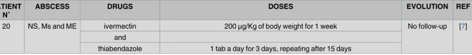
Table 1. (Continued)
PATIENT N˚
ABSCESS DRUGS DOSES EVOLUTION REF
20 NS, Ms and ME ivermectin 200μg/Kg of body weight for 1 week No follow-up [7]
and
thiabendazole 1 tab a day for 3 days, repeating after 15 days
CR = cervical region, NS = nasal sinuses, Ms = mastoid, ME = middle ear, TS = tonsil, TB = temporal bone, SMR = Sub Maxillary Region, CNS = central nervous system.
https://doi.org/10.1371/journal.pntd.0005510.t001
Top five papers
1. Sprent JF. Speciation and development in the genus Lagochilascaris.Parasitology. 1971;62(1):71–112.
2. Campos DMB, Freire Filha LG, Vieira MA, Pac¸oˆ JM, Maia MA. Experimental life cycle of Lagochilascaris minor Leiper, 1909.Rev Inst Med Trop São Paulo. 1992;34 (4):277–87.
3. Campos DMB, Barbosa AP. Lagochilascaris. In: Neves DP, Melo AL, Linardi PM, Vitor RWA, editors.Parasitologia humana. 13aed. São Paulo: Atheneu; 2016. p.514-23.
4. Leão RNQ, Fraiha-Neto H, Dias LB. Lagochilascarı´ase. In: Veronesi R, Focaccia R, editors,Tratado de Infectologia. 5a. ed. São Paulo: Atheneu, 2015. P.2107-11
Acknowledgments
The authors thank Prof. Dr. Maria Gonc¸alves da S. Barbalho for her assistance in creatingFig 1for this article.
References
1. Leiper RT. A new nematode worm from TrinidadLagochilascaris minor. Proc Zool Soc Lond. 1909; 4:742–3.
2. Draper JW. Infection withLagochilascaris minor. Brit Med J. 1963; 1:931–2. PMID:20789733
3. Chieffi PP, Proenc¸a NG, Pereira WA, Paschoalotti MA. Infecc¸ão cutaˆnea porLagochilascaris minor— tratamento e cura ra´pida pelo levamisol. An Bras Dermatol. 1981; 56(2):141–4.
4. Fraiha-Neto H, Leão RNQ. Lagochilascarı´ase. In: Coura JR, editors. Dinaˆmica das Doenc¸as Infeccio-sas e Parasita´rias. Rio de Janeiro: Guanabara-Koogan; 2005. p.1081–6.
5. Campos DMB, Barbosa AP. Lagochilascaris. In: Neves DP, Melo AL, Linardi PM, Vitor RWA, editors. Parasitologia humana. 13aed. São Paulo: Atheneu; 2016. p.514–23.
6. Orihuela R, Botto C, Delgado O, Ortiz A, Suarez JA, Arguello C. Human lagochilascariasis in Venezu-ela: description of a fatal case. Rev Soc Bras Med Trop. 1987; 20(4):217–21. PMID:3333879
7. Roig JL, Roig-Ocampos Forteza JL, Granato L, Poletti Serafini D. Otomastoidititis with right retroauricu-lar fistula byLagochilascaris minor. Braz J Otorhinolaryngol. 2010; 76(3):407. PMID:20658027
8. Bento RF, Mazza CC, Motti EF, Chan YT, Guimaraes JR, Miniti A. Human lagochilascariasis treated successfully with ivermectin: a case report. Rev Inst Med Trop Sao Paulo. 1993; 35(4):373–5. PMID: 8115799
9. Barrera-Pe´res M, Manrique-Saide P, Reyes-Novelo E, Escobedo-Ortegon J, Sanches-Moreno M, Sanches C.Lagochilascaris minorLeiper, 1909 (Nematoda: Ascarididae). In Mexico: three clinical cases from the Peninsula of Yucatan. Rev Inst Med Trop Sao Paulo. 2012; 54:315–7. PMID:23152314
10. Aquino RTR, Magliari MER, Vital Filho J, Silva MALG, Lima CAC, Rocha AJ. Lagochilascariasis leading to severe involvement of ocular globes, ears and meninges. Rev Inst Med Trop Sao Paulo. 2008; 50 (6):355–8. PMID:19082379
11. Botero D, Little MD. Two cases of humanLagochilascarisinfection in Colombia. Am J Trop Med Hyg. 1984; 33(3):381–6. PMID:6539572
12. Mondragon H, Cano M, Botero D. Primer caso de infeccio´n humana porLagochilascaris minoren Coloˆmbia. Antioquia Med. 1973; 23(9–10):463–4.
13. Veloso MG, Faria MC, de Freitas JD, Moraes MA, Gorini DF, de Mendonca JL. [Human lagochilascaria-sis. 3 cases encountered in the Federal District, Brazil]. Rev Inst Med Trop São Paulo. 1992; 34 (6):587–91. PMID:1342129
14. Oostburg BF, Varma AA.Lagochilascaris minorinfection in Surinam. Report of a case. Am J Trop Med Hyg. 1968; 17(4):548–50. PMID:5672783
15. Volcan GS, Ochoa FR, Medrano CE, de Valera Y.Lagochilascaris minorinfection in Venezuela. Report of a case. Am J Trop Med Hyg. 1982; 31(6):1111–3. PMID:6890772
Key learning points
• Lagochilascariasis is mostly a chronic human disease that can persist for several years, in which the parasite burrows into the subcutaneous tissues of the neck, paranasal sinuses, and mastoid. Other localizations of the parasite are the central nervous system, lungs, sacral region, eyeballs, and dental alveoli.
16. Moraes MA, Arnaud MV, de Macedo RC, Anglada AE. [Fatal pulmonary infection caused by Lagochi-lascaris sp., probablyLagochilascaris minorLeiper, 1909]. Rev Inst Med Trop São Paulo. 1985; 27 (1):46–52. PMID:4035207
17. Rosemberg S, Lopes MBS, Masuda Z, Campos R, Vieira-Bressan MCR. Fatal encephalopathy due to Lagochilascaris minorinfection. Am J Trop Med Hyg. 1986; 35:575–8. PMID:3706624
18. Pawan JL. A case of infection withLagochilascaris minor, Leiper, 1909. Ann Trop Med Parasitol. 1926; 20(2):201–2.
19. Pac¸oˆ JM, Campos DMB.Lagochilascaris minorLeiper, 1909: Nove de´cadas de revisão bibliogra´fica. Rev Patol Trop. 1998; 27(1):11–34.
20. Oostburg BF. Thiabendazole therapy ofLagochilascaris minorinfection in Surinam. Report of a case. Am J Trop Med Hyg. 1971; 20(4):580–3. PMID:5568126
21. Campos DMB, Freire Filha LG, Vieira MA, Pac¸oˆ JM, Maia MA. Experimental life cycle of Lagochilas-caris minorLeiper, 1909. Rev Inst Med Trop São Paulo. 1992; 34(4):277–87. PMID:1342084
22. Falcon-Ordaz J, Iturbe-Morgado JC, Rojas-Martı´nez AE, Garcı´a-Prieto L.Lagochilascaris minor (Nematoda: Ascarididae) from a wild cougar (Puma concolor) in Mexico. J Wildlife Dis. 2016; 52 (3):746–48.
23. Pawan JL. Another case of infection withLagochilascaris minorLeiper, 1909. Ann Trop Med Parasitol. 1927; 21(1):45–6.
24. Winckel WE, Treurniet AE. Infestation withLagochilascaris minor(Leiper) in man. Doc Med Geogr Trop. 1956; 8(1):23–8. PMID:13305481
25. Oostburg BF. The sixth case ofLagochilascariasis minorin Surinam. Trop Geogr Med. 1992; 44(1– 2):154–9. PMID:1496709
26. Vargas-Ocampo F, Alvarado-Aleman FJ. Infestation fromLagochilascaris minorin Mexico. Int J Derma-tol. 1997; 36(1):56–8. PMID:9071620
27. Brenes-Madrigal RR, Brenes AF. Lagochilascariasis humana en Costa Rica. Congreso Latinoameri-cano y Nacionale Microbiologia: Proceedings; Costa Rica. 1961; 35.
28. Orihuela R, Ortiz A, Delgado O, Botto C, Brown V, Marin G. Primer caso humano de Lagochilasarı´ase en Venezuela. Acta Cient Venez. 1982; 33:333.
29. Olle-Goig JE, Recacoechea M, Feeley T. First case ofLagochilascaris minorinfection in Bolivia. Trop Med Int Health. 1996; 1(6):851–3. PMID:8980600
30. Calvopina M, Guevara AG, Herrera M, Serrano M, Guderian RH. Treatment of human lagochilascaria-sis with ivermectin: first case report from Ecuador. Trans R Soc Trop Med Hyg. 1998; 92(2):223–4. PMID:9764339
31. Artigas PT, Araujo P, Romiti N, Ruivo M. [A new case of human parasitism, byLagochilascaris minor Leiper, 1909, observed in the State of Sao Paulo, Brasil]. Rev Inst Med Trop Sao Paulo. 1968; 10 (2):78–83. PMID:5679032
32. Leão RN, Leão Filho J, Bragadias L, Calheiros LB. [Human infection byLagochilascaris minorLeiper, 1909. Report of a case observed in the State of Para´ (Brazil)]. Rev Inst Med Trop São Paulo. 1978; 20 (5):300–6. PMID:725430
33. Moraes MA, Arnaud MV, de Lima PE. [New cases of human infection byLagochilascaris minorLeiper, 1909, found in the State of Para´, Brazil]. Rev Inst Med Trop São Paulo. 1983; 25(3):139–46. PMID: 6684323
34. Pac¸oˆ JM, Campos DM, Oliveira JA. Wild rodents as experimental intermediate hosts ofLagochilascaris minorLeiper, 1909. Mem Inst Oswaldo Cruz. 1999; 94(4):441–9. PMID:10445999
35. Fraiha H, Leão RQN, Costa FSA. Lagochilascarı´ase humana e dos animais dome´sticos. Zoon Rev Inst. 1989; 1(1):25–33.
36. Leão RNQ, Fraiha-Neto H, Dias LB. Lagochilascarı´ase. In: Veronesi R, Focaccia R, editors, Tratado de Infectologia. 5a. ed. São Paulo: Atheneu, 2015. P.2107–11
37. Campos DMB, Komma MD, Barbosa W, Santos MAQ, Pinto RNL, Barcelos M, et al. Notas Parasitolo´gi-cas sobre Lagochilascarı´ase humana em Goia´s. Rev Patol Trop. 1987; 16:129–42.
38. Guimarães VC, Barbosa AP, Camargo LA, Siqueira PH, Silva Filho JC, Castro VLS,et al. Otomastoidite porLagochilascaris minorem crianc¸a: relato de caso. Intl Arch Otorhinolaryngol. 2010; 14(3):373–6.
39. Barbosa AP, Campos DMB, Semerene AR, Teixeira ARL, Santana JM.Lagochilascaris minor third-stage larvae secrete metalloproteases with specificity for fibrinogen and native collagen. Microbes Infect. 2006; 8:2725–32.https://doi.org/10.1016/j.micinf.2006.08.001PMID:16979366
41. Freire-Filha LG, Campos DMB. Considerac¸ões sobre o desenvolvimento deLagochilascaris minor, 1909 em camundongos isogênicos da linhagem C57BL/6. Rev Patol Trop. 1992; 21(2):219–33.
42. Semerene AR, Lino Junior RS, Oliveira JA, Magalhães AV, Stefani MM, Barbosa AP, Campos DMB. Experimental lagochilascariosis: histopathological study of inflammatory response to larval migration in the Murine model. Mem Inst Oswaldo Cruz. 2004; 99(4):393–8. PMID:15322629
43. Smith JL, Bowman DD, Little MD. Life cycle and development ofLagochilascaris sprenti(Nematoda: ascarididae) from opossums (Marsupialia: didelphidae) in Louisiana. J Parasitol. 1983; 69(4):736–45. PMID:6685179
44. Odum Eugenio P. Fundamentos de ecologia. 5a. Ed. Lisboa: Fundac¸ão Calouste Gulbenkian, 1997, p.583.
45. Aguilar-Nascimento JE, Silva GM, Tadano T, Valadares Filho M, Akiyama AM, Castelo A. Infection of the soft tissue of the neck due toLagochilascaris minor. Trans R Soc Trop Med Hyg. 1993; 87(2):198. PMID:8337728
46. Campos DMB, Zanini LA, Santos ER, Pac¸o JM, Vieira MA. Observac¸ões parasitolo´gicas referentesà infecc¸ão croˆnica porLagochilascaris minor: resistênciaàdietilcarbamazina, levamisol, albendazol e ivermectin. Rev Soc Bras Med Trop. 1995; 28(suppl 1):21.
47. Barbosa CA, Campos DMB, Vieira MA, Oliveira JA. Avaliac¸ão in vitro da atividade ovicida e larvicida da ivermectina sobreLagochilascaris minor. Rev Patol Trop. 1997; 26(1):57–68.
48. Barbosa CA, Campos DM. [Assessment of ivermectin therapeutic efficacy on fourth-stage larvae of Lagochilascaris minorin experimentally infected cats]. Rev Soc Bras Med Trop. 2001; 34(4):373–6. PMID:11562732
49. Campos DMB, Barbosa AP, Oliveira JA, Barbosa CAL, Lobo TC, Silva LG, et al. Evaluation of the thera-peutic efficacy of levamisole hydrochloride on third-stage larvae ofLagochilascaris minorin experimen-tally infected mice. Rev Inst Med Trop Sao Paulo. 2016; 58(1):1–5.