SIV Infection of Lung Macrophages
Yue Li2,1, Guobin Kang1, Lijie Duan3, Wuxun Lu1, Michael G. Katze4,5, Mark G. Lewis6, Ashley T. Haase3, Qingsheng Li1*
1Nebraska Center for Virology, School of Biological Sciences, University of Nebraska-Lincoln, Lincoln, Nebraska, United States of America,2College of Life Sciences, Nankai University, Tianjin, People’s Republic of China,3Department of Microbiology, University of Minnesota Medical School, Minneapolis, Minnesota, United States of America,4Washington National Primate Research Center, Seattle, Washington, United States of America,5Department of Microbiology, School of Medicine, University of Washington, Seattle, Washington, United States of America,6BIOQUAL, Inc., 9600 Medical Center Drive, Rockville, Maryland, United States of America
Abstract
HIV-1 depletes CD4+ T cells in the blood, lymphatic tissues, gut and lungs. Here we investi-gated the relationship between depletion and infection of CD4+ T cells in the lung parenchy-ma. The lungs of 38 Indian rhesus macaques in early to later stages of SIVmac251 infection were examined, and the numbers of CD4+ T cells and macrophages plus the frequency of SIV RNA+ cells were quantified. We showed that SIV infected macrophages in the lung pa-renchyma, but only in small numbers except in the setting of interstitial inflammation where large numbers of SIV RNA+ macrophages were detected. However, even in this setting, the number of macrophages was not decreased. By contrast, there were few infected CD4+ T cells in lung parenchyma, but CD4+ T cells were nonetheless depleted by unknown mecha-nisms. The CD4+ T cells in lung parenchyma were depleted even though they were not pro-ductively infected, whereas SIV can infect large numbers of macrophages in the setting of interstitial inflammation without depleting them. These observations point to the need for fu-ture investigations into mechanisms of CD4+ T cell depletion at this mucosal site, and into mechanisms by which macrophage populations are maintained despite high levels of infec-tion. The large numbers of SIV RNA+ macrophages in lungs in the setting of interstitial in-flammation indicates that lung macrophages can be an important source for SIV persistent infection.
Introduction
Human immunodeficiency virus type 1 (HIV-1) depletes CD4+ T cells in blood, secondary lymphatic tissues, gut and lungs by mechanisms such as the cytopathic effects of infection, im-mune recognition and killing of infected cells. In the lung, CD4+ T cell count in bronchoalveo-lar lavage (BAL) quickly declines in acute HIV-1 and SIV infection [1–4], and indeed this
measurement is commonly used as a surrogate for mucosal CD4+ T cell depletion in acute HIV-1 infection. However, the relationship between infection in the lung parenchyma and de-creased CD4+ T cells in BAL has not been determined. To that end, we explored the role of
OPEN ACCESS
Citation:Li Y, Kang G, Duan L, Lu W, Katze MG, Lewis MG, et al. (2015) SIV Infection of Lung Macrophages. PLoS ONE 10(5): e0125500. doi:10.1371/journal.pone.0125500
Academic Editor:Cristian Apetrei, University of Pittsburgh Center for Vaccine Research, UNITED STATES
Received:December 18, 2014
Accepted:March 17, 2015
Published:May 1, 2015
Copyright:© 2015 Li et al. This is an open access article distributed under the terms of theCreative Commons Attribution License, which permits unrestricted use, distribution, and reproduction in any medium, provided the original author and source are credited.
Data Availability Statement:All relevant data are within the paper.
infection in depletion of CD4+ T cells in the lung parenchyma of SIV-infected macaques from the early to later stages of infection. We found that macrophages, but not CD4+ T cells, are the principal host cells that SIV infects. Relatively few macrophages or CD4+ T cells are infected except in the setting of lung interstitial inflammation where we found large numbers of SIV RNA+ macrophages. Even in this setting, SIV RNA+ CD4+ T cells were mainly localized out-side of the parenchyma and in broncho-alveolar lymphatic tissue (BALT). Despite productive infection of alveolar macrophages, macrophage populations were preserved, but CD4+ T cell populations were depleted even though they were not productively infected. Thus, mechanisms other than direct infection are responsible for the depletion of CD4+ T cells in BAL in acute SIV infection [2,5].
Materials and Methods
Ethics Statement
This study was reviewed and approved by the Institutional Animal Care and Use Committee (IACUC) at the University of Nebraska-Lincoln, BIOQUAL Inc., Washington National Pri-mate Research Center, and California National PriPri-mate Research Center. The care and hus-bandry of all rhesus macaque were provided in compliance with The Guide for the Care and Use of Laboratory Animals, the Animal Welfare Act, the PHS Policy on Humane Care and Use of Laboratory Animals, and applicable local, state, and federal laws. The comprehensive veteri-nary care program includes: disease detection and surveillance, prevention, diagnosis, treat-ment, and resolution; monitoring and promoting physical and psychological well-being of all animals. Monkeys were housed socially, if possible. Monkeys had access to food, water, light at 12-hour cycle and were provided with enrichment toys. Animals were sedated with ketamine for all technical procedures and were fully anesthetized for SIV inoculation and euthanasia under the directions of the attending veterinarians.
Virus and Animals Inoculation
The lungs of 44 adult Indian rhesus macaques (Macaca mulatta) were studied. The animals came from three studies and were housed at BIOQUAL Inc., Washington National Primate Re-search Center, and California National Primate ReRe-search Center. The animals were maintained in accordance with the Guide for the Care and Use of Laboratory Animals. Six macaques were SIV naïve, 29 were acutely infected, and 9 were chronically infected. All animals were free of simian retrovirus type D, simian T-lymphotrophic virus type 1, and herpes B virus. Macaques were inoculated SIVmac251 at a single dose of 3.4×10⁴TCID50intrarectally or 5.0×10⁵TCID50
intravaginally twice at four hours interval, respectively, and euthanized at designed time points. The lung tissues were collected, fixed in 4% paraformaldehyde for 4–6 hours and embedded
in paraffin.
Plasma Viral RNA Measurements
Virus load in EDTA-treated plasma was quantified using real-time RT-PCR assay based on amplification of conserved sequences in SIVgaggene [6]. The limit of detection for this assay was 30 viral RNA copy equivalents per ml plasma.
Detection of SIV RNA+ Cells in Lungs Using
In Situ
Hybridization
SIV RNA+ cells in the lungs were detected usingin situhybridization as previously described [7]. Briefly, 6-μm sections were cut and adhered to silanized slides. After de-paraffinization in
xylene, rehydration in PBS and permeabilization by treating the sections with HCl, digitonin
subcontract. BIOQUAL Inc. provided support in the form of salaries for author Dr. Mark Lewis, but did not have any additional role in the study design, data collection and analysis, decision to publish, or preparation of the manuscript. The specific roles of all the authors are articulated in the author contributions’
section.
and proteinase K, the sections were acetylated and hybridized to35S-labelled SIV-specific riboprobes. After washing and digestion with RNases, sections were coated with nuclear track emulsion, exposed, developed and counterstained with hematoxylin and eosin staining. SIV RNA+ cells were enumerated in sections of defined areas using Aperio ImageScope software (Aperio ePathology Solutions, CA).
Detection of CD4+ T Cells and Macrophages in Lungs Using
Immunohistochemical Staining
CD4+ T cells and CD68+ macrophages were detected using IHCS [7]. Briefly, 6-μm sections
were cut and adhered to silanized slides from 4% paraformaldehyde fixed and paraffin embed-ded tissues, and stained with mouse anti-human monoclonal antibody to CD4 (1F6, 1:60 dilution, Leica Microsystems) or CD68 (KP1, 1:200 dilution, DAKO North American, Inc., Carpinteria, CA) for macrophage assay. Sections on slides were pretreated in a Presto pressure cooker (National Presto Industries, Eau Claire, WI) in 1 mM EDTA (pH 8.0) for 35 seconds to unmask CD4 antigen or in 98°C water bath in 10 mM citrate buffer (pH 6.0) for 15 minutes to unmask CD68 antigen. The sections were sequentially treated with aqueous hydrogen perox-ide, blocked with normal horse serum, and incubated with primary antibody overnight at 4°C. The stained color was developed by using the Vectastain Mouse-IgG Peroxidase ABC kit (Vec-tor Labora(Vec-tories, Lumigen Inc. Southfield, MI) and diaminobenzidine (DAB) as substrate in DAB Enhancing Solution (Vector Laboratories, Burlingame, CA). The sections were counter-stained with hematoxylin.
Combined Immunohistochemistry Staining and
In Situ
Hybridization
Combined IHCS and ISH were performed as described previously [7]. Briefly, tissue sections on slides were incubated at 98°C for 15 minutes in 10mM citrate buffer (pH 6.0) for antigen re-trieval, hybridized, washed and digested with RNases, incubated with antibody against T cell marker (anti-CD3, SP7, 1:100 dilution, Lab Vision/NeoMarkers, CA) or macrophage marker (anti-CD163, NCI-Cd163, 1:100 dilution, Novocastra Reagents-Leica Biosystems, IL), and then stained with DAB with the Dako Envision and Peroxidase kit (Dako North America, Inc., Car-pinteria, CA). After washing, the sections were coated with nuclear track emulsion, exposed, developed and counterstained with hematoxylin.
Quantitation of CD4+ T cells, Macrophages, and SIV RNA+ Cells in
Lungs
Statistics
Statistical analysis was conducted with a nonparametric Mann-Whitney U test using Graphpad Prism software (Graphpad software, San Diego, CA, USA). P value<0.05 was
considered significant.
Results
SIV infected cells in the lungs in early and later stages of infection
We examined the lungs of 6 SIV-uninfected control rhesus macaques and 38 SIV-infected rhe-sus macaques (Table 1) to characterize SIV productive infection in the lungs from early to later stages of infection. The infected animals were inoculated via vaginal or rectal mucosal routes with high-dose SIVmac251 and sacrificed in the acute (1–14 days PI) (29 animals) or later
stages of infection (3–6 months PI) (9 animals). Plasma viral RNA (vRNA) in each animal was
determined by real-time RT-PCR and vRNA+ cells in the lung tissues were detected by using
in situhybridization (ISH,Table 1). Plasma viral load in these infected monkeys showed simi-lar kinetics as described previously [7], with the viral peak at 10–14 days PI. At 1–4 days PI, no
SIV RNA+ cells were detected in the lungs (Table 1) and other distal lymphatic tissues (data not shown), but by 6 days PI, small numbers of vRNA+ cells were detected in lungs of all six macaques, and SIV vRNA+ cells were detected in the lungs of 17 macaques sacrificed at 6–12
days PI (Table 1,Fig 1A1–1A2). The frequencies of vRNA+ cells were much higher in the lungs of 5 out of 9 macaques at 3–6 months PI (Table 1,Fig 1B1–1D2) as compared with all
the other macaques examined. Since mycobacterial co-infection can significantly increase HIV-1 and SIV replication [9,10], we examined but found no evidence of co-infection in these cases using acid fast staining (data not shown). Rather, the SIV RNA+ cells were associated with lung interstitial inflammation, as has been previously reported [11,12]
Lung macrophages are primary target cells for SIV
To define the cell types that SIV preferentially infects in lungs, vRNA+ cells were characterized using the combined IHCS and ISH. Macrophages were the main target cells in lung parenchy-ma in both acute (Fig 2A) and later stages of infections (Fig 2B), but were not the major cell type infected in broncho-associated lymphatic tissues (Fig 2C). The CD4+ T cells in the lung parenchyma were not infected in these cases, but CD4+ T cells were the major cell type infected with SIV in broncho-associated lymphatic tissue examined with the combined ISH and IHCS using CD3 antibody (data not shown).
CD4+ T cells but not macrophages are depleted in lungs
Despite the predominance of infection in macrophages, CD4+ T cells, but not macrophages were depleted in lungs. We quantified CD4+ and CD68+ cells in the lungs of uninfected ma-caques (as baseline controls) and mama-caques at various stages of SIV infection (Fig 3) by staining and quantitative image analysis (QIA) (Fig 3A–3C). In uninfected animals, there were 1941±371 CD4+ T cells/mm2(Fig 3B), but as early as 6 days PI, lung CD4+ T cells had decreased significantly (1056±364 cells/mm2, p<0.05, non-parametric Mann-Whitney U
test) and reached a nadir at 10 days PI (777±400 cells/mm2, p<0.05,Fig 3D) and remained
depleted through 12 weeks post infection (899±280 cells/mm2, p<0.01,Fig 3E and 3F). In
con-trast, we found no significant change in the number of alveolar and interstitial CD68+ macro-phages in acutely (p>0.05, day 3, 6 or 10 PI groups as compared with the control group,
respectively,) and chronically (p>0.05, 12 weeks PI group versus control group) infected
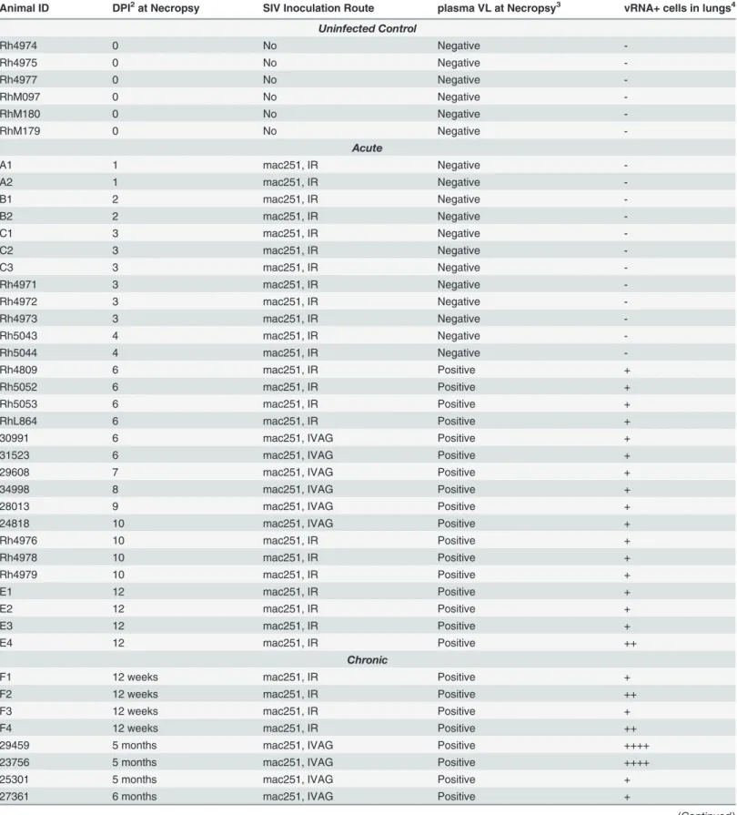
Table 1. SIV vRNA+ Cells in the Lungs of Macaques Infected with SIV1.
Animal ID DPI2at Necropsy SIV Inoculation Route plasma VL at Necropsy3 vRNA+ cells in lungs4
Uninfected Control
Rh4974 0 No Negative
-Rh4975 0 No Negative
-Rh4977 0 No Negative
-RhM097 0 No Negative
-RhM180 0 No Negative
-RhM179 0 No Negative
-Acute
A1 1 mac251, IR Negative
-A2 1 mac251, IR Negative
-B1 2 mac251, IR Negative
-B2 2 mac251, IR Negative
-C1 3 mac251, IR Negative
-C2 3 mac251, IR Negative
-C3 3 mac251, IR Negative
-Rh4971 3 mac251, IR Negative
-Rh4972 3 mac251, IR Negative
-Rh4973 3 mac251, IR Negative
-Rh5043 4 mac251, IR Negative
-Rh5044 4 mac251, IR Negative
-Rh4809 6 mac251, IR Positive +
Rh5052 6 mac251, IR Positive +
Rh5053 6 mac251, IR Positive +
RhL864 6 mac251, IR Positive +
30991 6 mac251, IVAG Positive +
31523 6 mac251, IVAG Positive +
29608 7 mac251, IVAG Positive +
34998 8 mac251, IVAG Positive +
28013 9 mac251, IVAG Positive +
24818 10 mac251, IVAG Positive +
Rh4976 10 mac251, IR Positive +
Rh4978 10 mac251, IR Positive +
Rh4979 10 mac251, IR Positive +
E1 12 mac251, IR Positive +
E2 12 mac251, IR Positive +
E3 12 mac251, IR Positive +
E4 12 mac251, IR Positive ++
Chronic
F1 12 weeks mac251, IR Positive +
F2 12 weeks mac251, IR Positive ++
F3 12 weeks mac251, IR Positive +
F4 12 weeks mac251, IR Positive ++
29459 5 months mac251, IVAG Positive ++++
23756 5 months mac251, IVAG Positive ++++
25301 5 months mac251, IVAG Positive +
27361 6 months mac251, IVAG Positive +
Discussion
In this study, we systematically examined the infected cell types and the depletion of CD4+ T cells in the lungs of SIV infected rhesus macaques in the very early to later stages of infection. We found that macrophages were the major target cells in the lung parenchyma in both early and later stages of infection, but SIV-infected macrophages were rare except in animals with lung interstitial inflammation defined by the increased infiltration of the pulmonary intersti-tium with a mixture of lymphocytes and plasma cells. This is also the case in HIV-1 infections where infected CD4+ T cells are rare outside of this inflammatory setting. The small number of infected macrophages in lung parenchyma, except in this setting, stands in stark contrast to the abundant SIV RNA+ cells in lymph nodes and gut where over 90% infected cells are CD4+ T cells as reported previously[13]. Moreover, SIV replication in CD4+ T cells peaks around 2 weeks post infection in secondary lymphatic tissues and gut mucosae [7], whereas virus replica-tion in lung macrophages was very limited in this time frame (Table 1,Fig 1A1–1A2), consis-tent with a previous report[14]. We did find high levels of virus replication in lungs in the
Table 1. (Continued)
Animal ID DPI2at Necropsy SIV Inoculation Route plasma VL at Necropsy3 vRNA+ cells in lungs4
25479 6 months mac251, IVAG Positive ++++
1Totally 38 infected and 6 uninfected rhesus macaques were used in this study, and vRNA+ cells were quantified by ISH. 2DPI = Days post infection.
3Virus load in plasma was detected by real-time RT-PCR.
4SIV vRNA+cells in lungs were detected using ISH with SIV-specific riboprobes:
−ith SIV-s+cells/mm2
+, 5–50 vRNA+cells/mm2
++, 51–100 vRNA+cells/mm2
++++,>200 vRNA+cells/mm2.
doi:10.1371/journal.pone.0125500.t001
Fig 1. SIV vRNA+ cells increase in the lungs of chronically infected macaques as compared with early infection.SIV vRNA+ cells were detected usingin situhybridization, and appeared as black dots in transmitted light and green dots under epipolarized light. There were a few vRNA+ cells in the lung
tissues during acute infection (A1, A2) and large numbers of vRNA+ cells in the lung during chronic infection (B1-D2).
Fig 2. Macrophages are the main SIV RNA+ cells in the lungs from very early (A) at 10 days PI to chronic infection at 5 months PI (B).SIV vRNA+ macrophages were distinguished from T cells by immunohistochemical staining for CD163. The red arrows indicate CD163+ vRNA+ cells identified by the overlying collection of silver grains (black dots); blue arrows indicate SIV RNA+ cells that are not macrophages in lung associated lymphatic tissue (C).
doi:10.1371/journal.pone.0125500.g002
Fig 3. The depletion of CD4+ T cells, but not macrophages, in lung tissues during SIV infection.CD4+ T cells (A-F) and CD68+ macrophages (G-I) in the lung tissues of uninfected and infected macaques detected using immunohistochemical staining and quantified using Aperio Spectrum Plus analysis program. The A-E are representative images of CD4+ T cells in uninfected lung tissues (A-C) and, infected at 10 days (D) and 12 weeks (E) PI respectively. (A) Digitized whole tissue section. (B) Magnified image from the box in theFig 3A, where the brown corresponds to the stained CD4+ T cells. (C) The red/ yellow marked-up regions correspond to the stained CD4+ T cells in theFig 3Bused for quantification with Aperio Spectrum Plus analysis program. The G-H are the representative images of pulmonary CD68+ macrophages in uninfected lung tissues (G) and infected at 12 weeks PI(H). TheFig 3F and 3Iare the histograms of CD4+ T cell and macrophage quantification in lung tissues, respectively. X-axis shows the time of infection at 0 (n = 6), 3 (n = 6), 6 (n = 6), 10 (n = 4) days and 12 weeks (n = 4) PI, and y-axis shows the cell number expressed as per square millimeter of lung tissue.*Indicates significant differences from controls (*P<0.05,**P<0.01). Statistical analysis of cell amount per mm2tissue was performed with non-parametric Mann-Whitney U test.
setting of lung interstitial inflammation, when virus replication in the lymphatic tissues and gut had already decreased. The increased infection of lung macrophages in this context cannot simply be attributed to target cell availability, as the macrophage population sizes were not dif-ferent in early and later stages of infection and did not differ from uninfected controls. Rather, the milieu of lung interstitial inflammation may provide more permissive macrophage targets for viral replication in this context. However, interstitial inflammation is not frequent in SIV infections, which clearly sets these primate lentivirus infections apart from ungulate lentivirus infections such as maedi where interstitial pneumonitis is a defining pathological feature of these infections [15,16]. Recently, lung macrophages have been classified into different subsets [17], and future studies are needed to test whether particular macrophage subset can preferen-tially support a high-level SIV replication in the setting of interstitial inflammation.
While severe CD4+ T cell depletion occurs in gut in the early HIV-1 infection [7,18,19], moderate CD4+ T cell depletion has been documented in bronchoalveolar lavage (BAL) [1–4],
but the interstitial CD4+ cells in lung have not been quantified. Here, we show significant inter-stitial CD4+ T cell depletion in the lungs during very early infection that is sustained through 12 weeks. In contrast, macrophage populations do not decline despite the predominance of in-fection of macrophages in the lungs. There is thus no direct link between productive inin-fection and population dynamics in the lungs. This disjunction between direct productive infection and depletion highlights the need for future studies to elucidate the mechanisms that account for CD4+ T cell depletion in the lungs and maintenance of macrophage populations even in the setting of direct infection. There are certainly precedents for redistribution of CD4 T cells as the mechanism underlying the apparent depletion in peripheral blood. Based on the recent demonstration that most CD8+ T cells isolated from mouse lung are actually confined to the pulmonary vasculature [20], we can envision altered dynamics of the flux between this pool and BAL as a mechanism accounting for apparent rapid CD4+ T cell depletion. Furthermore, the large numbers of SIV RNA+ macrophages in lungs in the setting of interstitial inflamma-tion indicates that lung macrophages can be an important source for SIV persistent infecinflamma-tion.
Acknowledgments
The authors wish to thank Dr. Christopher J. Miller for sharing rhesus lung tissues, Andrew Demers for his critical reading this manuscript, Timothy W. Schacker and Robert Palermo for discussions and suggestions.
Author Contributions
Conceived and designed the experiments: QL YL ATH. Performed the experiments: YL GK LD MGL. Analyzed the data: YL GK WL MGK QL. Wrote the paper: YL ATH QL.
References
1. Fernandez E, Leon P, Blanquer R, Marin M, Artero A, Pinilla A. A comparison of lymphocyte popula-tions of the blood and bronchoalveolar lavage in AIDS patients. Arch Bronconeumol. 1996; 32:271–
274. PMID:8814820
2. Brenchley JM, Knox KS, Asher AI, Price DA, Kohli LM, Gostick E, et al. High frequencies of polyfunc-tional HIV-specific T cells are associated with preservation of mucosal CD4 T cells in bronchoalveolar lavage. Mucosal Immunol. 2008; 1:49–58. doi:10.1038/mi.2007.5PMID:19079160
3. Knox KS, Vinton C, Hage CA, Kohli LM, Twigg HL, Klatt NR, et al. Reconstitution of CD4 T Cells in Bronchoalveolar Lavage Fluid after Initiation of Highly Active Antiretroviral Therapy. Journal of Virology. 2010; 84:9010–9018. doi:10.1128/JVI.01138-10PMID:20610726
5. Picker LJ, Hagen SI, Lum R, Reed-Inderbitzin EF, Daly LM, Sylwester AW, et al. Insufficient production and tissue delivery of CD4+ memory T cells in rapidly progressive simian immunodeficiency virus infec-tion. J Exp Med. 2004; 200:1299–1314. PMID:15545355
6. Cline AN, Bess JW, Piatak M Jr, Lifson JD. Highly sensitive SIV plasma viral load assay: practical con-siderations, realistic performance expectations, and application to reverse engineering of vaccines for AIDS. J Med Primatol. 2005; 34:303–312. PMID:16128925
7. Li Q, Duan L, Estes JD, Ma ZM, Rourke T, Wang Y, et al. Peak SIV replication in resting memory CD4+ T cells depletes gut lamina propria CD4+ T cells. Nature. 2005; 434:1148–1152. PMID:15793562
8. Wang LX, Kang G, Kumar P, Lu W, Li Y, Zhou Y, et al. Humanized-BLT mouse model of Kaposi's sar-coma-associated herpesvirus infection. Proc Natl Acad Sci U S A. 2014; 111:3146–3151. doi:10.1073/ pnas.1318175111PMID:24516154
9. Orenstein JM, Fox C, Wahl SM. Macrophages as a Source of HIV During Opportunistic Infections. Sci-ence. 1997; 276:1857–1861. PMID:9188531
10. Li Q, Mansfield KG, Lackner AA, Haase AT. Quantitative image analysis of simian immunodeficiency virus replication in macrophages coinfected with Mycobacterium avium complex. J Infect Dis. 2000; 181:867–871. PMID:10720506
11. Mankowski JL, Carter DL, Spelman JP, Nealen ML, Maughan KR, Kirstein LM, et al. Pathogenesis of simian immunodeficiency virus pneumonia: an immunopathological response to virus. Am J Pathol. 1998; 153:1123–1130. PMID:9777943
12. Das S, Miller RF. Lymphocytic interstitial pneumonitis in HIV infected adults. Sex Transm Infect. 2003; 79:88–93. PMID:12690125
13. Zhang Z-Q, Wietgrefe SW, Li Q, Shore MD, Duan L, Reilly C, et al. Roles of substrate availability and in-fection of resting and activated CD4+ T cells in transmission and acute simian immunodeficiency virus infection. Proceedings of the National Academy of Sciences of the United States of America. 2004; 101:5640–5645. PMID:15064398
14. Fuller CL, Choi YK, Fallert BA, Capuano S 3rd, Rajakumar P, Murphey-Corb M, et al. Restricted SIV replication in rhesus macaque lung tissues during the acute phase of infection. Am J Pathol. 2002; 161:969–978. PMID:12213725
15. Haase AT. Pathogenesis of lentivirus infections. Nature. 1986; 322:130–136. PMID:2425264
16. Haase AT. The role of active and covert infections in lentivirus pathogenesis. Ann N Y Acad Sci. 1994; 724:75–86. PMID:8030979
17. Cai Y, Sugimoto C, Arainga M, Alvarez X, Didier ES and Kuroda MJ. In vivo characterization of alveolar and interstitial lung macrophages in rhesus macaques: implications for understanding lung disease in humans. J Immunol. 2014; 192:2821–2829. doi:10.4049/jimmunol.1302269PMID:24534529
18. Veazey RS, DeMaria M, Chalifoux LV, Shvetz DE, Pauley DR, Knight HL, et al. Gastrointestinal Tract as a Major Site of CD4+ T Cell Depletion and Viral Replication in SIV Infection. Science. 1998; 280:427–431. PMID:9545219
19. Mattapallil JJ, Douek DC, Hill B, Nishimura Y, Martin M and Roederer M. Massive infection and loss of memory CD4+ T cells in multiple tissues during acute SIV infection. Nature. 2005; 434:1093–1097. PMID:15793563
20. Sage PT, Francisco LM, Carman CV, Sharpe AH. The receptor PD-1 controls follicular regulatory T cells in the lymph nodes and blood. Nat Immunol. 2013; 14:152–161. doi:10.1038/ni.2496PMID: