Sociedade Brasileira de Virologia
journal homepage: www.vrrjournal.org.br/ Review Article
ENTERIC VIRUSES AS CONTAMINANTS AND BIOINDICATORS IN
ENVIRONMENTAL SAMPLES
Fongaro, G.1; Silva, H.D.2; Elmahdy, E.M.1; Magri, M.E.1; Schissi, C.D.1; Moreira, M.3; Lanna, M.C.S.3; Silveira-Lacerda, E.P.2; Barardi, C.R.M.1*
1. Laboratório de Virologia Aplicada; Departamento de Microbiologia, Imunologia e Parasitologia; Universidade Federal de Santa Catarina, Florianópolis, Brazil. 2. Laboratório de Genética Molecular e Citogenética; Departamento de Biologia Geral Universidade Federal de Goiás.
3. Laboratório de Microbiologia; Departamento de Ciências Biológicas; Universidade Federal de Ouro Preto, Brazil
ABSTRACT
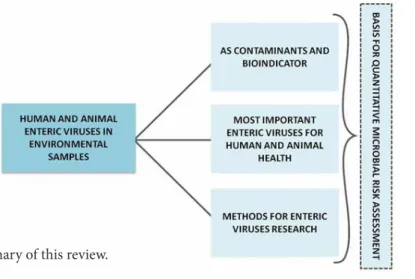
Gastroenteritis, diarrhea, and other diseases can be caused by enteric viruses transmitted by fecal-oral route. Human adenoviruses (HAdV), rotavirus A and C (RVA and RVC, respectively), hepatitis A and E virus (HAV and HEV, respectively), human astroviruses (HAstV), human noroviruses (HuNV) and enteroviruses (EV) are, among the enteric viruses, the most frequently detected in environment samples. These viruses are usually introduced into aquatic environments by human, industrial, or agricultural activities and are widely distributed all over the world. They have the common characteristics to be structurally stable and can also absorb to solid particles and biofilms, thereby protecting themselves from inactivating factors. This revision aimed to present and discuss: i) most relevant enteric viruses for human and animal health; ii) enteric viruses as contaminants and bioindicators in environmental samples; iii) molecular and cell culture methods for enteric virus detection; iv) use of enteric viruses for microbial risk assessment. Impacts of enteric viruses on environment and the potential use as bioindicators of the sanitary security, such as presence and infectivity studies were discussed as development of new tools for disinfection, monitoring, risk modeling and management, among other studies.
Keywords: enteric viruses; bioindicators; contaminants; environmental samples
Received in June 12, 2015 - Accepted in July 22, 2015 - Published online in July 30, 2015
1. ENTERIC VIRUSES AS CONTAMINANTS AND BIOINDICATORS IN DRINKING AND WASTEWATER
According to the World Health Organization (WHO), it is estimated that 663 million people worldwide
still use improper drinking water sources, including unprotected wells and springs and surface water (WHO, 2015).
World legislations for enteric virus vigilance in environment matrices are scarce. The Center for Disease Control and Prevention-(CDC-USA) recommends Hepatitis A virus surveillance in foods (CDC, 2005); The Centre for Environment, Fisheries & Aquaculture Science
* Corresponding author.
E-mail address: [email protected] doi - 10.17525/vrrjournal.v20i2.255
- EU (CEFAS-EU, 2015) recommends norovirus, hepatitis A virus and bacteriophage RNA virus surveillance in bivalve molluscs; Environmental Protection Agency US (USEPA, 1991), recommends 99.99% of enteric virus removal from drinking water in disinfection treatment; In Brazil, for agriculture recycle of human sludge, is necessary to evaluate enteric viruses and their quantity should be <0.25 plaque forming unit/g of sludge (National Committee of Environmental (CONAMA), 2006).
The research about viruses contamination on different environmental matrices is increasing all over the world. In Brazil there are research groups developing projects regarding virus’s behavior in drinking water, wastewater, sludge, seawater, bivalve molluscs and other kind of food (Miagostovich et al. 2008; Rigotto et al. 2010).
Enteric viruses can be introduced in aquatic environments by different human and animal activities, such as discharge of untreated or inadequately treated sewage; incorrect use of septic systems; agricultural activities and other factors (Fong et al. 2005).
Among the major enteric viruses that can be responsible for gastroenteritis or other clinical manifestations, the human adenoviruses (HAdV), the rotavirus species A (RVA), the hepatitis A and E virus (HAV and HEV, respectively), human astroviruses (HAstV), human noroviruses (HuNV) and enteroviruses (EV) are the most frequently detected in the aquatic environment (Chen et al., 2007; Fongaro et al. 2013). All these viruses are quite resistant to water and sewage treatment because they are able to quickly adsorb to solid particles and therefore protect themselves from inactivating factors (Hernroth et al. 2002). Among all the human enteric virus, HAdV is considered by some authors the most resistant to the treatments based on thermal treatment, chlorine and ozone and is often detected in water sources sources (Wyn-Jones et al. 2011; Katayama et al. 2008, USEPA, 2015).” So, HAdV can be, in principle, the most appropriated model to be used as bioindicator of viral contamination. This is true, when just the viral genomes are detected in the environmental samples. Nascimento et al (2014), using an infectivity assay proved that chlorine is very effective to inactivate HAdV from artificially seeded water samples and that the genome copies are maintained even when viral infectivity is lost. Therefore, they can be indicated as potential bioindicators of environmental contamination and surveillance (Fong et al., 2005).
In recent years, several researchers have proposed the use of human adenoviruses (HAdV) as bioindicators of other enteric contaminants (Wyn-Jones et al. 2011; Katayama et al. 2008). In Japan, AdV is the most widely bioindicator used to ensure the virological quality of water and it was recently included in the Draft Contaminant Candidate List 4-CCL (CCL4) by the Environmental Protection Agency (EPA), of the United
States (USEPA, 2015).
1.2 Use of endogenous and indigenous virus as internal biomarkers in environmental samples
Many wild types of enteric viruses, such as human noroviruses, hepatitis A and E viruses, human rotaviruses and enteric adenoviruses, are fastidious, cannot be or are difficult to adapt in conventional in vitro cell lines. This fact result on limitations for infectivity studies and so the use of bioindicators viruses or surrogates is essential (Sidhu et al. 2009).
Human norovirus (HNoV), is a common source of contamination (Wang et al. 2012), but there is still not totally established an in vitro cell culture system for the detection of infectious wild type norovirus due to the cellular tropism of human NoVs (HuNoVs) and also for the absence of stimulatory carbohydrate molecules because HuNoVs are well known to bind histo-blood group antigens (HBGAs) which are expressed by the host. ¬-Recently Jones et al., (2015) demonstrated a biologically substantial role for enteric bacteria during NoV infection leading to the development of an in vitro infection model for human noroviruses.
The feline calicivirus (FCV) and the murine norovirus (MNV) are common surrogate models used for HNoV (Bae and Schwab 2008). Mengovirus (avirulent genetically modified mengovirus M (vMC) is used as a processing control for HAV, HEV and HuNoV (Costafreda et al., 2006).
The bacteriophages are common models for pathogenic viruses because they are easy to enumerate compared with human or animal viruses that require living cells for cultivation, certain types of bacteriophages, specifically phages of Escherichia coli (coliphages) are pointed as candidate indicators of human enteric viruses in water (Love and Sobsey, 2007).
The coliphages are small, icosahedral and non-enveloped viruses, making them structurally similar to many human enteric viruses. There are two main types of coliphages: somatic and the male-specific (F+) (Love and Sobsey, 2007). The somatic coliphages are DNA viruses that infect E. coli through attachment to specific sites on the outer cell layer, such as lipopolysaccharide. The male-specific coliphages are single-stranded RNA and DNA viruses that infect the cell via the pili appendages present on the surface of male strains of the bacterium (Dore et al., 2000; Love and Sobsey, 2007).
2. IMPORTANT ENTERIC VIRUSES AFFECTING HUMAN AND ANIMAL HEALTH
2.1 Adenovirus
including avian, reptile and marsupial hosts. A possible fifth genera infecting fishes has been proposed to be called Ichtadenovirus (Benko et al., 2002).
The Adenoviruses (AdVs) are eliminated in feces, urine or respiratory excretions and the initial propagation may occur in the mucosa of the pharynx, in the conjunctiva, or the intestinal mucosa causing a variety of clinical features in the respiratory tract, eyes, gastrointestinal tract, and other organs (Wold & Horwitz, 2007; Mena 2009). Nowadays there are reported up to 68 HAdV serotypes classified into seven species: A to G, most of these new HAdV serotypes are results of homologous recombination within the same subgenus which can be considered a common evolutionary way; however, the mechanism of recombination and the potential hazards to human beings remain unknown. The proposal for update the classification of the HAdV from 51 to 68 serotypes are based on the genomic sequencing and bioinformatic analysis together with traditional serological methods and pathogenicity features (Jones et al., 2007; Buckwalter et al., 2012; Huang et al., 2013).
Among non-human adenoviruses that can cause serious health problems and lead to economic losses are the Porcine Adenoviruses (PAdV), that belongs to Mastadenovirus genera. Infections by PAdV are generally asymptomatic, but more severe symptoms, such as diarrhea and respiratory signs may occur (Jerman et al. 2014).
Regarding cattle, they are affected by BAdV species B and C consisting in BAdV types 1-10 (BAdV-1 -2, -3, -9, and -(BAdV-10) that belong to Mastadenovirus genera and the serotypes 4-8 (BAdV-4 -5, -6, -7, and -8) that belong to Atadenovirus genera D. BAdV-10 was isolated in vascular inclusions of cattles, providing strong evidences that adenoviral enteric vascular disease in cattle may be associated with this serotype (Estes & Greenberg 2013).
2.2 Rotavirus
Among the genres, the most interesting in relation to gastroenteritis and oro-fecal transmission is the genus rotavirus (RVs), formed by five species (A-E), 2 tentative species (F and G), and an unassigned specie (ADRV-N), referred as RVH (Santos & Hoshino 2005). The rotavirus genome contains 11 segments of dsRNA, which have a size range of 0.6 to 3.3 kilobase pairs each (Mattion et al., 1994).
The RV transmission occurs mainly by ingestion of food and water contaminated with human waste, and the source of infection for the young infant young children with gastroenteritis is usually acquired from an older sibling or parent with subclinical infection. Shedding of rotavirus from the intestinal tract prior to onset of diarrhea or following cessation of diarrhea has been well documented (Ludin et al., 2003).
Studies have reported the detection of these
pathogens in surface water and drinking water however, their occurrence in environmental samples is less frequent than adenovirus (Parashar et al. 2006; Spilki et al. 2013). The seasonal distribution of rotavirus infection has been extensively reported with a peak incidence in coldest months (Kapikian et al., 2001).
Rotavirus group A RV [RVA] is associated with the large majority of human RV infections and are the principal agents of infectious dehydrating diarrhea in infants. RVA genotypes G1P(8), G2 P(4), G3 P(8) and G9 P(8) undergo more easily genetic recombination and are responsible for most human gastroenteritis (Leite et al. 2008; Tate et al. 2010).
Analyses of the complete genome sequences of human rotavirus strains have identified two main genogroups, Wa-like (G1-P(8)-I1-R1-C1- M1-A1-N1-T1-E1-H1) and DS-1-like (G2-P(4)-I2-R2-C2-M2-A2-N2-T2-E2-H2) which contain the majority of wild-type HRV strains (Tate et al. 2010).
In Europe, Americas and some countries of Asia the G1P(8) strain is the most prevalent, being responsible for 69.4% of infections in Europe (Estes et al. 2007). In countries of the Americas, Europe and Australia that have adopted routine childhood immunization against rotavirus, significant reductions in the burden of severe childhood diarrhea have been observed, and besides protecting vaccinated children, disease rates also appear to be reduced in unvaccinated children, suggesting a herd protection from vaccination (Estes et al. 2007).
In Latin American countries a significant trend in declining mortality rates by RVA during the post-vaccine period from 2006 to 2009, whereas no decline was seen in control countries during these years. An estimated total of 1,777 of annual under-5 deaths were prevented in Brazil, El Salvador, Mexico, and Nicaragua during the post-vaccination period (Paternina-Caicedo et al., 2015).
Rotavirus D, E, F and G types are found in animals type A can also infect other mammals and birds and Rotavirus B and C have been detected in cattle, pigs, sheep, rats and humans (Kuga et al. 2009).
The presence of viral animal sequences in water, intended for human consumption, leads to the assumption that water can convey animal rotaviruses and therefore could participate in the occurrence of zoonotic viruses, suggesting a possible importance of an interspecies genetic reassortment phenomenon (Nakagomi 1991).
2.3 Enterovirus
Enterovirus E has a great impact on the health of the cattle herd and new serotypes/genotypes have been characterized. More recently, Zhang et al. (2015) characterized an enterovirus species E (HLJ-3531/2013) isolated from fecal samples of bovine with severe diarrhea and hemorrhagic intestinal mucosa in China.
Enterovirus F infections are typically asymptomatic, with healthy animals acting as carriers or symptomatic and associated with diarrhea and abortion. As these viruses are transmitted by fecal-oral route, it seemed reasonable to hypothesize that if enterovirus F could be found in the environment, it could serve as an indicator for the presence of feces derived from cattle farms. Recently, a molecular characterization of an enterovirus was described during an outbreak of respiratory infection in alpaca (McClenahan et al. 2013).
Enterovirus 71 (EV71) is a human enteric virus associated with hand, foot and mouth disease (HFMD) that can be excreted into aquatic environments and spread through water (Moon et al. 2012). This virus is more strongly correlated to Infection-Associated Acute Flaccid Paralysis (Lee et al. 2014).
2.4 Norovirus
Epidemiological studies have repeatedly shown that NoVs are widespread and that infection is common in the human population (GI, II and IV) as well as in pigs [GII], cattle and sheep (GIII), lion (GIV) and mice (GV) (Lucy et al. 2014). According to Wang et al. (2007) the NoVs are divided in five genogroups and 29 genetic clusters (genotypes) were suggested: eight genotypes in GI (GI.1–GI.8), nineteen in GII (GII.1–GII.19) two in GIII (GIII.1 and GIII.2), one in GIV and one in GV.
The human norovirus (HuNoV) causes an average of 570–800 deaths, 56,000–71,000 hospitalizations, 400,000 emergency department visits, 1.7–1.9 million outpatient visits, and 19–21 million total illnesses per year in the United States (Gregory et al. 2011). Thus, several studies have reported the detection of these pathogens in drinking water or different surface water. Norovirus GI, GII and GIV are infectious for humans and NV GII are more frequently detected in patients with acute gastroenteritis when compared with GI strains, all over the world (Koopmans et al. 2002).
In animals, the bovine NoV prototype strain Newbury agent 2 was discovered. It was first identified in the diarrheic feces of calves in 1978 (Woode et al. 1978). In cattle the signs most commonly observed are diarrhea and there seems to be a predilection for young animals. Calves were orally inoculated with ovine norovirus GIII.1 strain and developed severe watery diarrhea. The intestinal lesions were characterized by severe villus atrophy, together with loss and attenuation of villus epithelium (Otto et al. 2011).
Porcine NoVs belong to three distinct clusters in GII, which is also the most widely detected genogroup in
humans. In contrast, porcine NoVs have been exclusively detected in fecal samples of adult swine without clinical signs and the importance of NoV in development of diarrhea in pigs has not yet been established (Wang et al. 2007).
The norovirus genotype V, that infects mice, was first described in 2003 (Karst et al. 2003). It is feasible to cultivate Murine NoV in vitro in macrophages cell lines. This is a useful surrogate for HuNoV studies on environmental samples. These viruses exhibit an unexpected tropism for the hematopoietic cell lineages and dendritic cells (Mesquita et al. 2011).
2.5 Astrovirus
Astroviruses, first identified in 1975, are the third most common cause of gastroenteritis in humans. Astroviridae family is composed by two genera Mamastrovirus (MAstV) and Avastrovirus (AAstV). Astroviruses are small (28–30 nm), non-enveloped viruses with, single-stranded RNA (+ssRNA) genomes with an average of 6,400–7,900 nucleotides (nt) (Appleton and Higgins, 1975; De Benedicts et al., 2011).
Human astroviruses (HAstV) have been related to several epidemiologic outpatient studies and are an important cause of viral gastroenteritis in infants and young children. Its association with enteric diseases in animals is not well documented, with the exception of turkey and mink astrovirus infection (Benedicts et al. 2011).
Despite this, HAstV are important pathogens causing gastroenteritis. The data indicates that these viruses are less resistant to conventional disinfection processes. In one study about molecular detection of gastroenteritis viruses, HAstV showed a lower occurrence than EV, RV, NoV, AdV (Chitambar et al. 2012).
2.6 Hepatitis A and Hepatitis E virus
al. 2005).
HAV has fecal-oral transmission, being the major causative agent of non-parenteral hepatitis in developed countries and causing endemic infections in developing countries. The water supplies are major responsible for outbreaks of hepatitis A (Jothikumar et al. 2005).
Several outbreaks of hepatitis A have resulted from the consumption of raw or inadequately cooked oysters and clams harvested from water polluted with sewage (Sánchez et al., 2002). These bivalves (e.g., oysters or mussels) can filter up to 10 gallons of water per hour over a short period. During this process, HAV can be concentrated at least 100-fold and persist for about 7 days (Villar et al., 2006). However, the long incubation period of hepatitis A virus (HAV) infection may obscure the relationship between illness and its route of transmission. In addition, the lack of an easy and accessible test to detect HAV in water may turn this source of transmission difficult to recognize (Bosch, Pintó & Guix, 2014).).
The World Health Organization recommends vaccination campaigns in countries with intermediate endemicity, where a relatively large proportion of the adult population is susceptible to HAV and where hepatitis A represents a significant public health burden. The large-scale childhood vaccination may be considered as a supplement to health education and improved sanitation (Gupta et al. 2014).
Hepatitis E virus (HEV) is an important cause of viral hepatitis in many developing countries where it causes sporadic infections and large-scale epidemics. Nowadays it is recognized as zoonotic with swine and likely other animals serving as a reservoir for human infections (Ruggeri et al. 2013).
In developing countries, HEV strains belong to genotypes 1 and 2 and are responsible for most cases of hepatitis E (Ruggeri et al. 2013). In the zoonotic transmission cases it seems to be associated with genotypes 3 and 4 (Marek et al. 2010).
2.7 Aichivirus
Aichivirus (AiV) has also been proposed as a causative agent of human gastroenteritis transmitted by fecal-oral route through contaminated food or water. AiV is classified in Kobuvirus genus, Picornaviridae family and the virus has been recently renamed as Aichivirus A (Yamashita et al. 2000; Adams et al., 2013).
AiVconsists of human AiV (HAiV) 1, murine kobuvirus 1, and canine kobuvirus 1. HAiV (HAiV) were divided into three genotypes (A, B, and C) (Yamashita et al., 2000).
Clinical signs and symptoms of HAiV viruses infection include diarrhea, abdominal pain, vomiting and fever (Yamashita et al. 2000). AiV has been reported in several countries in Asia, Africa, South America, and Europe (Oh et al., 2006; Pham et al., 2007).
AiV are excreted in human feces directly or
after discharge of treated or untreated sewage (Lodder et al. 2005) and according to Kitajima et al. (2014) Aichi viruses could be used as potential indicators of wastewater reclamation system performance, with respect to virus occurrence and removal.
3. ENTERIC VIRUS SURVEILLANCE IN ENVIRONMENTAL SAMPLES: CONCENTRATION AND DETECTION METHODS
3.1 Virus Concentration
Virus concentration is the first stage in the analysis of viruses in the environment, aiming to recover and concentrate the largest possible number of viral particles, or even retain genetic material in suspension (Silva et al. 2011). Currently, there are a variety of methods for concentration and the choice of one or another depends on the type of sample, the type of virus to be isolated and the availability of financial resources for this purpose.
The microfiltration is based on the idea that enteric viruses have a broad range isoelectric point having predominantly negative electrostatic charge on pH close to neutral when in the environment (Hamaza et al. 2009). The electrostatic charge can be modified to positive reducing the pH of the water to a value below the isoelectric point of the capsid’s protein. Based on this property, viruses can be efficiently recovered using polarized micro membranes negatively or positively charged (Katayama et al. 2002; Silva et al. 2011).
The membranes used for microfiltration (0.2 0.45 μM pore size) typically consist of nylon, cellulose, nitrocellulose or fiber glass. The membranes can be simple (single membrane) or arranged in filter cartridges neutral, negatively or positively charged with differences on the elution protocols depending on the choice of the membrane. The filtration cartridge requires a large volume of water which may increase the effectiveness of the virological analysis. However, the methodology becomes laborious and costly when the environmental samples are rich in organic and inorganic contaminants that can quickly saturate the simple membranes and prevent the correct filtration of the sample. Electropositive filter (CUNO, Meriden, CT, USA) and elution method using 1.5% beef extract were recommended by US EPA as the viral concentration process for drinking water (USEPA, 2011).
Ultracentrifugation is usually employed as a second step in the process of viral concentration, particularly in samples of wastewater, where the concentration of pathogens is higher. However, Prata et al. (2012) recently reported that the ultracentrifugation associated with the microscopic enumeration of virus-like particles (VLP) was an adequate approach to concentrate viruses directly from environmental waters (recovery percentages between 66 and 72% in wastewaters and between 66 and 76% in recreational waters).
The next step of the concentration is the elution of the viral particles adsorbed into the membrane or in the concentrate, and typically uses an alkaline proteinaceous buffer, with glycine (~ 0.5 M) or beef extract (1.3%, pH 9 -11) and this promotes the recovery of viral particles adsorbed to solids (Puig et al. 1994). The elution with beef extract is the most used method; however, the beef extract can inhibit the PCR reactions and for this reason, for molecular detections, some researchers have proposed the use of NaOH (pH 10.8) as the most effective alternative for viral elution alone or associated with H2SO4 0.5 mM and 1 mM NaOH NaOH (Haramoto et al. 2007).
The methods for virus concentration from environmental samples must be selected, taking into account the sampling condition, and should include viral internal or external controls, to provide an accurate efficiency for virus recovery (as this is quite variable, according to salinity, pH, organic matter content and other specific parameters).
3.2 Virus detection using molecular methods
Polymerase Chain Reaction (PCR) and its variations are already well established and, when compared with cell culture methods are not indicated to predict viral infectivity especially on Quantitative Microbial Risk Assessment (QMRA) studies. However the molecular techniques are considered the gold techniques for monitoring viral contamination in environmental samples, because they are rapid, practical, very sensitive and cost effective (Watzinger et al. 2006). An obstacle is that environmental samples such as waters can contain some substances (humic and fulvic acids, ions and heavy metals) that can inhibit the enzymatic reactions causing false-negative results.
The Real Time PCR (qPCR) uses fluorescently probes or fluorescent DNA-binding dyes and primers to detect and quantify products generated during each amplification cycle. There are many ways to quantify viral genome copies by SYBR® Green DNA-binding dye and fluorogenic TaqMan probes or hydrolysis probes (Goyer et al. 2012). Hydrolysis probes such as TaqMan have the advantage to be more sensible when compared to SYBR Green in virus detection. However hydrolysis probes requires specific standards for each type of virus studied turning this assay more expensive and time-consuming.
SYBR Green assay has found to be more economical and readily available having the disadvantage to require a very standardized reaction and specific melting curves to avoid false positive results (Ciglenečki et al., 2008).
For environmental virus detection in different matrices, the TaqMan probe is widely preferred (Bofill-Mas et al., 2006; Rodríguez et al. 2009; Fongaro et al. 2012). However these techniques based on the detection of the genome, such as PCR, have the critical disadvantage of do not allow the discrimination between infectious and non-infectious viral particles and therefore, to answer this question, it is essential, when possible, to include cell culture methodologies (Rodríguez et al. 2009).”
Viral infectivity assays often include experiments in cell culture and such experiments are laborious. In addition, many viruses do not produce detectable cytopathic effects (Rodríguez et al. 2009).
Alternatively, to infer the presence of undamaged viral particles the samples can be treated with DNase or RNase, to check the integrity of the viral capsid as well as the presence of free viral genomes. The genetic material that is not protected by the viral capsid will be degraded by these nucleases (Nuanualsuwan et al. 2002). Briefly, the environmental samples are treated with nuclease (DNase or RNase). After nuclease inactivation, the nucleic acids protected by the viral capsids can be extracted and reverse transcribed (if the genome is RNA) for subsequent qPCR or PCR (Bofill-Mas et al., 2006).
3.3 Virus quantification using cell culture methods
3.3.1 Plaque assay
The plaque assay technique was first described by Dulbecco (1952), based on the methodology used to detect and calculate the titer of bacteriophage stocks. This assay is useful not only to titer virus stocks but also to detect infectious virus in environmental samples.
Plaque assay is a cell culture technique that relies on the ability of the virus to produce cytopathic effect (CPE) in an in vitro cell culture. An infected cell monolayer is monitored by partial immobilization of the viral spread by the use of thicker or semi-solid medium (such as agar or carboxymethyl cellulose) to prevent viral spread yielding punctual cell lysis. The virus effect will produce rounded spots in the cell monolayer and these plates are enumerated. The limitation of this methodology is the dependence and capacity of the virus to propagate in vitro in cell culture (Dulbecco 1952).
3.3.2 50% Tissue Culture Infective Dose (TCID50)
The TCID 50 is performed for measure the infectious virus titer and quantifies the amount of virus required to kill 50% of infected hosts or to produce a cytopathic effect in 50% of an inoculated tissue culture cell (Julian et al. 2012).
Alternatively, TCID 50 can be performed using the colorimetric thiazolyl blue (MTT) assay. This assay utilizes 3-(4, 5-dimethylthiazol-2-yl)-2, 5-diphenyl tetrazolium bromide to yield % values based on colorimetric (Lupini et al. 2009).
3.3.3 Immunofluorescence and Flow Cytometry
Immune detection methods also require cell infection and are based on the interaction of specific antibodies with the viral antigen (expressed by the infected cell). Thus, viable viruses that infect a permissive and susceptible cell can be detected by the use of specific antibodies labeled with fluorophores, which can be detected by a fluorescent microscopy using ultraviolet (UV) light with the proper wavelength needed to excite the fluorescent label. Immunofluorescence (IFA) is frequently the method of choice when sensitivity and specificity are required. The most usual fluorophore used is the fluorescein isothiocyanate (FITC) (excitation/ emission: 494 nm/519 nm), which emits a green fluorescence (Barardi et al. 1998).
IFA assay can be performed directly and indirectly. In the direct reaction, the viral antigen is detected by a specific antibody labeled with a fluorophore (primary antibody), while in the indirect reaction, the viral antigen is detected by a specific antibody which is detected by a secondary antibody labeled with a fluorophore and directed against the primary antibody. Although the direct reaction is faster, the main disadvantage is the need to conjugate each specific antibody with fluorophore. Especially for this reason, the indirect reaction is usually employed because only the secondary antibody has to be conjugated with the fluorescent label. In addition, the indirect IFA is slightly more sensitive and more versatile (Li et al. 2010).
Other usual method in virological study is flow cytometry (FC). FC is a method that provides a simple, rapid and efficient quantitative assay by using labeled antibodies (MAbs), fluorescent dyes or fluorescent proteins (GFP) to detect virus-infected cells in vitro. The cells are intercepted by lasers that detect their morphological characteristics and/or fluorescent signals (Li et al. 2010).
Despite the high sensitivity and accuracy of the IFA and FC to detect infectious virus, these methods are still limited by the cost of the equipment and trained operators, as well the cost of specific antibodies and cell culture laboratory facilities.
3.4 Virus detection by merge of molecular and cell
culture methods
3.4.1 Molecular beacons
The Molecular beacon (MB) usually is constructed with 25-35 nucleotides and a specific stem-loop conformation that allow the maintenance of the fluorescence by resonance energy transfer (RET) (Tyagi et al. 1996).
When the MB hybridizes with the target, there is a change in the loop conformation, resulting in an increase of the distance between the fluorophore and the quencher, breaking the FRET, allowing the fluorescence detection (Tyagi et al. 1996) The MB technology applied to environmental virology is still recent and limited, but this method was reported for virus detection, showing an specific binding on viral DNA in clinical samples (Dunams et al. 2012).
3.4.2 Cell Culture integrated with PCR or qPCR
The Cell Culture Integrated with PCR or qPCR (ICC-PCR) is a method that combines the high sensitivity of cell culture with the high specificity of PCR together. Initially, the method was based on the inoculation of the samples on cell monolayer, followed by extraction of cellular material released into the culture supernatant. The molecular reactions vary according to the genetic material of the virus, and may be a PCR, for virus DNA or RT-PCR, for RNA viruses (Li et al. 2002).
Strategies were required to confirm the virus infectivity and, for this, were used of viral mRNA transcribed into infected cells as RT-PCR templates (ICC-RT-PCR). Thus, the detection of viral mRNA in cell culture indicates the presence of infectious viral particles; specificity and sensitivity are also important aspects to consider, as the ICC-RT-qPCR relies on mRNA and thus avoids false negatives or positives (Ko et al. 2003; Rigotto et al. 2010). It is still possible after nucleic acids extraction; treat the extracted RNA with DNase in order to eliminate any DNA from non-viable virus which may overestimate the genomic copies detected by qPCR (Fongaro et al. 2012).
The use of ICC-RT-PCR or qPCR assays was reported as rapid and accurate for detection of HAdV in environmental monitoring (Ko et al. 2003; Rigotto et al. 2010).
4. USE OF ENTERIC VIRUSES FOR MICROBIAL RISK ASSESSMENT
The contamination of natural environments with microbial pathogens is related to health risks, and this poses one key factor when implementing environmental recommendations and regulations. In that context, the Quantitative Microbial Risk Assessment (QMRA) is becoming one useful and powerful tool for estimating risks.
in the 1940s and 1950s, paralleling the rise of the nuclear industry. Health risk assessments, however, had their beginnings in 1986 with the publication of the Guidelines for Carcinogenic Risk Assessment by the Environmental Protection Agency (EPA), and Microbial risk assessment is relatively new, beginning in the mid-1980s (Gerba 2008).
In brief, QMRA translates the pathogen dose that the consumer is exposed to for a particular scenario into probabilities of infection and illness through four steps: hazard identification, exposure assessment, human dose response effects, and final risk characterization (Haas et al. 1999).
It is preferable to use real data of the pathogen of interest when performing QMRA, but that is not always possible normally due to costs and technical issues. Another constrain is the lack of studies with pathogens other than bacteria and its indicators. In that context, studies with enteric viruses are of great importance in QMRA, especially if we consider their high persistence in the environment and high spreading rate (Carter 2005). Enteric viruses are also known to be responsible for several gastroenteritis outbreaks associated with consumption of contaminated water (Schvoerer et al. 1999; Kukkula et al. 1999; Greer et al. 2009).
Lately, several studies have been performed regarding risks estimations related to enteric viruses contaminating drinking water (Ter et al. 2010) recreational waters (Ter et al. 2010), wastewater to be reused in agriculture and wastewater for direct potable reuse (Barker et al. 2013).
5. FINAL CONSIDERATIONS
Studies of enteric viruses impact on environmental samples, such as presence and infectivity, is assisting in the development of new tools, such as development and validation of new methods for disinfection of environmental matrices (drinking water, reuse water, feces, sludge, food, biofertilizers), in monitoring of food contamination, in microbiology risk modeling, in risk management, among other studies, since the enteric viruses are potential bioindicators of the sanitary security.
REFERENCES
Adams MJ, King AM, Carstens EB. 2013. Ratification vote on taxonomic proposals to the International Committee on Taxonomy of Viruses 2013. Arch Virol
158:2023–2030.
Appleton, H., Higgins, P.G., 1975. Letter: viruses and gastroenteritis in infants. Lancet 1, p.1297.
Bae J & Schwab KJ 2008. Evaluation of murine norovirus, feline calicivirus, poliovirus and MS2 as surrogates for human norovirus in a model of viral persistence in surface water and groundwater. Applied and Environmental Microbiology, 74, 477-484.
Barardi CR, Emsile KR, Vesey G, et al. 1998. Development
of a rapid and sensitive quantitative assay for rotavirus based on flow cytometry. Journal of Virological Methods, vol74, pp.31-38.
Barker SF, Packer M, Scales PJ, Gray S, Snape I, Hamilton AJ, 2013. Pathogen reduction requirements for direct potable reuse in Antarctica: evaluating human health risks in small communities. Science of Total Environment, vol.461, p.723–733.
Benedicts P, Schultz-Cherry S, Burnham A, et al. 2011. Astrovirus infections in humansand animals – Molecular biology, genetic diversity, and interspecies transmissions. Infection, Genetics and Evolution
vol.11, pp.1529–1544.
Benko M, Elo P, Ursu K, et al. 2002. First molecular evidence for the existence of distinct fish and snake adenoviruses. Journal of Virology, vol.76, p. 10056– 10059.
Bofill-Mas S, Albinana-Gimenez N, Clemente-Casares P, et al. 2006. Quantification and stability of human adenoviruses and polyomavirus JCPyV in wastewater matrices. Applied and Environmental Microbiology, vol.72, pp.7894-7896.
Bosch, A., Pintó, R.M., Guix, S., 2014. Human astroviruses. Clin. Microbiol. Rev. 27, 393. 1048–1074. Buckwalter SP, Teo R, Espy MJ, Sloan LM, Smith TF,
Pritt BS: Real-time qualitative PCR for 57 human adenovirus types from multiple specimen sources. J Clin Microbiol 50, 766 –771 (2012).
Carter MJ. 2005. Enterically infecting viruses: pathogenicity, transmission and significance for food and waterborne infection. Journal of Applied Microbiology. vol.98 (6), pp.1354-1380.
CDC - Centers for Disease Control and Prevention. Guidelines for Viral Hepatitis Surveillance and Case Management. Atlanta, GA, 2005.
Cefas - The Centre for Environment, Fisheries & Aquaculture Science. Work programme for the eurl for bacteriological and viral contamination of bivalve molluscs. European Communities, 2015.
Chitambar S, Gopalkrishna V, Chhabra, P., et al. 2012. Diversity in the Enteric Viruses Detected in Outbreaks of Gastroenteritis from Mumbai, Western India. International Journal of Environmental Research and Public Health, vol.9, p.895-915. http://dx.doi. org/10.3390/ijerph9030895.
Ciglenečki UK, Grom Ja, , Toplak I, Jemeršićb L, Barlič-Maganjaa D. 2008. Real-time RT-PCR assay for rapid and specific detection of classical swine fever virus: Comparison of SYBR Green and TaqMan MGB detection methods using novel MGB probes. Jour Virol Methods, vol.147, p. 257–264.
CONAMA - National Committee of Environmental of Brazil. Resolution no 375, 2006.
for quantification of hepatitis A virus in clinical and shellfish samples. Applied and Environmental Microbiology, 72 (6), 3846–3855
Cuthbert JA. 2001. Hepatitis A: old and new. Clinical Microbiology Reviews, v.14, p.38-58.
Dore WJ, Henshilwood K & Lees DN 2000. Evaluation of F-specific RNA bacteriophages as a candidate human enteric virus indicator for Bivalve Molluscan shellfish.
Applied and Environmental Microbiology, vol.66(4), p. 1280-1285.
Dulbecco R 1952. Production of plaques in monolia yer tissue cultures by single particles of an animal virus”,
Pathology, vol.38, pp.747-752.
Dunams D, Sarkar P, Chen W, et al. 2012. Simultaneous detection of infectious human echoviruses and adenoviruses by an in situ nuclease-resistant molecular beacon assay. Applied and Environmental Microbiology, vol.78, pp.1584.
Estes M, Greenberg HB. 2007. Rotaviruses. In: Knipe D, Howley P, editors. Fields virology”, 5th ed. Philadelphia: Wolters Kluwer Health/Lippincott Williams & Wilkins; p. 1347–95.
Fongaro G, Nascimento MA, Viancelli A, et al. 2012. Surveillance of human viral contamination and physicochemical profiles in a surface water lagoon.
Water Science and Technology, vol.66, pp.2682-2687. Fongaro G, Nascimento MA, Rigotto C, ET AL. 2013.
Evaluation and molecular characterization of human adenovirus in drinking water supplies: viral integrity and viability assays. Virol J, vol. 10, p. 166.
Fongaro G, Viancelli A, Magri ME, et al. 2014. Utility of specific biomarkers to assess safety of swine manure for biofertilizing purposes. Science of the Total Environment, vol.479–480, pp.277–283.
Fong TT & Lipp EK 2005. Enteric Viruses of Humans and Animals in Aquatic Environments: Health Risks, Detection, and Potential Water Quality Assessment Tools. Microbiology and molecular biology reviews,
v.69, p.357–371.
Gao F, Wang YP, Mao QY, et al 2012. Enterovirus 71 viral capsid protein linear epitopes: identification and characterization. Journal of Virology, vol.pp26. Gerba CP 2008. Risk Assessment. In: MAIER RN, PEPPER
IL, GERBA CP. Environmental Microbiology. 2.ed. – Burlington: Academic Press/Elsevier, pp.598.
Gerba C. 1987. Phage as indicators of fecal pollution. In: Goyal SM, Gerba CP, Bitton G, editors.Phage ecology. New York (NY): Wiley-Interscience; p. 197–209. Girones R, Ferrús MA, Alonso JL, et al. 2010. Molecular
detection of pathogens in water - the pros and cons of molecular techniques. Water Research, vol.44, pp.4325-4339.
Goyer C & Dandie CD 2012. Quantification of microorganisms targeting conserved genes in complex environmental samples using qPCR. In: Quantitative real-time PCR in applied microbiology ed. Filion, M.
Norwich: Caister Academic Press pp.87-106.
Greer AL, Drews SJ, Fisman, DN 2009. Why “winter” vomiting disease? Seasonality, hydrology, and norovirus epidemiology in Toronto, Canada”,
EcoHealth, vol.6 (2), pp.192-199.
Gregory JB, Webster LF, Griffith JF, et al. 2011. Improved detection and quantitation of norovirus from water.
Journal of Virological Methods, vol.172, pp.38–45. Gupta E, Ballani N. 2014. State of the globe: Hepatitis
A virus - return of a water devil. Journal of Global Infectious Diseases, vol.6, pp.57-8.
Haas C, Rose J, Gerba C 1999. Quantitative Microbial Risk Assessment. John Wiley & Sons, New York.
Hamaza IA, Jurzik L, Stang L, et al. 2009. Detection of human viruses in rivers of a densly-populated area in Germany using a virus adsorption elution method optimized for PCR analyses. Water Research, vol.43 (10), pp.2657-2668.
Haramoto E, Katayama H, Oguma K & Ohgaki S 2007. Recovery of naked viral genomes in water by vírus concentration methods” Journal of Virological Methods, vol.142, pp.169-173.
Hernroth BE, Conden-Hansson AC, Rehnstam-Holm AS, et al 2002. Environmental factors influencing human viral pathogens and their potential indicator organisms in the blue mussel, Mytilus edulis: the first Scandinavian report. Applied and Environmental Microbiology, vol.68, pp. 4523–4533.
Huang GH & Xu WB 2013. Recent advance in new types of human adenovirus. Bing Du Xue Bao 29, 342–348. Jerman UD, Kolenc MM, Steyer A, et al. 2014. A Novel
Strain of Porcine Adenovirus Detected in Urinary Bladder Urothelial Cell Culture. Viruses. 6(6): 2505– 2518.
Jones MK, Watanabe M, Zhu S, et al. 2014. Enteric bacteria promote human and mouse norovirus infection of B cells. Science 346, 755–759.
Jothikumar N, Cromeans TL, Sobsey MD, et al 2005. Development and evaluation of a broadly reactive TaqMan assay for rapid detection of hepatitis A virus.
Microbiol, vol.37 (12), pp.3879–3882.
Julian TR & Schwab KJ 2012. Challenges in environmental detection of human viral pathogens. Current Opinion in Virology, vol.2, p.78-83.
Jones MS, Harrach B, Ganac RD, Gozum MM, Dela Cruz WP, Riedel B, Pan C, Delwart EL, Schnurr DP: New adenovirus species found in a patient presenting with gastroenteritis. J. Virol 81, 5978–5984 (2007).
Kapikian AZ, Hoshino Y & Chanock RM 2001. Rotaviruses. In: Fields Virology, 4th ed. by Knipe DM, Howley, PM Philadelphia: Lippincott, Williams and Wilkins, p. 1787-1833.
Karst, SM, Wobus, CE, Lay M, et al. 2003. STAT1-dependent innate immunity to a Norwalk-like virus.
Science 299, 1575–1578.
of a virus concentration method and it’s an application to detection of enterovirus and Norwalk virus from coastal seawater. Applied and Environmental Microbiology, vol.68 (3), pp.1033-1039.
Kitajima M, Iker BC, Pepper IL, Gerba CP 2014. Relative abundance and treatment reduction of viruses during wastewater treatment processes — Identification of potential viral indicators. Science of the Total Environment, vol.488–489, pp.290–296.
Ko A, Cromeans TL, Sobsey MD 2003. Detection of infectious adenovirus in cell culture by mRNA reverse transcription-PCR. Applied and Environmental Microbiology, vol.69, pp.7377–7384.
Koopmans M, Von Bonsdorff CH, Vinjé J, et al. 2002. Foodborne Viruses. FEMS Microbiol Review, vol.26, pp. 187-205.
Kuga K, Miyazaki A, Suzuki T, et al. 2009. Genetic diversity and classification of the outer capsid glycoprotein VP7 of porcine group B rotaviruses.
Archive of Virology, vol. 95, pp.154-1785.
Kukkula M, Maunula L, Silvennoinen E, et al. 1999. Outbreak of Viral Gastroenteritis Due to Drinking Water Contaminated by Norwalk-like Viruses”
Journal of Infectious Diseases, vol.180, pp.1771-1776. Lee HF & Chi CS 2014. Enterovirus 71
Infection-Associated Acute Flaccid Paralysis: A Case Series of Long-Term Neurologic Follow-Up. Journal of Child Neurology, Vol. 29 pp 1283-1290.
Leite JPG, Carvalho-Costa FA, Linhares AC 2008.Group A rotavirus genotypes and the ongoing Brazilian experience - A Review. Memórias do Instituto Oswaldo Cruz, vol.103, pp.745-753.
Li D, He M & Jiang SC 2010. Detection of Infectious Adenoviruses in Environmental Waters by Fluorescence-Activated Cell Sorting Assay. Applied and Environmental Microbiology, vol.76, pp.1442-1448.
Li JW, Wang XW & Chang-Qing Y 2002. Detection of enteroviruses and hepatitis a virus in water by consensus primer multiplex RT-PCR. World Journal of Gastroenterology, vol.15, pp.699-702.
Lodder WJ, Rutjes SA, Takumi K, et al.2013. Aichi Virus in Sewage and Surface Water, the Netherlands. Emerg Infect Dis, vol.19(8), p.1222–1230.
Lucy G & Ian G 2014. Norovirus gene expression and replication. Journal of General Virology, vol. 95, pp.278-291.
Ludin, CM, Radzi J & Maimunah, A. 2003. Rotavirus - A Retrospective Study of Incidence at the Hospital University Sains Malaysia (HUSM). Malaysian Journal of Medical Science, vol.10(2), p. 87–90.
Lupini C, Cecchinato M, Scagliarini A, et al. 2009. In vitro antiviral activity of chestnut and quebracho woods extracts against avian reovirus and metapneumovirus.
Research in Veterinary Science, vol.87, pp.482-7. Marek A, Bilic I, Prokofieva I, et al. 2010. Phylogenetic
analysis of avian hepatitis E virus samples from European and Australian chicken flocks supports the existence of a different genus within the Hepeviridae comprising at least three different genotypes, Vet. Microbiol, vol.145, pp.54–61.
Mattion N, Cohen J & Estes, MK 1994. The rotavirus proteins. In: Viral Infections of the Gastrointestinal Tract. by Kapikian, A.Z. New York: Marcel Dekker, p. 169–249.
McClenahan, SD, Scherba G, Borst L, et al. 2013.
Discovery of a Bovine Enterovirus in Alpaca. PLoS One. vol.8(8): e68777.
Mena KD & Gerba CP 2009. Waterborne Adenovirus.
Reviews of Environmental Contamination and Toxicology, 198, 133-167.
Meng XJ 2010. Hepatitis E virus: Animal Reservoirs and Zoonotic Risk. Veterinary Microbiology, Jan 27; vol.140(3-4): 256.
Mesquita JR, Vaz L, Cerqueira S, et al. 2011. Norovirus, hepatitis A virus and enterovirus presence in shellfish from high quality harvesting areas in Portugal. Food Microbiology, vol.28, pp.936-941.
Miagostovich MP, Ferreira FFM, Guimarães FR, et al. 2008. Molecular detection and characterization of gastroenteritis viruses occurring naturally in the stream waters of Manaus, Central Amazônia, Brazil.
Applied and Environmental Microbiology, vol.74, pp. 375-382.
Moon HJ, Song D, Seon BH, et al. 2012. Complete genome analysis of porcine enterovirus B isolated in Korea. Journal of Virology, 86: 10250.
Nakagomi O 1991. Genetic diversity and similarity in mammalian rotavirus in relation to interspecies transmission of rotavirus. Achieve Virology. Vol.120, pp.43–45.
Nascimento MA, Magri ME, Schissi CD, Célia RM Barardi.2015. Recombinant adenovirus as a model to evaluate the efficiency of free chlorine disinfection in filtered water samples. Virology Journal. DOI 10.1186/ s12985-015-0259-7.
Otto PH, Clarke IN, Lambden PR, et al. 2011. Infection of calves with bovine norovirus GIII.1 strain Jena virus: an experimental model to study the pathogenesis of norovirus infection. Journal of Virology, vol.85(22), pp.12013-21.
Oh DY, Silva PA, Hauroeder B, Diedrich S, Cardoso DD, Schreier E.2006. Molecular characterization of the first Aichi viruses isolated in Europe and in South America. Arch Virol 151:1199–1206.
Pham NT, Khamrin P, Nguyen TA, Kanti DS, Phan TG, Okitsu S,Ushijima H. 2007. Isolation and molecular characterization of Aichi viruses from fecal specimens collected in Japan,Bangladesh, Thailand, and Vietnam. J Clin Microbiol 45:2287–2288.
Recommendations and reports. CDC-MMWR 55 (RR12): 1-13.
Prata C, Ribeiro A, Cunha A, et al. 2012. Ultracentrifugation as a direct method to concentrate viruses in environmental waters: virus-like particle enumeration as a new approach to determine the efficiency of recovery. Journal of Environmental Monitoring, vol.12, pp.64-70.
Puig M, Jofre J, Lucena F, et al. 1994. Detection of adenoviruses and enteroviruses in polluted waters by Nested PCR amplification. Applied and Environmental Microbiology, vol.60, pp. 2963–2970.
Paternina-Caicedo A, Parashar UD, Alvis-Guzmán N, et a. 2015. Effect of rotavirus vaccine on childhood diarrhea mortality in five Latin American countries.
Vaccine. doi: 10.1016/j.vaccine.2015.06.058.
Rigotto C, Victoria M, Moresco M, et al. 2010. Assessment of adenovírus, hepatitis a vírus rotavirus presence in environmental samples in Florianopolis, South Brazil.
Journal of Applied Microbiology, vol.109, pp.1979-1987.
Rodríguez RA, Pepper IL & Gerba CP 2009. Application of PCR-based methods to assess the infectivity of enteric viruses in environmental samples. Applied and Environmental Microbiology, vol.75, pp.297–307. Ruggeri FM, Bartolo ID, Ponteiro E, et al. 2013. Zoonotic
transmission of hepatites E vírus in industrialized countries. New Microbiology, vol.33, pp.331-334. Sánchez G, Pintó, RM, Vanaclocha H, et al. 2002.
Molecular characterization of hepatitis A virus isolates from a transcontinental shellfish-borne outbreak.
Journal of Clinical Microbiology, vol.40: p.4148-4155. Santos N & Hoshino Y 2005. Global distribution of
rotavirus serotypes/genotypes and its implication for the development and implementation of an effective rotavirus vaccine. Journal of Medical Virology vol. 15, pp.29-56, 2005.
Schvoerer E, Bonnet F, Dubois V, et al. 1999. A hospital outbreak of gastroenteritis possibly related to the contamination of tap water by a small round structured virus. Journal of Hospital Infection, vol.43, pp.149–154.
Sidhu JPS & Toze SG, 2009. Human pathogens and their indicators in biosolids: A literature review.
Environment International, vol.35, pp.187–201. Silva HD, García-Zapata MTA & Anunciação CE 2011.
Why the use of adenoviruses as water quality virologic marker? Food and Environmental Virology, vol.3(3-4), pp. 138–140.
Spilki FR, Luz, RB, Fabres RB, et al. 2013. Detection of human adenovirus, rotavirus and enterovirus in water samples collected on dairy farms from Tenente Portela, Northwest of Rio Grande do Sul, Brazil.
Brazilian Journal of Microbiology, vol.44(3), pp.953-957.
Tate JE, Patel MM, Steele AD, et al. 2010. Global impact
of rotavirus vaccines. Expert Review of Vaccines, vol. 9, pp. 395-407.
Ter HLG, Waar N, Dukers-Muijrers H et al. 2010. Waterborne gastroenteritis outbreak at a scouting camp caused by two norovirus genogroups: GI and GII”, Journal of Clinical Virology, vol.47 (3), pp.268-272.
Tyagi S & Kramer FR 1996. Molecular beacons: probes that fluoresce upon hybridization. Natural Biotechnology, vol.14, p.303–308.
USEPA – United States Environmental Protection Agency. Contaminant Candidate List 4 - CCL, 2015. USEPA. United States Environmental Protection
Agency. Measurement of Enterovirus and Norovirus Occurrence in Water by Culture and RT-qPCR.
Washington, DC, 2014.
USEPA – United States Environmental Protection Agency. Guidance manual for compliance with the filtration and disinfection requirements for public water systems using surface water sources. Washington, DC: United States Environmental Protection Agency; 1991.
Villar LM, De Paula VS, Diniz-Mendes L, et al. 2006. Evaluation of methods used to concentrate and detect hepatitis A virus in water samples. Journal of Virology Methods, vol.137, p.169–176.
Wang J & Deng Z 2012. Detection and forecasting of oyster norovirus outbreaks: recent advances and future perspectives. Marine Environmental Research, vol.80, pp.62-69.
Wang QW, Costantini V & Saif LJ 2007. Porcine enteric caliciviruses: Genetic and antigenic relatedness to human caliciviruses, diagnosis and epidemiology.
Vaccine vol.25, pp.5453–5466.
Watzinger F, Ebner K & Lion T 2006. Detection and monitoring of virus infections by real-time PCR.
Molecular Aspects of Medicine, vol.27, pp.254-298. Wold WSM & Horwitz MS. 2007. Adenoviruses, p 2395–
2463. In Knipe DM, Howley PM, Griffin DE, Lamb RA, Martin MA, Roizman B, Straus SE (ed), Fields virology, 5th ed. Lippincott Williams & Wilkins, Philadelphia, PA.
Woode GN, & Bridger JC 1978. Isolation of small viruses resembling astroviruses and caliciviruses from acute enteritis of calves. Journal of Medical Microbiology
vol.11, pp441–452.
WHO - World Health Organization. 25 years – Progress on sanitation and drinking water. 2015.
Wyn-Jones AP & Sellwood J 2011. Enteric viruses in the aquatic environment. Journal of Applied Microbiology, vol.91, pp.945–962.
Microbiology, vol.38, p.2955–296.