PREVALENCE AND MOLECULAR ANALYSIS OF ANAPLASMA PLATYS IN DOGS IN LARA, VENEZUELA
Haibin Huang1; Ahmet Unver1; Miriam J. Perez2; Nelson G. Orellana2; Yasuko Rikihisa1*
1Department of Veterinary Biosciences, College of Veterinary Medicine, The Ohio State University, Columbus, Ohio 43210-1093,
U.S.A.; 2Departmento de Medicina Cirugía, Decanato de Ciencias Veterinarias, Universidad Centroccidental “Lisandro Alvarado”, Tarabana, Estado Lara, Venezuela
Submitted: October 13, 2004; Returned to authors for corrections: August 03, 2005; Approved: September 09, 2005
ABSTRACT
Blood specimens from clinically normal military dogs and their trainers, in Lara, Venezuela were screened for
Anaplasma platys, A. phagocytophilum, or Ehrlichia ewingii using 16S rRNA PCR tests. Sixteen percent (7/ 43) of dog specimens were positive by A. platys PCR test followed by sequencing of the PCR products, and all human blood specimens [25] were negative. All specimens from these dogs and humans were PCR negative for E. ewingii or A. phagocytophilum. Twelve Rhipicephalus sanguineus ticks removed from these dogs were negative for A. platys by reverse transcription PCR test. Almost the entire 16S rRNA gene (1,364 bp) and
groESL operon (1,646 bp) sequences of A. platys isolated from a dog were determined, revealing that both sequences were closely related to the sequences of an A. platys strain detected in R. sanguineus ticks from the Democratic Republic of Congo.
Key words: 16S rRNA, groESL operon, Anaplasma platys, dogs, Venezuela
INTRODUCTION
Anaplasma platys (formerly, Ehrlichia platys) is an obligatory intracellular bacterium of platelets and is the etiologic agent of canine infectious cyclic thrombocytopenia. The acute phase of infection is characterized by cyclic thrombocytopenia, but infected dogs are not severely ill and rarely show significant hemorrhage (11). Follicular hyperplasia of lymph nodes and plasmacytosis have been observed in the acute phase of infection, and some organs, such as spleen, may develop hemorrhage (3). Clinical cases of canine infectious cyclic thrombocytopenia have been reported throughout the world including the United States (11,17), Greece (15), France (4), Taiwan (8), Spain (21), Southern China (13), Australia (6) and Thailand and Venezuela (24). A. platys cannot be cultured and is even difficult to detect in vivo because of cyclic and often low levels of organisms (10). Currently, the bacterium is poorly known at a molecular level and estimates of prevalence in different areas are often made by
*Corresponding Author. Mailing address: Department of Veterinary Biosciences, College of Veterinary Medicine, The Ohio State University, 1925 Coffey Rd., Columbus, OH 43210-1093, USA. Tel.: (+1-614) 292-9677, Fax: (+1-614) 292-6473. E-mail: [email protected]
immunofluorescence, subject to considerable inaccuracy because of cross-reactions with other species. The PCR test confirmed by DNA sequencing or reverse line blot hybridization (23) is considered to be the most reliable laboratory diagnostic test for
A. platys infection. In Venezuela, while there was a report on microscopic observations of blood smear (2), prevalence of A. platys infection of dogs has not been determined. Furthermore, microscopic observations of A. platys-like organisms in human platelets were reported in Venezuela (1,25), and A. platys infection has been confirmed by PCR in one single dog only (24). The present study, therefore, examined the prevalence of A. platys
infection in populations at high risk of infection: military dogs, ticks removed from these dogs, and trainers of these dogs in Venezuela, and determined the molecular characteristics of the isolated strains. We also determined the prevalence of infection in these populations for Anaplasma phagocytophilum and
MATERIALS AND METHODS
Dog and human blood specimens
Blood specimens (5- to 10-mL) were collected in EDTA tubes from 23 and 20 military training dogs in Lara State, Venezuela, during December 1999 and April 2000, respectively. Also during December 1999 and April 2000, 20 and 5 blood specimens, respectively, were collected from men who had been involved in training these dogs.
DNA isolation
DNA was isolated from 200 µL of whole blood with a QIAamp blood kit (Qiagen Inc, Valencia, CA, USA), according to the manufacturer ’s instruction. DNA concentrations were determined by measuring the absorbance at 260 nm (A260) with a GeneQuant II RNA and DNA calculator (Pharmacia Biotech Inc., Cambridge, England).
Tick samples and cDNA synthesis
Rhipicephalus sanguineus ticks (eight males and four engorged females) were collected from military dogs in Lara State, Venezuela, during December 2000. These ticks were dissected with a sterile razor blade by dividing the body along the median plane under a dissecting microscope. The body halves pooled into three groups of four ticks each (two male groups and one female group) were homogenized with a glass homogenizer in TRIzol reagent (GIBCO-BRL, Grand Island, NY, USA), and the total RNA was extracted according to the manufacturer’s instruction. The final RNA pellet was resuspended in diethyl pyrocarbonate-treated, distilled deionized sterile water, heated at 70ºC for 10 min, and reverse transcribed in a 20-µL reaction mixture containing 10 mM random hexamer, 0.5 mM each deoxynucleoside triphosphate (dNTP) mixture, 1 U of RNase inhibitor BRL), and 200 U of SuperScript II RT (GIBCO-BRL) at 42ºC for 50 min. The synthesized cDNAs in the final solution were used as template in the PCR.
Detection of A. platys in dog and human blood, and tick samples
The hemi-nested PCR targeting the 16S rRNA gene was performed by using primers EP1, EP2, and EP3 (8). Distilled water was used as negative control. The positive control was
A. platys DNA purified from the blood of a dog infected with A. platys (kindly provided by Wen-Lan Chang, National Taiwan University, Taiwan). PCR reactions were accomplished on a GeneAmp 9700 thermocycler (Applied Biosystems, Foster City, CA, U.S.A.), using a 50-µL reaction mixture containing 1× PCR reaction buffer, 3 mM MgCl2, 0.2 mM each of dNTP mixture, 1.25 U of Taq DNA polymerase (GIBCO-BRL), primers, and template under the following conditions: 5 min of initial denaturation at 94ºC followed by 40 cycles of 1 min of denaturation at 94ºC, 1 min of annealing at 55ºC, and 1 min of extension at 72ºC, and 7 min of extension at 72ºC after the last cycle. For the first round
of PCR, 4-pmol portions each of EP1 and EP3 were used as primers, and 10 µL of DNA purified from each blood sample (containing 0.2 to 1.0 µg of whole DNA), or 1 µL of cDNA from each tick specimen was used as template. For the second round of PCR, 4-pmol each of EP2 and EP3 were used as primers, and 2 µL of PCR product from the first-round PCR was used as template. Second-round PCR products at 10 µL were electrophoresed in a 1.5% agarose gel, stained with ethidium bromide, and photographed by using Gel print 2000I (Biophotonics, Ann Arbor, MI, USA).
Detection of E. ewingii and A. phagocytophilum in dog and human blood samples
The nested PCR targeting the E. ewingii 16S rRNA gene (28) and a single round of PCR targeting A. phagocytophilum p44 multigene family (29) were used to detect E. ewingii and A. phagocytophilum infection, respectively. The PCR reaction condition was similar to that for A. platys detection.
16S rRNA gene and groESL operon cloning and sequencing
Two primer pairs EP1-EP3 and 750 (5’-TAGTCCACGCTGTA AACG-3’)-1400 (5’-CAGCTACCTTGTTACGAC-3’) were used to amplify overlapped fragments of the 16S rRNA gene. At the time when this work was being performed, no sequence information was available for A. platysgroESL gene. Therefore, two primer pairs, designed based on closely related groESL
sequence of A. phagocytophilum and A. marginale, GroEL-1F CATAGTGATGAAGGAGAGTG-3’)-GroEL-1R-2 (5’-CTTAACTGCTAGCTCGTC-3’) and GroEL-2F (5’-TGTAA AGGCGCCTGGTTTCG-3’)-GroEL-2R (5’-CGTTCTTA CTAGGAACATCAAC-3’), were used to amplify the overlapped fragments ofthe groESL operon gene. The reaction condition was similar to that for nested PCR except that 20 p-moL of each primer and DNA from dog 16 collected in December 1999 were used as template. The PCR products were cloned into the pCRII vector by using a TA cloning kit (Invitrogen, Co., San Diego, CA, USA) according to the manufacturer’s instruction. The recombinant DNAs were purified using the Concert rapid Miniprep system (GIBCO-BRL) and sequenced by a dideoxy termination method with a 373 DNA sequencer (Applied Biosystems). The 16S rRNA products of the second round PCR from A. platys-positive dog samples were also sequenced. The
A. platys 16S rRNA and groESL sequences have been deposited into GenBank Database under the accession number AF399917 and AF399916, respectively.
RESULTS
Prevalence of A. platys infection in dogs from Venezuela
PCR (Fig.1). These dogs were well groomed and no significant clinical signs were noted.
Since R. sanguineus ticks were reported to be infected with
A. platys (14,23) and RT-PCR based on 16S rRNA gene is 100-fold more sensitive than PCR based on 16S rRNA gene (9), R. sanguineus ticks were examined by A. platys 16S rRNA specific RT-PCR. All three pools of 4 ticks each were RT-PCR negative. In addition all the dog and human samples were PCR negative for either E. ewingii or A. phagocytophilum.
Analysis of A. platys strain from Venezuela
To compare A. platys strain from this geographic region with previously reported strains, sequences of 16S rRNA and
groESL genes were determined. Sequences of the second-round PCR products of 16S rRNA (348 bp) from all seven A. platys
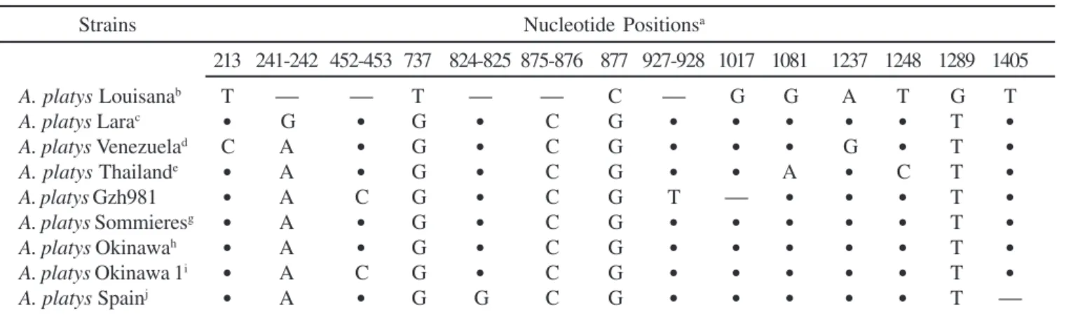
PCR-positive samples were identical. Thus, a nearly entire sequence (1,364 bp) of the 16S rRNA gene was obtained from the dog blood sample #16 (designated as A. platys Lara) collected in December 1999. Nucleotide differences between this new sequence and other A. platys 16S rRNA gene sequences currently available in the GenBank database are shown in Table 1. Strain polymorphisms were seen throughout the gene in 14/1,330 positions. The alignment of A. platys 16S rRNA gene sequences (1,330 bp) on database showed that A. platys
Lara 16S rRNA gene was closest to that of strain Sommieres from France and strain Okinawa from Japan (1 bp substitution). In addition, 16S rRNA gene sequences of strain Sommieres from France and strain Okinawa from Japan were identical. Lara’s
strain 16S rRNA sequence had 3 bp substitutions when compared to 1,330 bp of the strain Venezuela, obtained from a dog in Maracaibo, Venezuela.
A partial groESL operon sequence (1,646 bp) of A. platys
Lara was also obtained. The stop codon for GroES and the start codon for GroEL were deduced based on alignment of all groESL
operon sequence of other Anaplasma species available at GenBank database. The groESL sequence in the present study encoded the C-terminal 6 amino acids of GroES protein, a 51-bp intergenic space, and 524 amino acids of GroEL protein. The
Table 1. Nucleotide sequence differences among 16S rRNA gene from different strains of A. platys.
Strains Nucleotide Positionsa
213 241-242 452-453 737 824-825 875-876 877 927-928 1017 1081 1237 1248 1289 1405
A. platys Louisanab T ⎯ ⎯ T ⎯ ⎯ C ⎯ G G A T G T
A. platys Larac
•
G•
G•
C G•
•
•
•
•
T•
A. platys Venezuelad C A
•
G•
C G•
•
•
G•
T•
A. platys Thailande
•
A•
G•
C G•
•
A•
C T•
A. platys Gzh981
•
A C G•
C G T ⎯•
•
•
T•
A. platys Sommieresg
•
A•
G•
C G•
•
•
•
•
T•
A. platys Okinawah
•
A•
G•
C G•
•
•
•
•
T•
A. platys Okinawa 1i
•
A C G•
C G•
•
•
•
•
T•
A. platys Spainj
•
A•
G G C G•
•
•
•
•
T ⎯a The number represents the nucleotide position of the type strain (A. platysT Louisiana, GenBank accession number M82801); where two nucleotide positions are shown, some strains had an insertion;
•
, same base as the type strain; ⎯, deletion. The region (nucleotide position 70-1409 of the type strain) that can be aligned unambiguously (1,340 bp) were compared; bA. platys type strain from a dog in the U.S.A. (GenBank accession number: M82801); c Sequence obtained in this study from a Venezuelan dog (GenBank accession number: AF399917); dA. platys from dogs in Venezuela (GenBank accession number: AF287153); eA. platys from dogs in Thailand (GenBank accession number: AF286699); fA. platys from a dog in China (GenBank accession number: AF156784); gA. platys from a dog in France Sommieres (GenBank accession number: AF303467); h A. platys from a dog in Okinawa, Japan (GenBank accession number: AY077619); iA. platys from a dog in Okinawa, Japan (GenBank accession number: AF536828); jA. platys from a dog in Spain (GenBank accession number: AY530806).Figure 1. Agarose gel electrophoresis of the second round of
groEL sequence which we obtained, covered 95.3% of the complete groEL coding sequence of E. chaffeensis (GenBank accession no. AAB49805). Only four A. platys groESL operon sequences are currently available at GenBank. However, groEL
sequence from Louisana strain from the U.S.A. (GenBank accession no. AY008300) is 1070 bp, too short to be included in this comparison. The remaining three sequences were aligned with Lara groESL operon sequence and the differences between these sequences were summarized in Table 2. Lara strain groESL
sequence (1,646 bp) was identical to that of RDC strain from Democratic Republic of Congo. Compared to Sommieres strain from France, Lara groESL had 1 bp substitution and resulted in one amino acid change in GroEL (9th amino acid from the first methionine) from lysine in Sommieres strain to glutamic acid in Lara strain. Lara groESL had 3 bp substitutions when compared to that of Okinawa strain and resulted in one amino acid change at the same position. Sequences (51-bp) of groES and groEL
intergenic region were identical among four strains (RDC, Sommieres, Okinawa, and Lara).
products. The results indicate a high prevalence of subclinical infection of dogs. There have been only two PCR-based A. platys prevalence reports. In one kennel in North Carolina, USA, 33% of 27 dogs were A. platys PCR-positive (16). In Okinawa, Japan, 32% of 200 stray dogs were positive by A. platys-specific PCR (18). On the other hand, the indirect fluorescent antibody (IFA) test using blood from an experimentally infected dog as antigen in Florida, indicated that 5% of 100 clinically normal dogs were positive, while 35% of 26 thrombocytopenic dogs were seropositive (10). In Louisiana, USA, 50% of 86 dogs tested were IFA positive (12). In eastern North Carolina, USA, 68% of the 50 tested dogs had low platelet counts and 74% were serologically positive (5).
The 16S rRNA sequence of strain Lara was different from another Venezuela strain from Maracaibo, suggesting that there are at least two strains of A. platys in Venezuela. Interestingly, Lara strain’s groESL sequence was found to be identical to the sequence of the RDC strain from R. sanguineus ticks collected from dogs in Democratic Republic of Congo. The short (732 bp) partial 16S rRNA sequence of the RDC strain (GenBank accession no. AF478131) is available for comparison. The sequence of 16S rRNA gene of the RDC strain (from nucleotide position 1 to 726 bp of 16S rRNA sequence of the strain Lara) was 100% identical to that of Lara strain. The unusual 6 bp sequence at the 3’ end of the partial 16S rRNA gene of the RDC strain does not match with any 16S rRNA sequence in the
Anaplamaceae family. These data suggest that A. platys strains are not geographically segregated. Close similarity between the 16S rRNA gene and groESL sequences of strain Sommieres from France and strain Okinawa from Japan, also supports this hypothesis, although sequence analysis of additional genes is necessary to confirm this observation.
The dogs of the present study were previously tested for E. canis infection and the detected infection rate was 31% (26). Only two samples (dog 23 in December 1999 and dog 21 in April 2000) were found to be infected by E. canis and A. platys. Remaining five dogs were infected only with A. platys. The rate of A. platys-infected dogsthat were coinfected with E. canis
was 29% (2/7). Previously, a dog from Maracaibo, Venezuela was reported to be PCR positive for three pathogens: A. platys, A. phagocytophilum, and E. canis (24). Another case of a dog coinfected with A. platys and E. canis was reported in China (13). These data suggest that A. platys and E. canis may share the same vector. A. platys DNA was detected in R. sanguineus
ticks in Japan (14) and Spain (23). However, A. platys 16S rRNA was undetectable in three R. sanguineus tick pools in the present study, although the same three pools of tick specimens were all positive for E. canis 16S rRNA (26). Experimental A. platys
transmission by R. sanguineus ticks was reported to be unsuccessful (22). These results suggest that A. platys tick infection level is very low or R. sanguineus is not an effective vector for transmitting A. platys. It is also possible that other
Table 2. Nucleotide sequence differences among groESL from different strains of A. platys.
Strains Nucleotide Positionsa
117 341 1472
A. platys Sommieresb A G T
A. platys Larac G
•
•
A. platys RDCd G
•
•
A. platys Okinawae
•
A CA. platysTLouisianaf /
•
/a The number represents the nucleotide position of A. platys Sommieres strain, GenBank accession number AY0441621;
•
, same base with Sommieres strain; /, sequence not available. The region (nucleotide position 19-1664 of Sommieres strain) that can be aligned unambiguously (1,646 bp) were compared; bA. platys from a dog in France Sommieres (GenBank accession number: AY044161); c Sequence obtained in this study from a Venezuelan dog (GenBank accession number: AF399916); dA. platys from Rhipicephalus sanguineus ticks in Democratic Republic of Congo (RDC) (GenBank accession number: AF478129); e A. platys from a dog in Okinawa, Japan (GenBank accession number: AY077621); f A. platys from a dog in Louisiana (primer sequences: 20 bp at 5’ end and 23 bp at 3’ end, were removed) (GenBank accession number: AY008300).DISCUSSION
tick species are involved in A. platys transmission. In fact, a 16S rRNA gene sequence of Ehrlichia sp. Omatjenne (GenBank accession no. U54806) closely related to A. platys Lara strain (99.3% identity, 4 bp substitution and 1 bp deletion/1,330 bp in
Ehrlichia sp. Omatjenne) was found in Hyalomma truncatum
ticks in Africa. Alternatively, one of reasons for the A. platys
16S rRNA RT-PCR negative result may be due to very small number of R. sanguineus ticks collected and tested in the present study.
Dogs are reported to be infected with several rickettsial species in the family Anaplasmataceae: monocyte-tropic species (E. canis, E. chaffeensis, Neorickettsia helminthoeca, and
Neorickettsia risticii), granulocyte-tropic species (E. ewingii
and A. phagocytophilum), and platelet-tropic A. platys (20). In the USA, E. chaffeensis, E. ewingii, and A. phagocytophilum
are known to cause human ehrlichioses (20), and dogs may serve as sentinel of infection with these organisms. In Venezuela dogs are heavily infected with E. canis and infection of humans with the same strain of E. canis has been confirmed by culture isolation of E. canis of the identical 16S rRNA gene sequences from a human and a dog, respectively (19, 26). In the present study, however, all human and dog specimens tested were negative for E. ewingii or A. phagocytophilum infection. Thus, in Lara, Venezuela, these infections may not be prevalent. Despite the reports of rickettsiae-like organisms in human platelets in Venezuela (1, 25), there has been no evidence for human infection with A. platys in the present study. Further studies are needed to clarify potential human infection with A. platys in Venezuela.
ACKNOWLEDGEMENTS
This research was supported by a grant R01AI47407 from the National Institutes of Health and Ohio State University Faculty International Travel grant. We appreciate Venezuelan Military Center for Canine Training for collaboration. Authors appreciate Dr. Roger W. Stich at the Ohio State University, and Dr. James E. Keirans, Georgia Southern University for the tick species identification. We thank Wen-Lan Chang for providing the A. platys positive control DNA and A. platys nested PCR primers EP1, EP2 and EP3.
RESUMO
Prevalência e análise molecular de Anaplasma platys
em cães da Venezuela
Amostras de sangue coletadas de cães clinicamente sadios pertencentes ao exército da Venezuela e de seus treinadores foram analisadas pela técnica de PCR 16S rRNA específica para
Anaplasma platys, A. phagocytophilum ou Ehrlichia ewingii.
Dezesseis por cento (7/43) dos cães foram positivos, enquanto
que todas as amostras de origem humana [25] foram negativas para A. platys. Todas as amostras, tanto de humanos quanto de caninos, foram negativas para E. ewingii ou A. phagocytophilum. Doze carrapatos da espécie Rhipicephalus sanguineus, coletados dos cães, foram negativos para A. platys
pelo teste de PCR de transcrição reversa. As seqüênciasquase inteiras do gene 16S rRNA (1.364 pb) e do operon groESL (1.646 pb) de A. platys isolado de um cão foram determinadas, revelando que ambas as seqüências estão estreitamente relacionadas às seqüências de A. platys detectadas em carrapatos R. sanguineus na República Democrática do Congo.
Palavras-chave: 16S rRNA, groESL operon, Anaplasma platys, cães, Venezuela
REFERENCES
1 . Arraga-Alvarado, C.; Palmar, M.; Parra, O.; Salas, P. Fine structural characterisation of a Rickettsia-like organism in human platelets from patients with symptoms of ehrlichiosis. J. Med. Microbiol., 48, 991-997, 1999.
2 . Arraga-Alvarado, C.; Palmar, M.; Parra, O.; Salas, P. Ehrlichia platys (Anaplasma platys) in dogs from Maracaibo, Venezuela: an ultrastructural study of experimental and natural infections. Vet.
Pathol., 40,149-156, 2003.
3 . Baker, D.C.; Simpson, M.; Gaunt, S.D.; Corstvet, R.E. Acute Ehrlichia platys infection in the dog. Vet. Pathol., 24, 449-453, 1987. 4 . Beaufils, J.P. Un syndromes’ accompagnant d’inclusions plaquettaires
Chez le Chien, dans la region de montpellier. Description et comparaison aux affections engendrees par Ehrlichia canis et
Ehrlichia platys, 1985. (Ph.D. Thesis. Ecole Nationale Veter de Toulouse).
5 . Bradfield, J.F.; Vore, S.J.; Pryor, W.H. Jr. Ehrlichia platys infection in dogs. Lab. Anim. Sci., 46, 565-568, 1996
6 . Brown, G.K.; Martin, A.R.; Roberts, T.K.; Aitken, R.J. Detection of
Ehrlichia platys in dogs in Australia. Aust. Vet. J., 79, 554-558, 2001.
7 . Buller, R.S.; Arens, M.; Hmiel, S.P.; Paddock, C.D.; Sumner, J.W.; Rikhisa, Y.; Unver, A.; Gaudreault-Keener, M.; Manian, F.A.; Liddell, A.M.; Schmulewitz, N.; Storch, G.A. Ehrlichia ewingii, a newly recognized agent of human ehrlichiosis. N. Engl. J. Med., 341, 148-155, 1999.
8 . Chang, W.L.; Pan, M.J. Specific amplification of Ehrlichia platys
DNA from blood specimens by two-step PCR. J. Clin. Microbiol., 34, 3142-3146, 1996.
9 . Felek, S.; Unver A.; Stich, R.W.; Rikihisa, Y. Sensitive detection of
Ehrlichia chaffeensis in cell culture, blood, and tick specimens by reverse transcription-PCR. J. Clin. Microbiol., 39, 460-463, 2001. 10. French, T.W.; Harvey, J.W. Serologic diagnosis of infectious cyclic thrombocytopenia in dogs using an indirect fluorescent antibody test. Am. J. Vet. Res., 44, 2407-2411, 1983.
11. Harvey, J.W.; Simpson, C.F.; Gaskin, J.M. Cyclic thrombocytopenia induced by a Rickettsia-like agent in dogs. J. Infect. Dis., 137, 182-188, 1978.
12. Hoskins, J.D.; Breitschwerdt, E.B.; Gaunt, S.D.; French, T.W.; Burgdorfer, W. Antibodies to Ehrlichia canis, Ehrlichia platys, and spotted fever group rickettsiae in Louisiana dogs. J. Vet. Intern. Med., 2, 55-59, 1988.
13. Hua, P.; Yuhai, M.; Shide, T.; Yang, S.; Bohai, W.; Xiangrui, C. Canine ehrlichiosis caused simultaneously by Ehrlichia canis and
14. Inokuma, H.; Raoult, D.; Brouqui, P. Detection of Ehrlichia platys
DNA in brown dog ticks (Rhipicephalus sanguineus) in Okinawa Island, Japan. J. Clin. Microbiol., 38, 4219-4221, 2000.
15. Kontos, V.I.; Papadopoulos, O.; French, T.W. Natural and experimental canine infections with a Greek strain of Ehrlichia platys. Vet. Clin. Pathol., 20, 101-105, 1991.
16. Kordick, S.K.; Breitschwerdt, E.B.; Hegarty, B.C.; Southwick, K.L.; Colitz, C.M.; Hancock, S.I.; Bradley, J.M.; Rumbough, R.; McPherson, J.T.; MacCormack, J.N. Coinfection with multiple tick-borne pathogens in a Walker Hound kennel in North Carolina. J. Clin. Microbiol., 37, 2631-2638, 1999.
17. Mathew, J.S.; Ewing, S.A.; Murphy, G.L.; Kocan, K.M.; Corstvet, R.E.; Fox, J.C. Characterization of a new isolate of Ehrlichia platys
(Order Rickettsiales) using electron microscopy and polymerase chain reaction. Vet. Parasitol., 68, 1-10, 1997.
18. Motoi, Y.; Satoh, H.; Inokuma, H.; Kiyuuna, T.; Muramatsu, Y.; Ueno, H.; Morita, C. First detection of Ehrlichia platys in dogs and ticks in Okinawa, Japan. Microbiol. Immunol., 45, 89-91, 2001. 19. Perez, M.; Rikihisa, Y.; Wen, B. Ehrlichia canis-like agent isolated
from a man in Venezuela: antigenic and genetic characterization. J. Clin. Microbiol., 34, 2133-2139, 1996.
20. Rikihisa, Y. Diagnosis of emerging ehrlichial diseases of dogs, horses, and humans. J. Vet. Intern. Med., 14, 250-251, 2000.
21. Sainz, A.; Amusategui, I.; Tesouro, M.A. Ehrlichia platys infection and disease in dogs in Spain. J. Vet. Diagn. Invest., 11, 382-384, 1999. 22. Simpson, R.M.; Gaunt, S.D.; Hair, J.A.; Kocan, K.M.; Henk, W.G.; Casey, H.W. Evaluation of Rhipicephalus sanguineus as a potential biologic vector of Ehrlichia platys. Am. J. Vet. Res., 52, 1537-1541, 1991.
23. Sparagano, O.A.; de Vos, A.P.; Paoletti, B.; Camma, C.; de Santis, P.; Otranto, D.; Giangaspero, A. Molecular detection of Anaplasma platys in dogs using polymerase chain reaction and reverse line blot hybridization. J. Vet. Diagn. Invest., 15, 527-534, 2003.
24. Suksawat, J.; Pitulle, C.; Arraga-Alvarado, C.; Madrigal, K.; Hancock, S.I.; Breitschwerdt, E.B. Coinfection with three Ehrlichia species in dogs from Thailand and Venezuela with emphasis on consideration of 16S ribosomal DNA secondary structure. J. Clin. Microbiol., 39, 90-93, 2001.
25. Tami, I.; Martinez, I.; Tami, M.; Redondo, M.C.; Finol, H.; Simonovis, N. Identification of Ehrlichia species in blood smear.
Infect. Dis. Clin. Prac., 5, 555-557, 1996.
26. Unver, A.; Perez, M.; Orellana, N.; Huang, H.; Rikihisa, Y. Molecular and antigenic comparison of Ehrlichia canis isolates from dogs, ticks, and a human in Venezuela. J. Clin. Microbiol., 39, 2788-2793, 2001.
27. Unver, A.; Rikihisa, Y.; Kawahara, M.; Yamamoto, S. Analysis of 16S rRNA gene sequences of Ehrlichia canis, Anaplasma platys, and
Wolbachia species from canine blood in Japan. Ann. N. Y. Acad. Sci., 990, 692-698, 2003.
28. Warner, C.K.; Dawson, J.E. Genus- and species-level identification of Ehrlichia species by PCR and sequencing. In: Persing, D.H. (ed).
PCR protocols for emerging infectious diseases: a supplement to Diagnostic Molecular Microbiology: Principles and Applications. ASM Press, Washington, 1996, p.100-105.