R E V I E W
Open Access
Do schistosome vaccine trials in mice have
an intrinsic flaw that generates spurious
protection data?
R. Alan Wilson
1*, Xiao-Hong Li
2and William Castro-Borges
3Abstract
The laboratory mouse has been widely used to test the efficacy of schistosome vaccines and a long list of candidates has emerged from this work, many of them abundant internal proteins. These antigens do not have an additive effect when co-administered, or delivered as SWAP homogenate, a quarter of which comprises multiple candidates; the observed protection has an apparent ceiling of 40–50 %. We contend that the low level of maturation of penetrating cercariae (~32 % forSchistosoma mansoni) is a major limitation of the model since 68/100 parasites fail to mature in naïve mice due to natural causes. The pulmonary capillary bed is the obstacle encountered by schistosomula en route to the portal system. The fragility of pulmonary capillaries and their susceptibility to a cytokine-induced vascular leak syndrome have been documented. During lung transit schistosomula burst into the alveolar spaces, and possess only a limited capacity to re-enter tissues. The acquired immunity elicited by the radiation-attenuated (RA) cercarial vaccine relies on a pulmonary inflammatory response, involving cytokines such as IFNγand TNFα, to deflect additional parasites into the alveoli. A principal difference between antigen vaccine protocols and the RA vaccine is the short interval between the last antigen boost and cercarial challenge of mice (often two weeks). Thus, after antigen vaccination, challenge parasites will reach the lungs when both activated T cells and cytokine levels are maximal in the circulation. We propose that“protection”in this situation is the result of physiological effects on the pulmonary blood vessels, increasing the proportion of parasites that enter the alveoli. This hypothesis will explain why internal antigens, which are unlikely to interact with the immune response in a living schistosomulum, plus a variety of heterologous proteins, can reduce the level of maturation in a non-antigen-specific way. These proteins are “successful”precisely because they have not been selected for immunological silence. The same arguments apply to vaccine experiments withS. japonicumin the mouse model; this schistosome species seems a more robust parasite, even harder to eliminate by acquired immune responses. We propose a number of ways in which our
conclusions may be tested.
Keywords:Schistosoma mansoni,Schistosoma japonicum, Radiation attenuated vaccine, Antigen vaccine, Mouse, Hamster, Primate, Intravascular migration, Maturation, Pulmonary capillary, Inflammation
Background
The question posed in the title of this review was prompted by two recent sets of observations. Firstly, in 2014, publications from three Brasilian groups reported protection against schistosome challenge after vaccin-ation of mice using what are unquestionably intracellular proteins. The four proteins were Syntenin [1], Dynein
light chains DLC12 and DLC13 [2], and Y box protein [3]. Secondly, we undertook a quantitative shotgun proteomic analysis of the ubiquitous antigen preparation SWAP, the soluble cytosolic extract of adult worms [4]. We found that about one quarter of the protein mass comprised putative vaccine candidates identified by other researchers. In this situation why does SWAP, packed with multiple vaccine candidates, not have a dra-matic protective effect? Instead it performs no better than individual candidates administered alone, and sometimes worse. Do these antigens all trigger the same
* Correspondence:alan.wilson@york.ac.uk
1Centre for Immunology and Infection, Department of Biology, University of York, Heslington, York YO10 5DD, UK
Full list of author information is available at the end of the article
mechanism, which has a ceiling of approximately 40– 50 % protection, whether one or many are used?
In this review we ask whether the protective outcome of antigen vaccination is an artefact of the mouse model and not a measure of acquired immunity. This is not the same as saying that there are no protective anti-gens but simply that the mouse model may not be cap-able of discriminating between acquired immunity induced by antigens and an effect of vaccination on host physiology that diminishes schistosome matur-ation in some non-specific way.
The mouse as an animal model for schistosomes
Whilst the laboratory mouse is a convenient host for schistosomes, used in hundreds of vaccine testing stud-ies over the last 40 years, it does have a major drawback. It is described as a permissive host but in reality the actual numbers of penetrantSchistosoma mansoni para-sites that migrate from the skin to the portal system and mature into adult worms is quite low. The statistic can be obtained by counting the percentage of challenge control cercariae that reach maturity in mice exposed to the radiation-attenuated (RA) cercarial vaccine but re-ceiving no adjuvant or other treatment. In a sample of 26 experiments from the York group over a decade, using the Puerto Rican isolate, the maturation in C57Bl/ 6 mice was 32.3 % [5–9]. The maturation of cercariae of the same isolate in CBA mice in seven experiments by the London School of Hygiene group was an almost identical 32.5 % [10]. The precise values may differ with isolate and mouse strain but the stark fact from these data is that of 100 cercariae penetrating the skin of a naivemouse, 68 will fail to reach and mature in the site of parasitisation, the portal tract. Vaccine-induced pro-tection is defined as the reduction in burden between vaccinated and control groups and the low maturation in mice is dealt with by using the formula: % Protec-tion = (Control burden - Test burden)/Control burden × 100. For example with a ~40 % level of protection, in the control group 68 parasites will fail to mature, while vaccination of the test group eliminates only a further 13 parasites. Thus, of the 81 parasites that did not ma-ture in the test mouse, the vaccine treatment only accounted for (13/81 × 100 =) ~16 %; the rest died of natural causes. In truth, the way of calculating protec-tion in the mouse very effectively disguises only a small achievement in worm elimination.
A larger number of penetrant S. mansoni cercariae mature in the golden (Syrian) hamster with a mean of 56 % recorded in eight experiments [11–14], and values as high as 76 % in single experiments [15]. In non-human primates, the hosts evolutionarily closest to humans, there is comparable data available from chal-lenge control animals in vaccine experiments with RA
cercariae. A maturation of 82 % was recorded in a single vervet experiment (Cercopithecus aethiops [16]) and a mean of 80.5 % in three baboon experiments (Papio anubis [17]). In contrast, the laboratory rat presents an even greater obstacle to migration than the mouse, with 22–27 % of juvenile worms detected in the liver between days 11 and 21 [18]. We conclude that the laboratory mouse, viewed solely in the context of parasite matur-ation, does not seem the best choice for vaccine experi-ments, and rats are even worse.
Exactly what are the“natural causes”that limit maturation?
It is important to establish at the outset that the 68 % of non-maturing S. mansoni penetrants are not eliminated by immunological processes as their protracted migra-tion and slow development (~5 weeks) allow ample time for immune intervention. The best evidence is provided by experiments in which mice were exposed to whole body irradiation a few days before infection. The treat-ment severely depresses immunological responsive-ness without administration of chemicals or alteration of genotype. Most parameters decline by ~90 %, yet parasite migration and maturation are not enhanced [19–21]. This strongly supports a physical explanation for the low maturation.
the lungs. The principal feature revealed by the balance sheet is that no parasites are eliminated before 15 days post-infection (Fig. 1b), at which time < 10 % can be de-tected in the skin; this is the clearest possible demon-stration that parasite death in the skin is minimal. At 15 days the population is distributed around all organs of the mouse body, with the largest proportion in the lungs. The question is ‘what happens from 15 days onwards’?
When migrating schistosomula, recovered from the pulmonary vasculature of donor mice, are injectedviaa vein back to the lungs of naïve recipients, ~50 % reach maturity [26, 27]. However, > 80 % of the same batch, injected into the portal vessels, are recovered as mature worms four weeks later. Such experiments confirm that the mouse lungs are the real barrier on the migratory route. The reason why less than 100 % of injected schis-tosomula develop to maturity in the portal tract is that as many as 25–30 % actually negotiate the hepatic sinu-soids back to the venous circulation and lungs [26]. They must then traverse the lung obstacle again, as must those schistosomula returning from systemic organs. The magnitude of the lung barrier is underlined by ex-periments to determine the kinetics of larval migration
through different vascular beds [22, 23, 28]. The mean transit time of day 7 schistosomula through the lungs is 30–35 h. This contrasts with values of 16 h for systemic organs and 6.5 h for intestinal capillary beds [28] (esti-mates from an independent experiment with fewer time points gave values of 11.7 and 9.8 h, respectively [26].) Clearly negotiating the lung capillaries is hard work. It must be emphasised that schistosomula leaving the skin are short and stubby (100 to 190 μm long, Fig. 2b; [29])
and completely occlude pre-capillary arterioles of the lungs 10 to 20 μm in diameter, when they arrive [30].
They must then undergo a phase of development, be-coming longer (> 400 μm), thinner (~8 μm) and losing
mid body spines (Fig. 2c; [29, 30]) to facilitate transit along capillaries only 7μm wide. For the earliest arrivals,
the first transit through the lungs may take 78 h. In addition, the percentage maturation for Day 3 schistoso-mula delivered i.v. to the lungs is 30 % and for Day 4 schistosomula 41 %, compared with ~50 % for Day 7 lar-vae [15]. These data suggest that the first transit is the most difficult and protracted; indeed, it is quite possible that some schistosomula arriving from the skin never progress further [15]. Conversely, the percentage matur-ation of Day 12 and Day 17 schistosomula delivered to
the lungs i.v. is not significantly different from that of Day 7 larvae [27]. We should therefore consider the Day 7 lung schistosomulum as fully adapted to intravascular migration [29].
A combination of light and electron microscopy re-vealed that at day 7 post-infection all lung schistosomula were located in blood vessels of naïve mice [31]. At the next sampling time, day 11, a proportion of them were entirely or partially intra-alveolar, and this reached 80 % at day 20 and later. No cellular reactions were evident around intravascular parasites but at later sampling times alveolar parasites were associated with large in-flammatory foci, probably elicited as a response to non-specific tissue damage when they burst out. In spite of this inflammation, no damage to the schistosomula was observed. These observations were confirmed in an autoradiographic study which concluded that the alveo-lar alveo-larvae were unharmed and were likely to be coughed up, swallowed and digested [32]. Administration of schistosomulaviathe trachea to the pulmonary airspace revealed that a small number (~15 %) were able to re-enter blood vessels, migrate and mature in the liver, but this proportion diminished as the parasites aged [27]. Exit into alveoli appears to occur accidentally as
parasites attempt to traverse pulmonary blood vessels, and once there it becomes difficult to continue migration.
We can estimate the proportion of schistosomula becoming trapped and diverted to the airspaces on each circuit using a simple spreadsheet calculation (Additional file 1: Table S1A). An average P value of 0.36 for a schistosomulum getting stuck on each pas-sage through the lung vascular bed (including the first) creates a realistic migration profile. Only a few passages around the circulation, each taking about two days, are sufficient to generate the cumulative portal population. The remaining parasites blunder into the airspaces of the lungs or get stuck in other locations (Fig. 1). This phys-ical explanation of worm demise, with the averagePvalue of 0.36, accounts almost precisely for the ~32 % matur-ation in naïve mice. However, the maturmatur-ation data for Day 3 and Day 4 schistosomula indicate that they have Pvalues for getting stuck ofP= 0.415 and 0.28, respect-ively, while the 50 % maturation of day 7 (or older) lar-vae delivered i.v. to the lungs is achieved by P= 0.203 (Additional file 1: Table S1B). So what feature of the lungs, especially mouse lungs, presents a special obs-tacle to onward migration?
The first and most obvious aspect is that the pul-monary capillaries have very thin walls separated from the airspaces only by alveolar basement membrane and epithelium. The total thickness of the blood-gas barrier is ~0.3 μm with the alveolar epithelial cell in some
parts having a thickness of 0.1 μm [33]. Thus the
pul-monary capillary is only separated from the alveolar space by a layer of extracellular matrix and an epithe-lial cytoplasm, each 0.1μm thick. No other capillary in
the body is protected by such a thin layer of tissue, making the pulmonary capillaries fragile and vulnerable to failure [33]. The mechanical behaviour of alveolar capillaries is determined by the extracellular matrix layer, with type IV collagen the most important con-stituent determining the strength of the blood-gas bar-rier. The combined thickness of the three components of the blood-gas barrier correlates with the experimen-tal pressures required to damage to the pulmonary capillary wall in different species, the larger the animal, the thicker the blood-gas barrier (horse > dog > rabbit) [34]. Such measurements are not available for the mouse but it is plausible that the alveolar capillaries of this species may be particularly fragile due to its small size. Indeed, it is easy to appreciate how the vigorous and rhythmic extensions and contractions displayed by elongating schistosomula [29], especially the younger larvae, could provide the motive power to rupture al-veolar capillaries in the mouse lung.
A second difference is the nature of the pulmonary capillaries themselves. Those enveloping the alveoli are very short (~10 μm) forming a maze of vessels [35]
through which the schistosomulum must crawl (Fig. 2a). This contrasts with the network of longer bifurcating tubules that comprise the capillary bed of other organs and non-alveolar parts of the lungs (Fig. 2a, arrowed). Juxtaposing an image of an extended Day 6 lung schisto-somulum (Fig. 2c) next to that of the alveolar capillaries brings the task it faces in traversing the pulmonary vascu-lar bed into sharp focus. Unsurprisingly it must take a very convoluted path through the maze of short tubes with many options, as revealed by in situ electron micros-copy (Fig. 2d; [30]). Adding in the fact that it progresses along vessels in inchworm fashion using its anterior and posterior spines (Fig. 2e; [36]) everything converges to make pulmonary vascular transit a difficult propos-ition, with a significant probability of bursting into an alveolus.
The lungs are also subject to another potentially rele-vant phenomenon, the vascular leak syndrome (VLS). VLS was first reported following cytokine administration (Interleukin 2) to patients to treat certain cancers [37] and has been explored using the mouse as a model. It is characterised by leakage of vascular fluids into tissues, accompanied by hypotension; in the lungs this results in
pulmonary oedema. There is a general consensus that IL-2-induced VLS is caused by secondary release of in-flammatory cytokines such as IFNγ and TNFα [38, 39],
which may in turn increase production of vascular medi-ators such a nitric oxide (NO). It has even been sug-gested that VLS may be accompanied by modifications in the extracellular matrix [37], which could weaken the alveolar blood-air barrier to increase capillary fragility. The relevance of VLS to schistosome vaccines is that its causative agents are precisely those cytokines and medi-ators involved in the protective effector mechanism elic-ited by the RA vaccine (see below).
The radiation-attenuated (RA) cercarial vaccine in mice
The best studied model of acquired immunity to schis-tosomes is that induced by exposure of rodents and pri-mates to a dose of RA cercariae. We have a wealth of immunological and parasitological information about the actions of attenuated parasites in generating protec-tion and the fate of a normal challenge in vaccinated hosts (reviewed in [40–42]). In mice, optimally attenu-ated parasites undergo a truncattenu-ated migration as far as the lungs, priming the immune response via skin-draining lymph nodes. These attenuated larvae also “arm” the lungs by stimulating recruitment of effector T cells to the interstitial spaces and airways [43]. Mul-tiple vaccinations have an additive effect and almost sterile immunity can be achieved by co-administration of attenuated larvae and the cytokine IL-12 [44, 45]. However, we now know that subcutaneous administra-tion of IL-12 can produce adverse systemic effects [46]. Initial priming has a strong Th1 component but subse-quent exposures to RA cercariae amplify Th2 cells and en-hance antibody involvement. A striking feature is that a period of five weeks is left after application of the attenu-ated parasites, before challenge with normal cercariae. This allows any non-specific inflammatory events to subside before challenge parasite entry; vaccination and challenge are normally performed on separate sites such the abdominal skin and the tail, to avoid residual inflammation left by the initial exposure.
immunological events or mechanisms, it is clear that the major site of parasite loss is the lungs. This organ, difficult to negotiate in naïve mice, becomes more so in vaccinated animals. Increasing the chance of getting stuck on each circuit from P= 0.36 toP= 0.53 predicts the 40 % reduc-tion in worm burden (Addireduc-tional file 1: Table S1), as an additional 13 parasites fail to mature beyond the 68 in naïve animals (Fig. 3). The corresponding values for a 70 % reduction in burden areP= 0.72 with an additional 22 parasites prevented from maturing, still less than a quarter of the total.
What is the evidence that the protection induced by the RA vaccine has an immunological basis? Adoptive transfer of the immunity elicited by the RA vaccine has been achieved using a parabiotic union between mice [47]. Subsequent parabiotic experiments where the naïve and vaccinated partners were separated before challenge demonstrated two key facts about the model [9]. Firstly, recruitment of effector cells to the lungs was a crucial
pre-arming component of the protective response. Sec-ondly, an anamnestic response was elicited by percutan-eous challenge and could act against migrating larvae in the lungs. However, when the challenge was provided by day seven schistosomula administered directly to the lung vasculature, no protection was evident. Passive transfer of protection with homologous serum from vaccinated mice proved difficult to demonstrate [48]. However, it was eventually achieved using serum from multiply vaccinated mice [49], especially high-titre serum from IFNγR
-/-knock-donors [50]; in both studies the serum could be administered as late as day 4 or 7 post-challenge, revealing that the target was the lung, not the skin schistosomulum. To our knowledge, the passive or adoptive transfer of pro-tection to naïve mice has not been achieved using serum or cells respectively, from any donors reportedly exhibit-ing antigen-induced acquired immunity.
How are the extra challenge schistosomula arrested in the lungs? A focal inflammatory response develops
to each larva over several days [51]. The mass of cells around the parasite hinders onward migration and in-creases the probability of deflection into an alveolus. IFNγis a key cytokine [52, 53] and production of TNFα
also appears to be central to protection; mice lacking the TNF receptor 1 (TNFRI-/-) were not protected by exposure to the RA vaccine [8]. However, pulmonary inflammation alone is not sufficient since the cell infil-tration around migrating larvae in the lungs of IFNγR
-/-and TNFRI-/- mice is actually greater, but is ineffective at increasing the proportion of schistosomula deflected into the alveoli; this implies that there are other down-stream mediators. Strikingly, the protection does not involve direct immune damage to schistosomula. Al-most no evidence of cytological damage inflicted by the enveloping inflammation was observed in trapped chal-lenge larvae in situ [31] and lung schistosomula were not susceptible to ADCC mechanisms in vitro [54]. Most telling, transfer experiments with challenge schis-tosomula trapped in the lungs (which would not ma-ture if left in situ) showed that they could migrate and develop normally if introduced into the pulmonary or portal circulation of naïve animals [27]. This durability is a testament to the anti-oxidant capabilities of migrat-ing larvae [55]. In summary, the inflammatory effector mechanism of the RA vaccine apparently alters the pul-monary environment to decrease the probability that a schistosomulum can successfully traverse the vascular beds, but it does not cause it direct harm.
Although the effector mechanism in RA vaccinated mice operates in the lungs, the effector T cells were generated in the lymph nodes draining the skin vaccin-ation site. They exited via the efferent lymphatics to join the circulation from where they were available for
recruitment to the lungs by the attenuated vaccinating schistosomula [56]. Such circulating effector T cells can also be recruited to other sites like the footpad or pinna by an inflammatory stimulus and this forms the basis of a delayed-type hypersensitivity (DTH) assay to estimate their level in the circulation. In mice exposed to the RA vaccine, DTH T cells are high by ten days post-exposure, peak at day 17, are in decline by day 21 and almost down to background by day 35 when the cercar-ial challenge is administered (Fig. 4 [57]). Exposure to the challenge rapidly stimulates a recall response with a surge of DTH T cells into the circulation, peaking at day 7, declining by day 14 and well towards background by day 21. These data provide the clear justification for the five-week interval between vaccination and chal-lenge, and also key evidence for an anamnestic re-sponse to challenge larvae. The same kind of peripheral responsiveness has been observed in chimpanzees ex-posed to repeated doses of the RA vaccine. There was a progressively increasing reactivity of peripheral blood mononuclear cells to schistosome antigens with each successive vaccination; reactive cells were still circulat-ing four weeks after challenge [58].
Vaccine antigen testing in the mouse How does it differ from RA vaccine experiments?
In testing of vaccine antigens in the mouse a common design is employed. A recombinant protein, less fre-quently a parasite fraction or a purified protein, is formulated with an adjuvant for subcutaneous or intra-peritoneal administration. Freund’s complete adjuvant is a frequent choice for priming followed by two doses of Freund’s incomplete adjuvant as the booster, spaced two or three weeks apart. Mice that will serve as
challenge controls receive the adjuvant alone formu-lated with saline; these are not equivalent. An irrele-vant antigen of similar immunogenicity would be a better control; very seldom is a no-adjuvant group in-cluded. (Vaccination with DNA constructs follows a similar pattern, but without extraneous adjuvants). The mice are all challenged with the same pool of cer-cariae at an interval after the last boost, and then per-fused 5–8 weeks later to recover and count adult worms.
A major, and we suggest significant, difference from the RA vaccine is the much shorter interval between the last boost and the day of challenge. The preferred time appears to be around 14–15 days but it may be as short as 10 days and less commonly three or occasionally four weeks. Seldom is the interval five weeks as is standard with the RA vaccine in mice, and we can find few in-stances where it is longer than that. Recent experiments with two tegument vaccine candidates appear to support our contention. When C57Bl/6 mice were vaccinated with Sm22.6 and challenged 15 days after the last boost, they showed a mean 34.5 % protection [59]. In contrast, when the antigen was administered to Balb/c mice that were challenged 30 days after the last boost, the protec-tion elicited was 0 and 18 % [60]. A similar pattern was observed with Sm29 where 51 % protection was ob-served after a challenge of C57Bl/6 mice at 15 days after the last boost [61] and 0 % protection after a challenge of Balb/c mice at 30 days [60]. Of course it can be ar-gued that mouse strain was the key determinant of pro-tection, not the interval between boost and challenge, a point that can only be resolved by further experiments.
A key feature of our hypothesis is that the short time interval between boost and challenge may provide an ex-planation for the apparent immunity elicited by some very unlikely vaccine candidates. At the end of the anti-gen vaccine schedule, is the level of inflammatory cyto-kines in the circulation sufficient alone to modify pulmonary vascular physiology? Alternatively, do migrat-ing challenge schistosomula elicit an inflammatory stimulus in the lungs that recruits vaccine-activated cells from the periphery, unrelated to their own antigen spe-cificity? Remember that only a small additional number of larvae, above and beyond those that will not make it anyway, need to be arrested by the lung obstacle for 40 or 50 % protection to be generated. It is not 40 or 50 out of 100 penetrants, it is 13 to 16 extra larvae. We suggest that only a small additional effect on the pul-monary capillaries would be needed to impede this extra fraction of schistosomula.
Are the vaccine antigens plausible candidates?
Over the last three decades quite a long list of schisto-some candidates has been put forward, primarily based
on the results of protection experiments in mice. The candidates divide into two categories, internal and sur-face exposed/secreted. The first is larger and well repre-sented in SWAP [4]. It includes TPI, GST, Sm14 fatty acid binding protein (FABP), Aldolase, GAPDH, Calpo-nin, Sm20.8, Sm22.6, paramyosin, myosin heavy chain, 14-4-3 chaperone, and stomatin. The shorter list of ex-posed proteins, present in small amounts in SWAP, in-cludes: the tetraspanins Sm23 and TSP-2, tegument Sm29 and calpain, and gut-secreted Cathepsin B.
The list encompasses the six WHO candidates that have acquired almost mythical status. Two of them are localised in muscle (paramyosin and myosin heavy chain) and three are cytosolic (TPI, GST, FABP). They did not perform well in independent murine trials but acquired a life of their own such that today they can still be described as the most promising candidates [62]. A goal of 40 % protection for a usable vaccine was set by WHO in the early 1990s so it is evident that, in spite of much work with different adjuvants and live vectors over the intervening 20+ years, a real and perhaps insuper-able obstacle to improvement remains. Multiple expo-sures of mice to irradiated parasites drive protection towards 100 %, whereas co-administration of two vac-cine antigens [63, 64] or multiple epitope constructs [65, 66] do not markedly increment protection. The real test in this respect is the administration of SWAP with its multiplicity of candidates, where protection seldom reaches or exceeds 50 % [67, 68]. There has always been a conceptual problem with the cytosolic and cytoskeletal constituents as vaccine antigens. How could an immune effector mechanism “recognise” these internal proteins in a live parasite and interact with them to cause its demise? It is a question that has never received a satis-factory answer from the internal candidate enthusiasts.
Live BCG as an adjuvant
For one candidate, paramyosin, there a comprehensive body of relevant immunological data. However, the puri-fied protein was only effective when administered with live BCG as an adjuvant. The production of IFNγ and
already known to reduce worm burden non-specifically by generating pulmonary inflammation to interfere with migration [73]. In the paramyosin experiments the BCG was given intradermally (i.d.) with the assumption that it would not enter the circulation and be disseminated, al-though this does seem not to have been formally tested. However, i.d. administration of BCG, with subsequent detection in internal tissues does occur [74] so it may not be possible to disentangle vaccine antigen and BCG effects, making pulmonary involvement more plausible.
What can we deduce from the properties of the vaccine candidates?
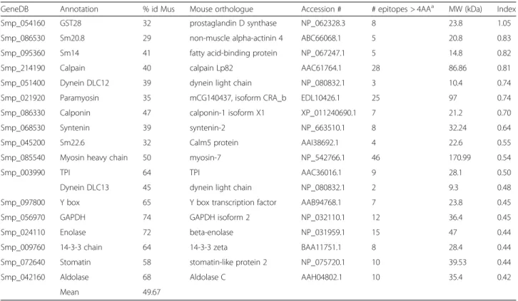
It is a reasonable assumption that the closer the iden-tity of a schistosome protein to its nearest ortholog in the murine host, the lower the probability that a unique epitope will exist. The candidates range in percentage homology between 29 and 74 % (mean 50 %; Table 1). Bepipred (http://tools.immuneepito-pe.org/bcell/), the B cell epitope predictor indicates that the larger schistosome proteins have higher num-bers of predicted epitopes, with myosin, paramyosin and calpain the winners. The likely immunogenicity of each candidate can be gauged using a simple formula where the number of epitopes is divided by the propor-tional identity of sequence times the Mw in kDa. On that basis, the leaders are GST-28, Sm20.8 EF hand Ca2+ bind-ing protein, Sm14 FABP and calpain (mean 36 %
homology). The least immunogenic are Enolase, 14-3-3, Stomatin and Aldolase (mean 66 % homology). (Indeed enolase is only included in the list to make the point that when we cloned and expressed it for vaccine experiments we could achieve no protection at all.) Our contention is that in the mouse model pure immunogenicity is likely to be the best predictor of an effect, simply because it will provide the strongest stimulation to the immune system.
A different way of examining the vaccine potential of a protein is to determine the effects that evolutionary pres-sures might exert to alter the nucleotide sequence of the encoding gene. This can be estimated by analysing the rates of non-synonymous to synonymous substitutions (dN/dS) between orthologs from different schistosome species. Such a study was recently performed with the micro-exon (MEG) and venom allergen-like (VAL) genes of schisto-somes [75]. The proteins they encode are associated with the glands and secretions of different life cycle stages [76– 79], positioned at the parasite-host interface where they will be exposed to selection pressure from the immune system. The two classes of genes revealed significantly higher dN/ dS values when compared with a set of control genes cod-ing for secreted proteins, and for other proteins previously localized to the tegument. Analyses of paralog genes indi-cated that exposure of the protein to the definitive host im-mune system was indeed a determining factor leading to the higher dN/dS values. In addition, two other proteins (Sm29 and TSP-2) exposed at the tegument surface, and
Table 1Molecular properties of vaccine candidates
GeneDB Annotation % id Mus Mouse orthologue Accession # # epitopes > 4AAa MW (kDa) Index
Smp_054160 GST28 32 prostaglandin D synthase NP_062328.3 8 23.8 1.05
Smp_086530 Sm20.8 29 non-muscle alpha-actinin 4 ABC66068.1 5 20.8 0.83
Smp_095360 Sm14 41 fatty acid-binding protein NP_067247.1 5 14.8 0.82
Smp_214190 Calpain 40 calpain Lp82 AAC61764.1 28 86.86 0.81
Smp_051400 Dynein DLC12 39 dynein light chain NP_080832.1 3 10.4 0.74
Smp_021920 Paramyosin 35 mCG140437, isoform CRA_b EDL10426.1 25 97 0.74
Smp_086330 Calponin 47 calponin-1 isoform X1 XP_011240690.1 7 21.2 0.70
Smp_068530 Syntenin 39 syntenin-2 NP_663510.1 8 32.24 0.64
Smp_045200 Sm22.6 32 Calm5 protein AAI38692.1 4 22.6 0.55
Smp_085540 Myosin heavy chain 50 myosin-7 NP_542766.1 46 170.99 0.54
Smp_003990 TPI 64 TPI AAC36016.1 9 28.1 0.50
Dynein DLC13 45 dynein light chain NP_080832.1 2 9.3 0.48
Smp_097800 Y box 65 Y box transcription factor AAB94768.1 7 23.8 0.45
Smp_056970 GAPDH 74 GAPDH isoform 2 NP_032110.1 12 36.4 0.45
Smp_024110 Enolase 72 beta-enolase NP_031959.1 15 47 0.44
Smp_009760 14-3-3 chain 64 14-3-3 zeta BAA11751.1 8 28.4 0.44
Smp_072640 Stomatin 58 stomatin-like protein 2 NP_075720.1 10 39.53 0.44
Smp_042160 Aldolase 68 Aldolase C AAH04802.1 10 35.4 0.42
Mean 49.67
several lipid-processing proteins present in the worm vom-itus displayed dN/dS values similar to those observed for MEGs and VAL genes. This provides further evidence of an additional selective pressure from the immune system on exposed proteins rather than a phenomenon specific for MEG and VAL genes. In complete contrast, almost all previously proposed vaccine candidates display very low rates of non-synonymous changes (Table 2). This is en-tirely consistent with their internal location and inaccess-ibility to immune effector responses in the live parasite. It also explains their strong immunogenicity when finally presented to the immune system upon release from the damaged or dead parasite. They have not been selected to be immunologically silent. This makes them the ideal agents to elicit a strong acquired response when adminis-tered multiple times with an adjuvant. Furthermore, when cercarial challenge is given only 10 to 15 days after the last boost, associated host responses to these reactive proteins will be maximal in the circulation, and so well placed to interfere non-specifically with an already fraught parasite migratory process.
What can protection experiments with heterologous antigens tell us?
Our hypothesis is that protection in the mouse model may be a bystander effect caused by high levels of circulating cy-tokines or the presence of activated T cells, macrophages and other leucocytes in the circulation, coincident with ar-rival of challenge larvae in the lungs. A corollary is that vac-cination with irrelevant antigens should produce the same effect. The extensive experiments with live BCG are evi-dence for an“irrelevant” antigen effect on parasite matur-ation [73] but other heterologous proteins have been tested. Crude Fasciola hepatica extracts protected mice against schistosome challenge [80] with the major activity attributed to an abundant and immunogenic 12 kDa FABP [81]. Inde-pendently theS. mansonihomologue of thisFasciolaprotein was cloned, shown to elicit protection in mice and proposed as a dual Fasciola/Schistosoma vaccine [82, 83]. Does the cross-protection result from shared epitopes between the two proteins? The sequence homology is only 49 %, spread evenly throughout the polypeptide, and BepiPred does not identify stretches of amino acids in common that might serve as shared epitopes. This evolutionary distance is confirmed by a comparison with S. bovis FABP which has also been the subject of Fasciola cross-protection studies [84]. It differs from its S. mansoni equivalent at only two amino acid positions but is again only 49 % identical to theF. hepaticaprotein. Only epitope mapping of the respective fatty acid binding proteins with specific antisera can resolve this point.
Protection experiments have also been performed using heterologous proteases as adjuvants to boost pro-tective responses to vaccine candidates such as GAPDH and 14-4-3 via a Th2-mediated immune response. It is remarkable that papain alone, from the plantCarica pa-paya, could induce a > 50 % reduction in worm burden when mice were challenged 14 days after a second ad-ministration of the enzyme subcutaneously [85]. The logic of this approach was that papain can activate ba-sophils to secrete Th2-type cytokines in the absence of antigen-specific IgE [86]. These cells normally com-prise ~1 % of circulating leucocytes and it would be pertinent to discover if they had any effect on circulating cytokines or pulmonary inflammation in papain-treated mice. In a similar way, functionally active F. hepatica cathepsin L1, which shares 47 % identity with itsS. man-soniorthologue, is capable alone of inducing up to 50 % reduction in worm burden when injected 14 days before cercarial challenge [87]. When co-administered with schistosome proteins, a reduction in burden of up to 73 % may ensue. (Remember that this is 73 % of 32 %, i.e., 23 worms stoppedviathe immune response against 68 from natural causes, so still only a quarter.) A plausible alterna-tive to an adjuvant role for these proteases, and the re-activity of F. hepatica FABP is that we are witnessing a
Table 2dN/dSmeasurements for genes encoding vaccine
candidates
Vaccine candidate Sma × Sha Sma × Sja
Sm29 0.83 0.41
TSP-2 0.34 (0.65a) 0.12 (0.33a)
GST-28 0.32 0.16
SmTOR 0.3 0.2
Sjserpin 0.29 0.41
SjTGR 0.29 0.09
SjVLDL 0.27 0.11
Sm22.6 0.21 0.25
StoLP-2 0.2 0.05
Calpain 0.18 0.15
Sm21.7 0.18 0.53
SjCathepsin 0.15 0.13
GAPDH 0.14 0.06
SmRho 0.11 0.13
TPI 0.11 0.13
Sm14 0.11 0.06
Sm23 0.08 0.1
SOD 0.07 0.14
Aldolase 0.05 0.02
Sj22.7 0.04 0.004
Sm14-3-3 0.03 0.19
Myosin heavy chain 0.02 0.03
Paramyosin 0.01 0.04
Sma, Schistosoma mansoni; Sha,S. haematobium; Sja,S. japonicum aValue for the portion of the gene encoding the exposed
bystander effect in the circulation impacting on the lungs, not specific acquired immunity.
Do vaccine antigens identified in mice protect non-human primates?
A perplexing feature of the candidates identified in mouse vaccine trials is that they do not translate well to a permis-sive primate host like the baboon (Papio anubis). Note that since maturation in baboons can be > 80 % of penetrant cercariae, an acquired immune response has much more work to do than in a mouse to achieve a high level of pro-tection. Nevertheless, > 80 % protection has been induced in primates like the baboon by multiple doses of the RA vaccine [17]. The > 80 % level of maturation means that only 20 out of 100 parasites die of“natural causes”during migration and a further 64 die due to immune effector mechanisms, the exact reverse of the mouse situation. The protection elicited in the baboon by the RA vaccine is also additive, in proportion to the number of exposures to attenuated cercariae, and a clear saw-tooth pattern of boost and decline in specific antibody production is ob-served with each successive vaccination [88]. The duration of protection has also been tested; it declines from 72 to 53 % when the interval between last boost and challenge is extended for three weeks to three months. Thus we contend that the baboon provides a robust test of vaccine potential. How well have vaccination experiments per-formed using the candidate antigens first trialled in mice?
A trial with SmGST28 produced contradictory results [89], depending on the adjuvant used with -26 and 38 % protection achieved. Another trial with myosin heavy chain (IrV5) gave 25 and 26 % protection in two differ-ent adjuvant formulations [90]. More recdiffer-ently, the tegu-ment antigen Sm29 proved to be immunogenic but failed to induce protection (Kariuki, personal communi-cation) while three anti-oxidant enzymes (two super-oxide dismutases and glutathione peroxidase) in a DNA vaccine formulation all failed to reduce worm burden significantly, but did depress faecal egg excretion [91]. Only one vaccine candidate, Sm-p80 calpain has achieved a good level of protection in baboon: 48 % with a DNA construct [92], then 52–58 % with a recombinant protein [93] (there was a 4 week gap between last boost and challenge). Overall, it appears harder to elicit pro-tection with single antigen formulations in baboons than in mice against a S. mansoni cercarial challenge: only calpain passes the primate test but persistence of protec-tion induced does not yet appear to have been tested.
AreS. japonicumvaccine experiments in mice subject to the same constraints?
In comparison withS. mansonithere is much less detailed information available about the mouse model infected withS. japonicum. However, the percentage maturation in
naïve mice appears to be higher at ~50 % ([94]; Chinese strain), while that in hamsters (~53 %) is about the same [95], and in rabbits it is 53.5 % [96]. The pattern of schis-tosomulum migration and the site of elimination of non-maturing parasites are less well understood but an early study concluded that migration was entirely in the blood-stream, with passive transport between organs and active crawling through capillary barriers [97]. A more rapid mi-gration ofS. japonicumwas noted with peak numbers in the lungs at 3–4 days and the first arrivals in the portal vein from 3.5 days. This qualitative pattern was later con-firmed by mincing and incubation of tissues to recover migrating parasites [98]. The single quantitative auto-radiographic tracking study reported that the skin was not a site of attrition after primary infection but that parasite elimination occurred after migration to the lungs and con-tinued up to the liver stage [99]. The presence of large numbers of petechial haemorrhages on the lung surface has been recorded in several studies [97, 99, 100]; they are seldom observed in S. mansoni-infected hosts in spite of numerous schistosomula entering the alveoli. Most strik-ing, petechiae have been recorded on other organs like the kidney, and on the walls of the stomach [97]. These obser-vations confirm that migration occurs through systemic organs and that schistosomula arrive in the portal system via the capillary beds of splanchnic organs. Based on minimum recorded length, S. japonicum schistosomula are apparently one third larger than those of S. mansoni [97] so may cause greater vascular damage. In addition, given the > 50 % maturation rate, they may also have a greater capacity to re-enter the tissues to continue migrating.
presumably containing multiple candidates, has been re-ported to elicit > 40 % protection [109]. Some unexpected antigens also induced protection; for example, hypoxanthine-guanine phosphoribosyltransferase con-ferred over 40 % protection against challenge [110] and mitochondrial succinic dehydrogenase was also reported to induce significant protection [111]. The S. japonicum counterparts of surface proteins reported to elicit good protection againstS. mansoni, (SjTSP2 [112], Sj29 [113]), were not as effective when used to vaccinate mice before challenge with S. japonicumcercariae. Finally, protection has been induced in mice against S. japonicumchallenge using heterologous antigens as diverse as Lumbricus ter-restris(earthworm; [114]) andTrichinella spiralisextracts [115]. Although the evidence is more fragmentary, we sug-gest that our bystander hypothesis is equally applicable to S. japonicumvaccine experiments in mice.
Conclusions
In the title of this review we ask if there is a flaw in the mouse model. We have built a case that indeed there is, namely the fragility of the pulmonary capillaries. The consequence is that during lung transit in a naïve mouse, schistosomula burst into alveoli and have only a limited capability to continue migration thereafter; this is very much the dominant determinant of parasite mat-uration. There is good evidence that the RA vaccine makes pulmonary migration more difficult viaa specific acquired response to surface and secreted antigens of the schistosomulum; additional worms are deflected but they are not harmed by the inflammatory responses. Conversely, our hypothesis is that for many of the proposed vaccine antigens the vaccination protocol in-creases the difficulty of pulmonary migration in a non-antigen-specific way. The kinetics of T cell production after vaccination with an antigen will likely mirror those after exposure to the RA vaccine. As the interval between last boost and challenge is usually short, acti-vated cells will still be available in the circulation for re-cruitment to the lungs when the challenge parasites arrive. Note, such recruitment is not antigen-specific, but activation-specific. It is also likely that the level of pro-inflammatory cytokines in the circulation will be high, perhaps sufficient to modify the pulmonary ves-sels. This would be independent of the accessibility of the vaccine antigen in the live parasite. Our explanation also encompasses the protective effects of the various heterologous antigens - they are simply very immunoreactive.
How can we probe the mouse model to test the assertions made in this review?
A major point of this overview of vaccine testing in the mouse was to provide a series of pointers for
experiments to bring clarity to the situation. This is a plea to stop treating the mouse as a black-box test bed and make immunological measurements strictly in the parasitological context of challenge parasite migration and elimination. The onus is on vaccine researchers to show that their specific antigen model has a solid knowledge base in acquired immunity. Some key points are:
(i) What happens to the level of protection if the interval between the last boost and cercarial challenge is extended at least to five weeks and preferably longer? The very short interval of 10–15 days is a severe criticism of many antigen vaccine experiments.
(ii) What is the profile of activated T cells and of cytokines in the circulation after vaccination? Has it declined to background levels before cercarial challenge? The emphasis here is on circulation, not spleen or lymph nodes. Proliferation of peripheral blood mononuclear cells, cytokine production with and without antigen restimulation, detection of a DTH response by footpad or pinna swelling, are all appropriate assays. Key signatures would be IFNγ,
TNFαor nitric oxide production, or the presence
of activated monocytes/macrophages.
(iii)Does percutaneous cercarial challenge at 5 weeks or later elicit a detectable secondary response to the target antigen in the circulation of the antigen-vaccinated mice in the days immediately after challenge, measured as above? This is vital missing evidence that the living challenge larvae can trigger a recall response to vaccine antigens, especially internal ones.
(iv) Where are challenge parasites eliminated in antigen-vaccinated animals? Skin, lungs or later, we simply do not know. This was crucial to understand-ing how the RA vaccine operated. Although
75Se-Methionine is no longer commercially available, 35S Methionine and Cysteine provide an intensity
of radioactive label that is still sufficient to allow detection of parasites in the skin and lungs by auto-radiography so the question of elimination in those sites can be explored.
(v) Continuing the possibility of lung involvement, is there evidence for cell recruitment to the lungs after challenge that might interfere with migration in a non-antigen specific way? This is testable by broncho-alveolar lavage, and flow cytometric phenotyping of recovered populations. (vi) Are there physiological or pharmacological
administration of pro-inflammatory cytokines such as IFNγor TNFαdiminish schistosome maturation
as implied by the RA vaccine? Alternatively, can we emulate the protection achieved with the RA vaccine using pharmacological interventions? Molecules that disrupt pulmonary hemodynamics could in theory modify the barrier to parasite migration. In this context, would nitric oxide-generating molecules, by promoting vasodilation, significantly alter vascular tonus in the lungs? This would be difficult to test due to the short-lived nature of these molecules; perhaps their effects could be sustained by the use of phosphodiesterase inhibitors. Other molecules capable of altering lung fluid homeostasis (e.g., leukotriene D4, platelet-activating factor, and thromboxane A2 mimetics) could represent potential drugs to be tested.
A recent issue of Frontiers in Immunology was entitled “The Schistosomiasis Vaccine - It is Time to Stand up”. We doubt on present evidence that many claims of efficacy are plausible. The reservations we raise about the mouse as a test-bed for schistosome vaccine antigens require thorough scrutiny. It is our contention that due to the poor level of maturation ofS. mansoni parasites, almost any laboratory host would be a better option (only the rat is worse). The possibility of bystander effects drastically altering migration and maturation should diminish in hosts where a greater proportion of penetrants mature. On that basis, the ham-ster would be a better choice for large scale tests with S. mansoni and the rabbit with S. japonicum. The baboon provides the ultimate choice as a permissive primate. Its body mass, typically 6–10 kg, coupled with a high percent-age maturation of penetrant cercariae, point to a robust pulmonary blood/air barrier keeping accidental parasite loss to the alveoli to a minimum. The baboon’s ability to tolerate a large cercarial challenge without developing severe of le-thal pathology and its phylogenetic proximity toHomo sa-piens are further positive attributes. We suggest that the results of antigen trials in baboons should be in place be-fore the expensive scale-up to human trials is ever contemplated.
Additional file
Additional file 1:Spreadsheet calculations documenting the predicted migration profile of schistosomula when the probablility of getting stuck in the lungs on each circuit of the vascular system is modified. aCercarial challenge, maturation in the naive mouse = 32 %.bIntravenous injection of day 7 schistosomula to the pulmonary vasculature, maturation = 50 %.cCercarial challenge of an antigen-vaccinated mouse, 40 % protection.dDitto, 70 % protection. (XLSX 31.2 kb)
Abbreviations
i.v.:intravenous; IFNγ: interferon gamma; TNFα: tumour necrosis factor alpha.
Competing interests
The authors declare that they have no competing interests.
Authors’contributions
This review is the result of tripartite discussions between the three authors. The section onSchistosoma japonicumis primarily the work of XHL. All authors read and approved the final version of the manuscript.
Acknowledgements
This work was supported by Special Visiting Researcher Program (Coordenação de Aperfeiçoamento de Pessoal de Nível Superior - CAPES) grant number 170/ 2012, Ministry of Education, Brazilian Federal Government to WCB that allowed RAW to work in his laboratory. XHL is supported by grant number 15ZR1444330 from Natural Science Foundation of Shanghai, China.
Author details
1Centre for Immunology and Infection, Department of Biology, University of York, Heslington, York YO10 5DD, UK.2National Institute of Parasitic Diseases, Chinese Center for Disease Control and Prevention, Shanghai 200025, People’s Republic of China.3Departamento de Ciências Biológicas, Universidade Federal de Ouro Preto, Campus Morro do Cruzeiro, Ouro Preto, Minas Gerais, Brasil.
Received: 30 November 2015 Accepted: 9 February 2016
References
1. Figueiredo BC, Assis NR, Morais SB, Ricci ND, Pinheiro CS, Martins VP, et al. Schistosome syntenin partially protects vaccinated mice against
Schistosoma mansoniinfection. PLoS Negl Trop Dis. 2014;8(8), e3107. 2. Diniz PP, Nakajima E, Miyasato PA, Nakano E, de Oliveira RM, Martins EA.
Two SmDLC antigens as potential vaccines against schistosomiasis. Acta Trop. 2014;140:193–201.
3. Dias SR, Boroni M, Rocha EA, Dias TL, de Laet SD, Oliveira FM, et al. Evaluation of theSchistosoma mansoniY-box-binding protein (SMYB1) potential as a vaccine candidate against schistosomiasis. Front Genet. 2014;5:174. 4. Neves LX, Sanson AL, Wilson RA, Castro-Borges W. What’s in SWAP?
Abundance of the principal constituents in a soluble extract of Schistosoma mansoni revealed by shotgun proteomics. Parasit Vectors. 2015;8:337. 5. Aitken R, Coulson PS, Wilson RA. Pulmonary leukocytic responses are linked
to the acquired immunity of mice vaccinated with irradiated cercariae of
Schistosoma mansoni. J Immunol. 1988;140(10):3573–9.
6. Mountford AP, Coulson PS, Saunders N, Wilson RA. Characteristics of protective immunity in mice induced by drug-attenuated larvae of
Schistosoma mansoni.Antigen localization and antibody responses. J Immunol. 1989;143(3):989–95.
7. Smythies LE, Pemberton RM, Coulson PS, Mountford AP, Wilson RA. T cell-derived cytokines associated with pulmonary immune mechanisms in mice vaccinated with irradiated cercariae ofSchistosoma mansoni. J Immunol. 1992;148(5):1512–8.
8. Street M, Coulson PS, Sadler C, Warnock LJ, McLaughlin D, Bluethmann H, et al. TNF is essential for the cell-mediated protective immunity induced by the radiation-attenuated schistosome vaccine. J Immunol. 1999;163(8):4489–94. 9. Coulson PS, Wilson RA. Recruitment of lymphocytes to the lung through
vaccination enhances the immunity of mice exposed to irradiated schistosomes. Infect Immun. 1997;65(1):42–8.
10. Long E, Harrison R, Bickle Q, Bain J, Nelson G, Doenhoff M. Factors affecting the acquisition of resistance againstSchistosoma mansoniin the mouse. The effect of varying the route and the number of primary infections, and the correlation between the size of the primary infection and the degree of resistance that is acquired. Parasitology. 1980;81(2):355–71.
11. El Ridi R, Tallima H, Salah M, Aboueldahab M, Fahmy OM, Al-Halbosiy MF, et al. Efficacy and mechanism of action of arachidonic acid in the treatment of hamsters infected withSchistosoma mansoniorSchistosoma haematobium. Int J Antimicrob Agents. 2012;39(3):232–9.
12. Xiao SH, Keiser J, Chollet J, Utzinger J, Dong Y, Endriss Y, et al. In vitro and in vivo activities of synthetic trioxolanes against major human schistosome species. Antimicrob Agents Chemother. 2007;51(4):1440–5.
14. Xiao SH, Chollet J, Weiss NA, Bergquist RN, Tanner M. Preventive effect of artemether in experimental animals infected withSchistosoma mansoni. Parasitol Int. 2000;49(1):19–24.
15. Miller P, Wilson RA. Migration of the schistosomula ofSchistosoma mansoni
from the lungs to the hepatic portal system. Parasitology. 1980;80(2):267–88. 16. Yole DS, Reid GD, Wilson RA. Protection againstSchistosoma mansoniand
associated immune responses induced in the vervet monkeyCercopithecus aethiopsby the irradiated cercaria vaccine. Am J Trop Med Hyg. 1996;54(3):265–70. 17. Yole DS, Pemberton R, Reid GD, Wilson RA. Protective immunity to
Schistosoma mansoni induced in the olive baboonPapio anubisby the irradiated cercaria vaccine. Parasitology. 1996;112(Pt 1):37–46.
18. Knopf PM, Cioli D, Mangold BL, Dean DA. Migration ofSchistosoma mansoni
in normal and passively immunized laboratory rats. Am J Trop Med Hyg. 1986;35(6):1173–84.
19. Cheever AW, Duvall RH. Variable maturation and oviposition by female
Schistosoma japonicumin mice: the effects of irradiation of the host prior to infection. Am J Trop Med Hyg. 1987;37(3):562–9.
20. Vignali DA, Bickle QD, Taylor MG. Studies on immunity toSchistosoma mansoniin vivo: whole-body irradiation has no effect on vaccine-induced resistance in mice. Parasitology. 1988;96(Pt 1):49–61.
21. Aitken R, Wilson RA. The growth and development ofSchistosoma mansoni in mice exposed to sublethal doses of radiation. J Parasitol. 1989;75(6):958–63. 22. Wilson RA. The saga of schistosome migration and attrition. Parasitology.
2009;136(12):1581–92.
23. Wilson RA. Cercariae to liver worms: development and migration in the mammalian host. In: Rollinson D, Simpson AJG, editors. The biology of schistosomes: from genes to latrines. London: Academic; 1987. p. 115–46. 24. Dean DA, Mangold BL, Georgi JR, Jacobson RH. Comparison ofSchistosoma
mansonimigration patterns in normal and irradiated cercaria-immunized mice by means of autoradiographic analysis. Evidence that worm elimination occurs after the skin phase in immunized mice. Am J Trop Med Hyg. 1984;33(1):89–96. 25. Wilson RA, Coulson PS, Dixon B. Migration of the schistosomula of
Schistosoma mansoniin mice vaccinated with radiation-attenuated cercariae, and normal mice: an attempt to identify the timing and site of parasite death. Parasitology. 1986;92(Pt 1):101–16.
26. Mangold BL, Dean DA, Coulson PS, Wilson RA. Site requirements and kinetics of immune-dependent elimination of intravascularly administered lung stage schistosomula in mice immunized with highly irradiated cercariae ofSchistosoma mansoni. Am J Trop Med Hyg. 1986;35(2):332–44. 27. Coulson PS, Wilson RA. Examination of the mechanisms of pulmonary
phase resistance toSchistosoma mansoniin vaccinated mice. Am J Trop Med Hyg. 1988;38(3):529–39.
28. Wilson RA, Coulson PS.Schistosoma mansoni: dynamics of migration through the vascular system of the mouse. Parasitology. 1986;92(Pt 1):83–100. 29. Wilson RA, Draskau T, Miller P, Lawson JR.Schistosoma mansoni: the activity
and development of the schistosomulum during migration from the skin to the hepatic portal system. Parasitology. 1978;77(1):57–73.
30. Crabtree JE, Wilson RA.Schistosoma mansoni: an ultrastructural examination of pulmonary migration. Parasitology. 1986;92(Pt 2):343–54.
31. Crabtree JE, Wilson RA. The role of pulmonary cellular reactions in the resistance of vaccinated mice toSchistosoma mansoni. Parasite Immunol. 1986;8(3):265–85.
32. Dean DA, Mangold BL. Evidence that both normal and immune elimination ofSchistosoma mansonitake place at the lung stage of migration prior to parasite death. Am J Trop Med Hyg. 1992;47(2):238–48.
33. West JB. Fragility of pulmonary capillaries. J Appl Physiol (1985). 2013;115(1):1–15. 34. Birks EK, Mathieu-Costello O, Fu Z, Tyler WS, West JB. Comparative aspects
of the strength of pulmonary capillaries in rabbit, dog, and horse. Respir Physiol. 1994;97(2):235–46.
35. Guntheroth WG, Luchtel DL, Kawabori I. Pulmonary microcirculation: tubules rather than sheet and post. J Appl Physiol Respir Environ Exerc Physiol. 1982;53(2):510–5.
36. Crabtree JE, Wilson RA.Schistosoma mansoni: a scanning electron microscope study of the developing schistosomulum. Parasitology. 1980; 81(Pt 3):553–64.
37. Baluna R, Vitetta ES. Vascular leak syndrome: a side effect of immunotherapy. Immunopharmacology. 1997;37(2–3):117–32.
38. Dubinett SM, Huang M, Lichtenstein A, McBride WH, Wang J, Markovitz G, et al. Tumor necrosis factor-alpha plays a central role in interleukin-2-induced pulmonary vascular leak and lymphocyte accumulation. Cell Immunol. 1994;157(1):170–80.
39. Park KY, Kim SJ, Oh E, Heo TH. Induction of vascular leak syndrome by tumor necrosis factor-alpha alone. Biomed Pharmacother. 2015;70:213–6. 40. Coulson PS. The radiation-attenuated vaccine against schistosomes in animal
models: paradigm for a human vaccine? Adv Parasitol. 1997;39:271–336. 41. Hewitson JP, Hamblin PA, Mountford AP. Immunity induced by the
radiation-attenuated schistosome vaccine. Parasite Immunol. 2005;27(7–8):271–80. 42. Bickle QD. Radiation-attenuated schistosome vaccination–a brief historical
perspective. Parasitology. 2009;136(12):1621–32.
43. Coulson PS, Wilson RA. Pulmonary T helper lymphocytes are CD44hi, CD45RB- effector/memory cells in mice vaccinated with attenuated cercariae ofSchistosoma mansoni. J Immunol. 1993;151(7):3663–71. 44. Wynn TA, Jankovic D, Hieny S, Cheever AW, Sher A. IL-12 enhances
vaccine-induced immunity toSchistosoma mansoniin mice and decreases T helper 2 cytokine expression, IgE production, and tissue eosinophilia. J Immunol. 1995;154(9):4701–9.
45. Anderson S, Shires VL, Wilson RA, Mountford AP. In the absence of IL-12, the induction of Th1-mediated protective immunity by the attenuated schistosome vaccine is impaired, revealing an alternative pathway with Th2-type characteristics. Eur J Immunol. 1998;28(9):2827–38.
46. Portielje JE, Kruit WH, Eerenberg AJ, Schuler M, Sparreboom A, Lamers CH, et al. Subcutaneous injection of interleukin 12 induces systemic inflammatory responses in humans: implications for the use of IL-12 as vaccine adjuvant. Cancer Immunol Immunother. 2005;54(1):37–43. 47. Dean DA, Bukowski MA, Clark SS. Attempts to transfer the resistance of
Schistosoma mansoni-infected and irradiated cercaria-immunized mice by means of parabiosis. Am J Trop Med Hyg. 1981;30(1):113–20.
48. Bickle QD, Andrews BJ, Doenhoff MJ, Ford MJ, Taylor MG. Resistance against
Schistosoma mansoniinduced by highly irradiated infections: studies on species specificity of immunization and attempts to transfer resistance. Parasitology. 1985;90(Pt 2):301–12.
49. Mangold BL, Dean DA. Passive transfer with serum and IgG antibodies of irradiated cercaria-induced resistance againstSchistosoma mansoniin mice. J Immunol. 1986;136(7):2644–8.
50. Wilson RA, Coulson PS, Mountford AP. Immune responses to the radiation-attenuated schistosome vaccine: what can we learn from knock-out mice? Immunol Lett. 1999;65(1–2):117–23.
51. Smythies LE, Betts C, Coulson PS, Dowling MA, Wilson RA. Kinetics and mechanism of effector focus formation in the lungs of mice vaccinated with irradiated cercariae ofSchistosoma mansoni. Parasite Immunol. 1996;18(7):359–69. 52. Smythies LE, Coulson PS, Wilson RA. Monoclonal antibody to IFN-gamma
modifies pulmonary inflammatory responses and abrogates immunity to
Schistosoma mansoniin mice vaccinated with attenuated cercariae. J Immunol. 1992;149(11):3654–8.
53. Wilson RA, Coulson PS, Betts C, Dowling MA, Smythies LE. Impaired immunity and altered pulmonary responses in mice with a disrupted interferon-gamma receptor gene exposed to the irradiatedSchistosoma mansonivaccine. Immunology. 1996;87(2):275–82.
54. Dessein A, Samuelson JC, Butterworth AE, Hogan M, Sherry BA, Vadas MA, et al. Immune evasion bySchistosoma mansoni: loss of susceptibility to antibody or complement-dependent eosinophil attack by schistosomula cultured in medium free of macromolecules. Parasitology. 1981;82(Pt 3):357–74. 55. Nare B, Smith JM, Prichard RK.Schistosoma mansoni: levels of antioxidants
and resistance to oxidants increase during development. Exp Parasitol. 1990; 70(4):389–97.
56. Mountford AP, Coulson PS, Pemberton RM, Smythies LE, Wilson RA. The generation of interferon-gamma-producing T lymphocytes in skin-draining lymph nodes, and their recruitment to the lungs, is associated with protective immunity toSchistosoma mansoni. Immunology. 1992;75(2):250–6. 57. Ratcliffe EC, Wilson RA. The magnitude and kinetics of delayed-type
hypersensitivity responses in mice vaccinated with irradiated cercariae of
Schistosoma mansoni. Parasitology. 1991;103(Pt 1):65–75.
58. Eberl M, Langermans JA, Frost PA, Vervenne RA, van Dam GJ, Deelder AM, et al. Cellular and humoral immune responses and protection against schistosomes induced by a radiation-attenuated vaccine in chimpanzees. Infect Immun. 2001;69(9):5352–62.
59. Pacifico LG, Fonseca CT, Chiari L, Oliveira SC. Immunization with
Schistosoma mansoni22.6 kDa antigen induces partial protection against experimental infection in a recombinant protein form but not as DNA vaccine. Immunobiology. 2006;211(1–2):97–104.
response in mice previously exposed to aSchistosoma mansoniinfection. PLoS Negl Trop Dis. 2015;9(2):e0003537.
61. Cardoso FC, Macedo GC, Gava E, Kitten GT, Mati VL, de Melo AL, et al.
Schistosoma mansonitegument protein Sm29 is able to induce a Th1-type of immune response and protection against parasite infection. PLoS Negl Trop Dis. 2008;2(10), e308.
62. Stephenson R, You H, McManus DP, Toth I. Schistosome vaccine adjuvants in preclinical and clinical research. Vaccines (Basel). 2014;2(3):654–85. 63. Ewaisha RE, Bahey-El-Din M, Mossallam SF, Amer EI, Aboushleib HM, Khalil
AM. Combination of the two schistosomal antigens Sm14 and Sm29 elicits significant protection against experimentalSchistosoma mansoniinfection. Exp Parasitol. 2014;145:51–60.
64. Pinheiro CS, Ribeiro AP, Cardoso FC, Martins VP, Figueiredo BC, Assis NR, et al. A multivalent chimeric vaccine composed ofSchistosoma mansoni
SmTSP-2 and Sm29 was able to induce protection against infection in mice. Parasite Immunol. 2014;36(7):303–12.
65. Argiro L, Henri S, Dessein H, Kouriba B, Dessein AJ, Bourgois A. Induction of a protection againstS. mansoniwith a MAP containing epitopes of Sm37-GAPDH and Sm10-DLC. Effect of coadsorption with GM-CSF on alum. Vaccine. 2000;18(19):2033–8.
66. Yang W, Jackson DC, Zeng Q, McManus DP. Multi-epitope schistosome vaccine candidates tested for protective immunogenicity in mice. Vaccine. 2000;19(1):103–13.
67. Murrell KD, Dean DA, Stafford EE. Resistance to infection withSchistosoma mansoniafter immunization with worm extracts or live cercariae: role of cytotoxic antibody in mice and guinea pigs. Am J Trop Med Hyg. 1975;24(6 Pt 1):955–62. 68. James SL, Pearce EJ. The influence of adjuvant on induction of protective
immunity by a non-living vaccine against schistosomiasis. J Immunol. 1988; 140(8):2753–9.
69. James SL. Activated macrophages as effector cells of protective immunity to schistosomiasis. Immunol Res. 1986;5(2):139–48.
70. James SL, DeBlois LA. Induction of protective immunity againstSchistosoma mansoniby a nonliving vaccine. II. Response of mouse strains with selective immune defects. J Immunol. 1986;136(10):3864–71.
71. James SL. Induction of protective immunity againstSchistosoma mansoniby a non-living vaccine. V. Effects of varying the immunization and infection schedule and site. Parasite Immunol. 1987;9(5):531–41.
72. Pearce EJ, James SL, Hieny S, Lanar DE, Sher A. Induction of protective immunity againstSchistosoma mansoniby vaccination with schistosome paramyosin (Sm97), a nonsurface parasite antigen. Proc Natl Acad Sci U S A. 1988;85(15):5678–82. 73. Civil RH, Warren KS, Mahmoud AA. Conditions for bacille
Calmette-Guerin-induced resistance to infection withSchistosoma mansoniin mice. J Infect Dis. 1978;137(5):550–5.
74. Waeckerle-Men Y, Bruffaerts N, Liang Y, Jurion F, Sander P, Kundig TM, et al. Lymph node targeting of BCG vaccines amplifies CD4 and CD8 T-cell responses and protection againstMycobacterium tuberculosis. Vaccine. 2013;31(7):1057–64. 75. Philippsen GS, Wilson RA, DeMarco R. Accelerated evolution of schistosome
genes coding for proteins located at the host-parasite interface. Genome Biol Evol. 2015;7(2):431–43.
76. DeMarco R, Mathieson W, Manuel SJ, Dillon GP, Curwen RS, Ashton PD, et al. Protein variation in blood-dwelling schistosome worms generated by differential splicing of micro-exon gene transcripts. Genome Res. 2010;20(8):1112–21. 77. Parker-Manuel SJ, Ivens AC, Dillon GP, Wilson RA. Gene expression patterns
in larvalSchistosoma mansoniassociated with infection of the mammalian host. PLoS Negl Trop Dis. 2011;5(8), e1274.
78. Li XH, de Castro-Borges W, Parker-Manuel S, Vance GM, Demarco R, Neves LX, et al. The schistosome oesophageal gland: initiator of blood processing. PLoS Negl Trop Dis. 2013;7(7), e2337.
79. Chalmers IW, McArdle AJ, Coulson RM, Wagner MA, Schmid R, Hirai H, et al. Developmentally regulated expression, alternative splicing and distinct sub-groupings in members of theSchistosoma mansonivenom allergen-like (SmVAL) gene family. BMC Genomics. 2008;9:89.
80. Hillyer GV, del Llano de Diaz A, Reyes CN.Schistosoma mansoni: acquired immunity in mice and hamsters using antigens ofFasciola hepatica. Exp Parasitol. 1977;42(2):348–55.
81. Hillyer GV, Garcia Rosa MI, Alicea H, Hernandez A. Successful vaccination against murineSchistosoma mansoniinfection with a purified 12 KdFasciola hepaticacross-reactive antigen. Am J Trop Med Hyg. 1988;38(1):103–10. 82. Moser D, Tendler M, Griffiths G, Klinkert MQ. A 14-kDaSchistosoma mansoni
polypeptide is homologous to a gene family of fatty acid binding proteins. J Biol Chem. 1991;266(13):8447–54.
83. Tendler M, Brito CA, Vilar MM, Serra-Freire N, Diogo CM, Almeida MS, et al. A
Schistosoma mansonifatty acid-binding protein, Sm14, is the potential basis of a dual-purpose anti-helminth vaccine. Proc Natl Acad Sci U S A. 1996;93(1):269–73. 84. Abane JL, Oleaga A, Ramajo V, Casanueva P, Arellano JL, Hillyer GV, et al.
Vaccination of mice againstSchistosoma boviswith a recombinant fatty acid binding protein fromFasciola hepatica. Vet Parasitol. 2000;91(1–2):33–42. 85. El Ridi R, Tallima H. Vaccine-induced protection against murine
schistosomiasis mansoni with larval excretory-secretory antigens and papain or type-2 cytokines. J Parasitol. 2013;99(2):194–202.
86. Sokol CL, Medzhitov R. Role of basophils in the initiation of Th2 responses. Curr Opin Immunol. 2010;22(1):73–7.
87. El Ridi R, Tallima H, Selim S, Donnelly S, Cotton S, Gonzales Santana B, et al. Cysteine peptidases as schistosomiasis vaccines with inbuilt adjuvanticity. PLoS One. 2014;9(1), e85401.
88. Kariuki TM, Farah IO, Yole DS, Mwenda JM, Van Dam GJ, Deelder AM, et al. Parameters of the attenuated schistosome vaccine evaluated in the olive baboon. Infect Immun. 2004;72(9):5526–9.
89. Boulanger D, Reid GD, Sturrock RF, Wolowczuk I, Balloul JM, Grezel D, et al. Immunization of mice and baboons with the recombinant Sm28GST affects both worm viability and fecundity after experimental infection with
Schistosoma mansoni. Parasite Immunol. 1991;13(5):473–90.
90. Soisson LA, Reid GD, Farah IO, Nyindo M, Strand M. Protective immunity in baboons vaccinated with a recombinant antigen or radiation-attenuated cercariae ofSchistosoma mansoni is antibody-dependent. J Immunol. 1993; 151(9):4782–9.
91. Carvalho-Queiroz C, Nyakundi R, Ogongo P, Rikoi H, Egilmez NK, Farah IO, et al. Protective potential of antioxidant enzymes as vaccines for schistosomiasis in a non-human primate model. Front Immunol. 2015;6:273. 92. Zhang W, Ahmad G, Torben W, Noor Z, Le L, Damian RT, et al.
Sm-p80-based DNA vaccine provides baboons with levels of protection against
Schistosoma mansoniinfection comparable to those achieved by the irradiated cercarial vaccine. J Infect Dis. 2010;201(7):1105–12. 93. Ahmad G, Zhang W, Torben W, Ahrorov A, Damian RT, Wolf RF, et al.
Preclinical prophylactic efficacy testing of Sm-p80-based vaccine in a nonhuman primate model ofSchistosoma mansoniinfection and
immunoglobulin G and E responses to Sm-p80 in human serum samples from an area where schistosomiasis is endemic. J Infect Dis. 2011;204(9):1437–49. 94. Ho YH. On th host specificity ofSchistosoma japonicum. Chin Med J. 1963;
82(7):403–14.
95. Xiao SH, Mei JY, Jiao PY.Schistosoma japonicum-infected hamsters (Mesocricetus auratus) used as a model in experimental chemotherapy with praziquantel, artemether, and OZ compounds. Parasitol Res. 2011;108(2):431–7. 96. Xiao SH, Jiqing Y, Jinying M, Huifang G, Peiying J, Tanner M. Effect of
praziquantel together with artemether onSchistosoma japonicumparasites of different ages in rabbits. Parasitol Int. 2000;49(1):25–30.
97. Tang CC, Tang CT, Tang C. Studies on the migratory route ofSchistosoma japonicumin its final host. Acta Zool Sin. 1973;19:323–36.
98. Gui M, Kusel JR, Shi YE, Ruppel A.Schistosoma japonicumandS. mansoni:
comparison of larval migration patterns in mice. J Helminthol. 1995;69(1):19–25. 99. Laxer MJ, Tuazon CU. Migration of 75Se-methionine-labeledSchistosoma
japonicumin normal and immunized mice. J Infect Dis. 1992;166(5):1133–8. 100. Moloney NA, Webbe G. The host-parasite relationship ofSchistosoma
japonicumin CBA mice. Parasitology. 1983;87(Pt 2):327–42.
101. Li XH, Liu SX. Progress on attenuated vaccines against schistosomiasis. Int J Parasit Dis. 2003;30(3):97–102.
102. Dunne DW, Jones FM, Cook L, Moloney NA. Passively transferable protection againstSchistosoma japonicuminduced in the mouse by multiple vaccination with attenuated larvae: the development of immunity, antibody isotype responses and antigen recognition. Parasite Immunol. 1994;16(12):655–68. 103. Lin D, Tian F, Wu H, Gao Y, Wu J, Zhang D, et al. Multiple vaccinations with
UV- attenuated cercariae in pig enhance protective immunity against
Schistosoma japonicuminfection as compared to single vaccination. Parasit Vectors. 2011;4:103.
104. Hsu SY, Hsu HF, Xu ST, Shi FH, He YX, Clarke WR, et al. Vaccination against bovine schistosomiasis japonica with highly X-irradiated schistosomula. Am J Trop Med Hyg. 1983;32(2):367–70.
105. Li XH, Cao JP, Liu SX. Recent progess on candidate antigens for a vaccine against schistosomiasis japonicum in China. Chinese J Zoonoses. 2005;21(10):901–5. 106. Zhu YC, Ren JG, Si J, Harn DA, Yu CX, Liang YS, et al. Protective immunity with