The genetic and molecular basis of congenital cataract
Base genética e molecular da catarata congênita
ALESSANDRO SANTANA1, MAURO WAISWOL1
Submitted for publication: September 5, 2010 Accepted for publication: February 28, 2011
Study carried out at the Department of Ophthalmology of Irmandade da Santa Casa de Misericórdia de São Paulo.
1Physician, Seção de Catarata Congênita, Departamento de Oftalmologia, Irmandade da Santa Casa de Misericórdia de São Paulo - São Paulo (SP) - Brasil.
Funding: No specific financial support was available for this study.
Disclosure of potential conflicts of interest: A.Santana, None; M.Waiswol, None.
Correspondence address: Alessandro Santana. Rua Dona Veridiana, 107/74 São Paulo SP -01238-010 - Brazil - E-mail: [email protected]
Editorial Note: After completing the confidential analysis of the manuscript, ABO discloses, with his agreement, the name Dr. Paulo Pierre Filho as a reviewer. We thank his effort and expertise in participating in this process.
INTRODUCTION
The identification of the mutations causing childhood cataract should lead to a greater understanding of the mechanisms impli-cated in cataractogenesis and provide further insights into normal lens development and physiology. Moreover, gene mapping is an important step in understanding the molecular defects of age-related cataract, which also has a strong genetic component to its etiology, and in longer term may lead to the development for a medical therapy to slow lens opacification.
C
HILDHOODBLINDNESSThere are approximately 1.5 million blind children in the world; 90% of them live in developing countries(1-2). The control of blindness in children is considered a high priority within the World Health Organization’s (WHO’s) VISION 2020 - The Right to Sight pro-gram(3). There are several reasons for this: the greatly emotional, social, and economic costs to the child, the family, and society; in addition, many of the causes of blindness in children are either preventable or treatable(4). Although the total number of blind chil-dren is lower than of adults, the number of “blind years” due to childhood blindness is estimated to be similar to the number of “cataract blind years” in adults.
Congenital cataract is the leading cause of reversible blindness in childhood. Its occurrence, depending on the regional socioeco-ABSTRACT
Congenital cataracts are one of the most treatable causes of visual impairment and blindness during infancy, with an estimated prevalence of 1 to 6 cases per 10,000 live births. Approximately fifty percent of all congenital cataract cases may have a genetic cause. All three types of Mendelian inheritance have been reported for cataract; however, autosomal dominant transmission seems to be the most frequent. The transparency and high refractive index of the lens are achieved by the precise architecture of the fiber cells and the homeostasis of the lens proteins in terms of their concentration, stability, and supramolecular organization. Research on heredi-tary congenital cataract led to the identification of several classes of candidate genes that encode proteins such crystallins, lens specific connexins, aquaporine, cytoskeletal structural proteins, and developmental regulators. The purpose of this study was to review the literature on the recent advances made in understanding the molecular genetic basis of congenital cataracts.
Keywords: Cataract/congenital; Blindness/etiology; Crystallins/genetics; Gamma-crystallins/genetics; Molecular biology; Genes; Mutation
RESUMO
A catarata congênita é uma das principais causas tratáveis de cegueira na infância, com prevalência estimada em 1 a 6 casos por 10.000 nascidos vivos, sendo a causa hereditária responsável por até metade dos casos. Dentre os padrões de herança já descritos para a catarata, a transmissão autossômica dominante é a mais frequente. A transparência e o alto índice refrativo do cristalino são resultados da disposição regular das fibras lenticulares e do equilíbrio homeostático; além da estabilidade e da organização supramolecular das proteínas do cristalino. Pesquisas sobre catarata congênita hereditária têm levado à identificação de várias classes de genes responsáveis pela codificação das proteínas do cristalino, tais como: cristalinas, conexinas, aquaporinas, proteínas do citoesqueleto e reguladores do desenvolvimento. O objetivo deste estudo foi a revisão da literatura sobre os recentes avanços na compreensão da base genética e molecular da catarata congê-nita.
Descritores: Catarata/congênito; Cegueira/etiologia; Cristalinas/genética; Gama-cris-talinas/genética; Biologia molecular; Genes; Mutação
nomic development, is of 1 to 6 cases per 10,000 live births in indus-trialized countries(3-5), and of 5 to 15 per 10,000 in the poorest areas of the world(2). Congenital cataract is visible at birth or during the first decade of life. About 20,000 to 40,000 new cases of bilateral congenital cataract are diagnosed each year(2). In Brazil, congenital cataract accounts for 12.8% of the cases of blindness in childhood(6-8). Due to different causes, including metabolic disorders (galactose-mia), infections during embryogenesis(5), gene defects and chromo-somal abnormalities(9). Cataract may be an isolated anomaly, seen in association with another ocular developmental abnormality, or part of a multisystem syndrome, such as Down’s syndrome, Wilson’s disease, and myotonic dystrophy(10).
E
MBRIOLOGYLens formation is the result of a series of inductive processes. Studies of the embryology and morphogenesis of the ocular lens in animal models and humans provide an insight into the temporal and spatial disturbances that may result in the different ocular phe-notypes found in inherited congenital cataract(16). The lens forms from surface ectodermal cells overlying the optic vesicle. The lens placode appears on the optic vesicle which protrudes from the forebrain, around the 25th day of gestation, it is a thickening of the surface ectoderm, a single layer of cuboidal cells, which invaginate into the neural ectoderm of the optic vesicle as the lens pit, becoming free from the surface by the 33rd day. The anterior cells remain as a single layer of cuboidal epithelial cells whereas the posterior cells elongate to form primary lens fiber cells and obli-terate the lumen of the vesicle. These cells are called primary lens fibers. The epithelial cells on the anterior surface of the lens vesicle then migrate laterally to the equatorial region and form the lens bow. During the third gestational month the cells in the bow region form secondary lens fibers which elongate until they encap-sulate the primary lens fibers(14).
Lens sutures appear in the second month at the anterior and posterior poles of the spherical embryonal nucleus and occur as a result of the terminal ends of the secondary lens fibers abutting each other. The anterior lens suture has an upright Y-configuration while the posterior suture has an inverted Y-configuration. The secondary lens fibers are laid down in a strictly ordered manner, such that a diffraction grating is set up with destructive interfe-rence to minimize scattering of light(17). The secondary lens fibers are continually produced after birth and these post-natal fibers form the lens cortex. The adult lens is aneural, avascular and alym-phatic. The lens contains large concentrations of proteins, known as crystallins. Up to 90% of the total soluble protein in the ocular lens is contributed by crystallins, which account for about 38% of the wet weight of the lens. High concentrations of protein in the fiber cells lead to higher refractive indices, giving the lens its functional phenotype, namely transparency. The center of the lens is usually somewhat dehydrated and compacted and, therefore, contains higher protein concentration(18).
G
ENESIMPLICATEDINCATARACTOGENESISCongenital cataracts are also genetically heterogeneous. It is known that different mutations in the same gene can cause simi-lar cataract patterns, while the highly variable morphologies of cataracts within some families suggest that the same mutation in a single gene can lead to different phenotypes(15). To date, more than 25 loci and genes on different chromosomes have been associated with congenital cataract(19). Mutations in distinct genes, which encode the main cytoplasmic proteins of human lens, have been associated with cataracts of various morphologies(20), inclu-ding genes encoinclu-ding crystallins (CRYA, CRYB, and CRYG)(21), lens specific connexins (Cx43, Cx46, and Cx50)(22), major intrinsic pro-tein (MIP) or aquaporine(23), cytoskeletal structural proteins(24), pai-red-like homeodomain transcription factor 3 (PITX3)(25), avian mus-culoaponeurotic fibrosarcoma (MAF)(26), and heat shock trans-cription factor 4 (HSF4)(27).
1. Crystallin proteins
Crystallin proteins represent more than 90% of lens soluble proteins in humans, encompassing almost 35% of its mass, and accounting for its optical transparency and high refractive index(14). Crystallins are subdivided into α, β-, and ϒ-crystallins according to the order of their elution on gel exclusion chromatography. They are thus presumed to be inherently stable and are synthesized in the fiber cells, which lack nuclei; therefore, there is no chance of their renewal(21).
The α-crystallins make up 40% of human lens crystallin and com-prise of two related proteins, αA and αB-crystallin, with molecular weights around 20 kDa. These proteins are encoded by separate genes; respectively, CRYAA and CRYAB genes(22). Both genes consist of three exons (coding regions) separated by two introns (non-coding regions). The αA is 173 amino acids long and αB is 175 amino acids long. The αA is slightly more acid than αB. These two proteins exist in a ratio of three (αA) to one (αB) in the lens(21). A number of reports have suggested that αA and αB are independent proteins and not subunits of the same protein. However, others observations have indicated structural and functional similarities, between these pro-teins, in the lens(28). The occurrence of mutations in the CRYAA gene, for example, would have a similar effect to the αB-crystallin func-tion. Experimental studies have supported these findings and showed accumulation of insoluble residues and inclusion bodies of
αB-crystallin protein associated with the presence of mutations in
CRYAA(29).
The α-crystallins are related to the small heat-shock protein family, and are found in high levels in the brain, muscle, and lung. However, they are essentially a lens-specific protein. The impor-tance of α-crystallins in the maintenance of lens transparency is in its ability to inhibit the precipitation of denatured protein, inclu-ding the β- and ϒ-crystallins(28). It acts as a molecular chaperone and is thereby stabilizing, and maintains the integrity of lens fiber cells and their homeostasis from various insults. Molecular chaperons facilitate the correct folding of proteins invivo and are of extreme importance in keeping these proteins properly folded and in a functional state. Ultimately, α-crystallins contribute to cellular ar-chitecture by interacting with and regulating the cytoskeleton(16).
Currently, eight mutations in CRYAA gene were described(22) (Table 1). First mutation (R116C) have been associated with conge-nital nuclear cataract, microcornea, and microphthalmia, arginine is replaced by cysteine at position 116. This substitution resulted in abnormal oligomerization of α- and β-crystallins resulting in opa-cification of the lens(29-30). Second mutation is characterized for substitution of a threonine by a premature stop codon (W9X), with recessive inheritance, resulting in a truncated protein, the three affected members in this family were found to be homozygous for this substitution(31). Others three mutations, associated with autoso-mal dominant inheritance were described, R21L, R49C, and G98R, this associated with a total cataract phenotype in an Indian family(32). Recently, new mutations were related (R12C, R21W, and R116H), respectively associated with posterior polar, lamellar, and nuclear cataract(22). Autosomal dominant isolated posterior polar cataract has been mapped to the CRYAB gene locus on 11q22 and a deletion mutation (450delA) identified in all affected family members(33).
The β- and ϒ-crystallins polypeptides are recognized as members of a related β/ϒ-crystallin superfamily. The ϒ-crystallins are found as monomers, consisting of 173-174 residues, with a molecular mass of about 20 kDa(21). The β-crystallins are polymeric structures com-prised of a family of seven subunits of 22-33 kDa. The β-crystallin family consists of four acidic and three basic forms depending on the isoelectric point and the terminal extensions. The basic (βB) members of this group contain C-terminal extensions, while the acidic members (βA) do not have this extension(34).
The β/ϒ-crystallins proteins are proteins that share a “Greek Key” (GK) motif unit base. They contain two domains, an N-terminal domain and a C-terminal domain as the core. Each domain contains two GK motifs; each GK motif is composed of four antiparallel β-strands. The two domains are connected by a distinct connecting peptide(21).
The β-crystallins are divided into seven subgroups, three basic (βB1, βB2, and βB3-crystallins) located on chromosome 22q11 and four acidic forms; βA1/A3-crystallin located on chromosome 17q11;
and c.271-273delGGA). The IVS3 + 1G → A mutation has been descri-bed in two unrelated families and cause different cataract pheno-types; lamellar, and sutural cataract in an Indian family, and nuclear cataract in an Australian family(34). To the CRYBB2 gene three mu-tations were identified (Table 1), Q155X, D128V, and W151C, the first mutation is predicted to remove the final 51 amino acids, resulting in an unstable molecule(35).
Biochemically, the ϒ-crystallins are characterized as monomers with a molecular mass of 21 kDa, and 173-174 amino acid residues long. Mutations associated with a clinical phenotype have been found up to now only in CRYGC and CRYGD genes (located on chro-mosome 2q), which encode respectively, the ϒC and ϒD-crystallin proteins(21). Although the resulting phenotypes can vary significantly, mutations in ϒ-crystallins tend to produce nuclear or lamellar ca-taracts, consistent with their high level of expression in the lens nucleus. The ϒ-crystallins encoding genes (CRYG genes) in all mammals consist of three exons: the first one codes only three amino acids, and the subsequent two are responsible for two Greek Key motifs each.
An increasing number of mutations in the CRYG genes have been described in association with human congenital cataract(32). To date, five mutations in the CRYGC gene (T5P, 225-226insGCGGC, C109X, W157X, and R168W) were reported(36). Of the missense mutations, T5P is associated with nuclear cataract and R168W have
been reported to cause lamellar and nuclear cataract in Mexican and Indian families(37). Others ten mutations in the CRYGD gene have been described(11) (Table 2). Hansen et al.(22) related that the me-chanisms through which protein abnormalities cause loss of lens transparency are still speculative. Functional studies on the mutant
CRYGD gene have shown that the R36S and R58H not alter the protein fold, but rather to alter the surface characteristics of the protein, turned out them less soluble and more prone to crystalli-zation. In the R14C mutation the protein also maintains a normal protein fold, but is susceptible to aggregation. Consequently, the crystallins do not need to undergo denaturation or other major changes in their protein folds to cause cataracts(20).
2. Connexin proteins
Connexin proteins are constituents of gap-junctions, especially important for nutrition and intercellular communication in the avascular lens. They mediate the intercellular transportation of small biomolecules, including ions, nutrients, and metabolites. These functions of connexins play an important role in lens metabolic homeostasis and maintenance of transparency of fibers within the ocular lens. Because of its unique function and anatomy, the mammalian lens is critically dependent on the proper functioning
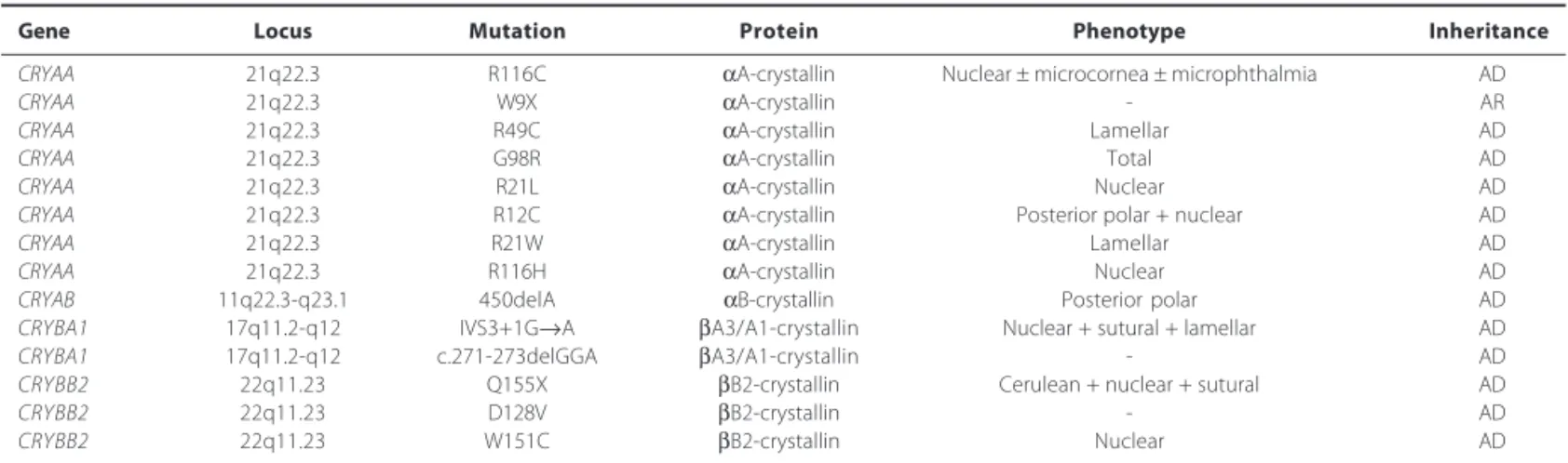
Table 1. Mutations identified in CRYAA, CRYAB, CRYBA1, and CRYBB2 genes in association with congenital cataract
Gene Locus Mutation Protein Phenotype Inheritance
CRYAA 21q22.3 R116C αA-crystallin Nuclear ± microcornea ± microphthalmia AD
CRYAA 21q22.3 W9X αA-crystallin - AR
CRYAA 21q22.3 R49C αA-crystallin Lamellar AD
CRYAA 21q22.3 G98R αA-crystallin Total AD
CRYAA 21q22.3 R21L αA-crystallin Nuclear AD
CRYAA 21q22.3 R12C αA-crystallin Posterior polar + nuclear AD
CRYAA 21q22.3 R21W αA-crystallin Lamellar AD
CRYAA 21q22.3 R116H αA-crystallin Nuclear AD
CRYAB 11q22.3-q23.1 450delA αB-crystallin Posterior polar AD
CRYBA1 17q11.2-q12 IVS3+1G→A βA3/A1-crystallin Nuclear + sutural + lamellar AD
CRYBA1 17q11.2-q12 c.271-273delGGA βA3/A1-crystallin - AD
CRYBB2 22q11.23 Q155X βB2-crystallin Cerulean + nuclear + sutural AD
CRYBB2 22q11.23 D128V βB2-crystallin - AD
CRYBB2 22q11.23 W151C βB2-crystallin Nuclear AD
R= arginine; C= cysteine; W= tryptophan; X= stop codon; G= glycine; L= leucine; H= histidine; Q= glutamine; D= aspartic acid; V= valine; del= deletion; AD= autosomal dominant; AR= autosomal recessive
Table 2. Mutations identified in CRYGC and CRYGD genes in association with congenital cataract
Gene Locus Mutation Protein Phenotype Inheritance
CRYGC 2q33-q35 T5P ϒC-crystallin Lamellar pulverulent AD
CRYGC 2q33-q35 225-226insGCGGC ϒC-crystallin Nuclear pulverulent AD
CRYGC 2q33-q35 R168W ϒC-crystallin Lamellar AD
CRYGC 2q33-q35 C109X ϒC-crystallin Nuclear AD
CRYGC 2q33-q35 W157X ϒC-crystallin Nuclear + microcornea AD
CRYGD 2q33-q35 R58H ϒD-crystallin Lamellar AD
CRYGD 2q33-q35 R14C ϒD-crystallin Punctate juvenile progressive AD
CRYGD 2q33-q35 Y134X ϒD-crystallin Total AD
CRYGD 2q33-q35 E107A ϒD-crystallin Nuclear AD
CRYGD 2q33-q35 W156X ϒD-crystallin Nuclear AD
CRYGD 2q33-q35 P23T ϒD-crystallin Lamellar AD
CRYGD 2q33-q35 R36S ϒD-crystallin Coraliform AD
CRYGD 2q33-q35 Y56X ϒD-crystallin Nuclear AD
CRYGD 2q33-q35 G61C ϒD-crystallin Coralliform AD
CRYGD 2q33-q35 R140X ϒD-crystallin Nuclear AD
of gap-junction proteins(16). Each gap-junction channel is composed of two hemi-channels, or connexons, which dock in the extracellular space between adjacent cells, and each connexon is comprised of six integral transmembrane protein subunits known as connexins(38). Connexins are a multigene family consisting of >20 members, three of which are expressed in the lens (Cx43, Cx46, and Cx50). The lens epithelial cells show a predominant expression of connexin 43 (Cx43). During differentiation into fibers, Cx43 expression is down regulated and replaced by connexin 46 (Cx46) and connexin 50 (Cx50)(14).
Mutations of specific connexin genes have been associated with several disease including genetic deafness, skin disease, peripheral neuropathies, heart defects and cataracts. Mutations in both Cx46
and Cx50 genes have produced phenotypically similar autosomal dominant lamellar pulverulent cataracts. Cx50 on chromosome 1q22 is constituted of two exons, which resulted in a protein with 433 residues. Shiels et al.(39) reported a missense mutation in codon 88 of the Cx50 gene, leading to the substitution of proline by serine (P88S), associated with autosomal dominant lamellar pulverulent cataract in an English family. To date, there are other thirteen mutations on Cx50 found in different hereditary cataract pedigrees; they are V44E, V64G, V79L, P88Q, Q48K, P189S, R198Q, R23T, W45S, D47N, D47Y, S276F, and I274M. This last mutation, located in the cytoplasmic COOH-terminus, was found in a Russian family in 2001, and it was related to a lamellar pulverulent cataract(40). Cx46 consists of a single exon encoding a 435 amino acid protein in humans and is localized on chromosome 13 (13q11-q13). Fourteen mutations in
Cx46 gene involving the different domains so far been reported to be associated with autosomal dominant congenital cataract in humans (Table 3).
3. Major intrinsic protein (MIP) or Aquaporine-0 (AQP0)
MIP is an integral membrane protein member of the aquaporine family of water transporters and small selected molecular of plas-matic membrane. It is the most highly expressed membrane protein in the lens, almost 80% of transport protein. Lamellar, cortical and polymorphic cataracts have been associated with missense muta-tions in the AQP0 gene. Recently, two mutations have been related; the first mutation, T138R, is associated with a progressive congenital lamellar and polar cataract, and the second E134G is associated with lamellar cataract(23). Both of these mutations appear to act by inter-fering with normal trafficking of AQP0 to the plasma membrane and thus with water channel activity (Table 3).
4. Cytoskeletal proteins
The architecture of lens cells is resulted of the interaction of the cytoskeleton, crystallin proteins, and cytoplasm. The cytoskeletal is a network of varied cytoplasmatic proteins which are involved in providing structural support, cell motility, and determination and maintenance of cell volume and shape. Lens cells present three different filaments, which are differentiated by diameter, types of subunits, and molecular organization: microfilaments, microtubes, and intermediate filaments(16). Microfilaments and microtubes fa-cilitate changes of ions, while intermediate filaments aid lens cells in overcomping physical stresses including lens accommodation and changes of temperature(41).
Beaded Filament Structural Proteins (BFSP) are a type of inter-mediate filament unique eye-lens-specific cytoskeletal structure, which contains two core components of BFSP1(also called filensin)
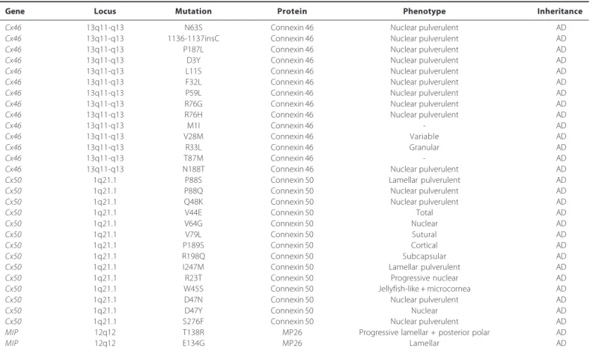
Table 3. Mutations identified in Cx46, Cx50, and MIP genes in association with congenital cataract
Gene Locus Mutation Protein Phenotype Inheritance
Cx46 13q11-q13 N63S Connexin 46 Nuclear pulverulent AD
Cx46 13q11-q13 1136-1137insC Connexin 46 Nuclear pulverulent AD
Cx46 13q11-q13 P187L Connexin 46 Nuclear pulverulent AD
Cx46 13q11-q13 D3Y Connexin 46 Nuclear pulverulent AD
Cx46 13q11-q13 L11S Connexin 46 Nuclear pulverulent AD
Cx46 13q11-q13 F32L Connexin 46 Nuclear pulverulent AD
Cx46 13q11-q13 P59L Connexin 46 Nuclear pulverulent AD
Cx46 13q11-q13 R76G Connexin 46 Nuclear pulverulent AD
Cx46 13q11-q13 R76H Connexin 46 Nuclear pulverulent AD
Cx46 13q11-q13 M1I Connexin 46 - AD
Cx46 13q11-q13 V28M Connexin 46 Variable AD
Cx46 13q11-q13 R33L Connexin 46 Granular AD
Cx46 13q11-q13 T87M Connexin 46 - AD
Cx46 13q11-q13 N188T Connexin 46 Nuclear pulverulent AD
Cx50 1q21.1 P88S Connexin 50 Lamellar pulverulent AD
Cx50 1q21.1 P88Q Connexin 50 Nuclear pulverulent AD
Cx50 1q21.1 Q48K Connexin 50 Nuclear pulverulent AD
Cx50 1q21.1 V44E Connexin 50 Total AD
Cx50 1q21.1 V64G Connexin 50 Nuclear AD
Cx50 1q21.1 V79L Connexin 50 Sutural AD
Cx50 1q21.1 P189S Connexin 50 Cortical AD
Cx50 1q21.1 R198Q Connexin 50 Subcapsular AD
Cx50 1q21.1 I247M Connexin 50 Lamellar pulverulent AD
Cx50 1q21.1 R23T Connexin 50 Progressive nuclear AD
Cx50 1q21.1 W45S Connexin 50 Jellyfish-like + microcornea AD
Cx50 1q21.1 D47N Connexin 50 Nuclear pulverulent AD
Cx50 1q21.1 D47Y Connexin 50 Nuclear AD
Cx50 1q21.1 S276F Connexin 50 Nuclear pulverulent AD
MIP 12q12 T138R MP26 Progressive lamellar + posterior polar AD
MIP 12q12 E134G MP26 Lamellar AD
and BFSP2 (also called CP49), highly divergent intermediate fila-ment proteins that combine in the presence of α-crystallin to form the appropriate beaded structure. The gene encoding BFSP2 are located on human chromosome 3q21. Actually, two mutations in
BFSP2 gene have been demonstrated with the occurrence of con-genital cataracts. The first mutation (R278W) had associated with juvenile-onset cataract(41), while the second mutation (delE233), had identified in a large family, which affected members had congenital nuclear, sutural, and cortical cataracts that varied in severity among different individuals (Table 4).
5. Developmental regulators
Embryonic lens development is predicted by spatial and tem-poral interactions of several genes and their products. The genes involved in this complex process encoding growth factors and transcription factors. These proteins regulate transcription of a number of tissue-specific genes during differentiation, and play a crucial role in lens plan specification. Depending on the nature of the genetic defect involved, mutations in genes that define de-velopment can result in multiple abnormalities of the eye, parti-cularly congenital cataract. Mutations in transcription factors genes have been implicated in anterior segment dysgenesis, but only three: PITX3, MAF, and HSF4, have been associated with isolated cataract (Table 4).
Mutations PITX3 gene result predominantly in autosomal domi-nant congenital cataract associated with dysgenesis of the anterior segment, including corneal opacity, iris adhesions, microcornea, and microphthalmia. Two mutations had been described, a 17-bp inser-tion of the coding sequence (656-657ins17bp), resulting in a frame shift, and a G→A transition in exon 2 of PITX3 gene, resulting in a serine to asparagine substitution at codon 13 (S13N)(25). Jamieson etal.(42) related a R288P substitution within the MAF gene on chromosome 16q22-q23 in a family with autosomal dominant juvenile pulveru-lent cataract, iris coloboma, and microcornea. In addition to MAF
gene, another missense mutation (K297R) was found in twelve affec-ted members in an Indian family, associaaffec-ted with autosomal domi-nant cerulean cataract and microcornea(26). HSF4 gene regulates the expression of heat-shock proteins (HSPs), which may be important components of lens development. Currently, six mutations of this gene had been associated with congenital cataracts, which resulted in lamellar progressive and nuclear phenotype(27).
C
RYSTALLINGENEMUTATIONSASSOCIATEDWITH CONGENITALCATARACTINBRAZILIANFAMILIESA recent study in Brazil, Santana et al.(43) have demonstrated a novel nonsense mutation (Y56X) in the CRYGD gene and a
pre-Table 4. Mutations identified in BFSP2, PITX3, MAF, and HSF4 genes in association with congenital cataract
Gene Locus Mutation Protein Phenotype Inheritance
BFSP2 3q21.2-q22.3 R287W BFSP2 Nuclear + juvenile sutural AD
BFSP2 3q21.2-q22.3 delE233 BFSP2 Cortical + nuclear + sutural AD
PITX3 10q24-q25 S13N PITX3 Total AD
PITX3 10q24-q25 656-657ins17pb PITX3 Cataract + anterior dysgenesis AD
MAF 16q22-q23 R288P MAF Pulverulent + microcornea + iris coloboma AD
MAF 16q22-q23 K297R MAF Cerulean + microcornea AD
HSF4 16q22 A19D HSF4 Lamellar AD
HSF4 16q22 R73H HSF4 Total AD
HSF4 16q22 I86V HSF4 Lamellar AD
HSF4 16q22 L114P HSF4 Lamellar AD
HSF4 16q22 R119C HSF4 Lamellar AD
HSF4 16q22 R175P HSF4 Lamellar AR
R= arginine; W= tryptophan; del= deletion; S= serine; N= asparagine; ins= insertion; P= proline; K= lysine; A= alanine; D= aspartic acid; H= histidine; I= isoleucine; V= valine; L= leucine; C= cysteine; AD= autosomal dominant; AR= autosomal recessive
viously reported missense mutation (R12C) in the CRYAA gene asso-ciated with nuclear congenital cataract in Brazilian families. Addi-tionally, they also observed a new polymorphism (S119S) in the
CRYGC gene. In this study, eleven families with autosomal dominant childhood cataracts were identified, seven families presenting with nuclear phenotype and remaining families with lamellar phenotype. A total of thirty-four affected members and forty-four unaffected members were evaluated. DNA sequencing analysis of
CRYGD gene showed a novel heterozygous nonsense mutation (TAC→TAG) within the second exon (Figure 1). Cataract was most likely caused by this point mutation that led to the replacement of a tyrosine by a premature stop codon at position 56 (Y56X). The congenital bilateral nuclear cataract in another family was associa-ted with a mutation in CRYAA, a point mutation in exon 1 (CGC→
TGC), which led to the replacement of an arginine at position 12 for a cysteine (R12C) (Figure 2). A variety of sequence variations re-ferred as single nucleotide polymorphisms was observed in the probands for the CRYAA, CRYGC and CRYGD genes. None of these sequences changed in the coding regions led to amino acids alte-rations. Three known polymorphisms were observed in the sequen-cing analysis: D2D (rs872331) polymorphism (GAC→GAT) in the
CRYAA gene (exon 1), observed in ten probands, Y17Y (rs2242074) polymorphism (TAT→TAC) observed in the CRYGD gene(exon 2), in seven probands, and R95R (rs2305430) polymorphism (AGA→AGG) in the CRYGD gene (exon 3), in nine probands. A new polymor-phism in the third exon of the CRYGC gene (S119S) was observed in a family (Figure 3).
CONCLUSION
Figure 1 . A) The DNA sequencing chromatograms of the PCR product encompasses exon 2 of CRYGD gene (5’→3’) of an unaffected individual; B) The DNA sequencing chromatograms of the PCR product encompassing exon 2 of CRYGD gene shows a heterozygous TAC→TAG transition that replaced a tyrosine (Y) by a premature stop codon (X) at amino acid 56 (Y56X) in affected individual. Modified from: Santana A. Avaliação estrutural dos genes CRYAA, CRYGC e CRYGD em pacientes portadores de catarata congênita autossômica dominante [doutorado]. São Paulo: Santa Casa de Misericórdia de São Paulo; 2009.
S= serine; G= glycine; L= leucine; Q= glutamine; Y= tyrosine; F= phenylalanine; R= arginine
A B
A B
Figure 2 . A) Direct sequencing of the PCR product encompasses exon 1 of CRYAA gene (5’→3’) of an unaffected individual; B) Direct sequencing of the PCR product encompassing exon 1 of CRYAA gene of an affected individual shows a heterozygous CGC→TGC transition that replaced arginine (R) by cysteine (C) at amino acid 12 (R12C). Modified from: Santana A. Avaliação estrutural dos genes CRYAA, CRYGC e CRYGD em pacientes portadores de catarata congênita autossômica dominante [doutorado]. São Paulo: Santa Casa de Misericórdia de São Paulo; 2009.
P= proline; W= tryptophan; F= phenylalanine; K= lysine; T= threonine; L= leucine; G= glycine
Figure 3. A) The DNA sequencing chromatograms of the PCR product encompasses exon 3 of CRYGC gene (5’→3’) shows homozygous for allele C; B) The DNA sequencing chromatograms of the PCR product encompassing exon 3 of CRYGC gene shows a heterozygous polymorphism AGC→AGT transition, which does not result in the substitution of serine (S) at amino acid 119 (S119S). Modified from: Santana A. Avaliação estrutural dos genes CRYAA, CRYGC e CRYGD em pacientes portadores de catarata congênita autossômica dominante [doutorado]. São Paulo: Santa Casa de Misericórdia de São Paulo; 2009.
A B
R= arginine; F= phenylalanine; H= histidine; L= leucine; S= serine; E= glutamic acid; I= isoleucine
markedly inhibition of the DNA synthesis and repair has also been observed in experimental studies. Oxidative stress has been asso-ciated expression changes (increase or decrease) of genes in the lens epithelium. Known genetic and environmental factors might direct the application of preventive measures, such as
REFERENCES
1. Foster A, Gilbert C, Rahi J. Epidemiology of cataract in childhood: a global perspective. J Cataract Refract Surg. 1997;23 Suppl 1:601-4.
2. Apple DJ, Ram J, Foster A, Peng Q. Elimination of cataract blindness: a global perspective entering the new millennium. Surv Ophthalmol. 2000;45 Suppl 1:S1-196.
3. Gilbert C, Foster A. Childhood blindness in the context of VISION 2020 - The right to sight. Bull World Health Organ. 2001;79(3):227-32.
4. Foster A. Worldwide blindness, increasing but avoidable. Semin Ophthalmol. 1993; 8(3):166-70.
5. Rahi JS, Scripathi S, Gilbert C, Foster A. Childhood blindness in India: causes in 1318 blind school students in nine states. Eye (Lond). 1995;9(5):545-50.
6. Tartarella MB, Kawakami LT, Scarpi MJ, Hayashi S. Aspectos cirúrgicos em catarata congê-nita. Arq Bras Oftalmol. 1995;58(1):24-8.
7. Carvalho KM, Minguini N, Moreira Filho DC, Kara-José N. Characteristics of a pediatric low-vision population. J Pediatr Ophthalmol Strabismus. 1998;35(3):162-5.
8. Haddad MA, Lobato FJ, Sampaio MW, Kara-José N. Pediatric and adolescent population with visual impairment: study of 385 cases. Clinics (São Paulo). 2006;61(3):239-46. 9. Eckstein M, Vijayalakshmi P, Killerdar M, Gilbert C, Foster A. Aetiology of childhood
cataract in south India. Br J Ophthalmol. 1996;80(7):628-32.
10. He W, Li S. Congenital cataracts: gene mapping. Hum Genet. 2000;106(1):1-13. 11. Messina-Baas OM, Gonzalez-Huerta LM, Cuevas-Covarrubias SA. Two affected siblings
with nuclear cataract associated with a novel missense mutation in the CRYGD gene. Mol Vis. 2006;12:995-1000.
12. Rahi JS, Dezateux C. National cross sectional study of detection of congenital and infantile cataract in the United Kingdom: role of childhood screening and surveillance. The British Congenital Cataract Interest Group. BMJ. 1999;318(7180):362-5.
13. Oliveira ML, Di Giovanni ME, Porfírio Neto Jr, Tartarella MB. Catarata congênita: aspectos diagnósticos, clínicos e cirúrgicos em pacientes submetidos à lensectomia. Arq Bras Oftalmol. 2004;67(6):921-6.
14. Beby F, Morle L, Michon L, Bozon M, Edery P, Burillon C, et al. [The genetics of hereditary cataract]. J Fr Ophtalmol. 2003;26(4):400-8. French.
15. Gill D, Klose R, Munier FL, McFadden M, Priston M, Billingsley G, et al. Genetic hete-rogeneity of the Coppock-like cataract: a mutation in CRYBB2 on chromosome 22q11.2. Invest Ophthalmol Vis Sci. 2000;41(1):159-65.
16. Reddy MA, Francis PJ, Berry V, Bhattacharya S, Moore AT. Molecular genetic basis of inherited cataract and associated phenotypes. Surv Ophthalmol. 2004(3);49:300-15. 17. Bettelheim FA, Chylack LT Jr. Light scattering of whole excised human cataractous lenses.
Relationships between different light scattering parameters. Exp Eye Res. 1985;41(1):19-30. 18. Kannabiran C, Balasubramanian D. Molecular genetics of cataract. Indian J Ophthalmol.
2000;48(1):5-13.
19. Guleria K, Sperling K, Singh D, Varon R, Singh JR, Vanita V. A novel mutation in the connexin 46 (GJA3) gene associated with autosomal dominant congenital cataract in an Indian family. Mol Vis. 2007;13:1657-65.
20. Hejtmamcik JF, Smaoui N. Molecular genetics of cataract. Dev Ophthalmol. 2003;37:67-82. 21. Bhat SP. Crystallins, genes and cataract. Prog Drug Res. 2003;60:205-63.
22. Hansen L, Yao W, Eiberg H, Kjaer KW, Baggesen K, Hejtmancik JF, et al. Genetic hete-rogeneity in microcornea-cataract: five novel mutations in CRYAA, CRYGD, and GJA8. Invest Ophthalmol Vis Sci. 2007;48(9):3937-44.
23. Berry V, Francis P, Kaushal S, Moore A, Bhattacharya S. Missense mutations in MIP underlie autosomal dominant “polymorphic” and lamellar cataracts linked to 12q. Nat Genet. 2000;25(1):15-7.
24. Jakobs PM, Hess JF, FitzGerald PG, Kramer P, Weleber RG, Litt M. Autosomal-dominant congenital cataract associated with deletion mutation in the human beaded filament protein gene BFSP2. Am J Hum Genet. 2000;66(4):1432-6.
25. Semina EV, Ferrell RE, Mintz-Hittner HA, Bitoun P, Alward WL, Reiter RS, et al. A novel homeobox gene PITX3 is mutated in families with autosomal-dominant cataract and ASMD. Nat Genet. 1998;19(2):167-70.
26. Vanita V, Singh D, Robinson PN, Sperling K, Singh JR. A novel mutation in the DNA-binding domain of MAF at 16q23-1 associated with autosomal dominant “cerulean cataract” in an Indian family. Am J Med Genet A. 2006;140(6):558-66.
27. Forshew T, Johnson CA, Khaliq S, Pasha S, Willis C, Abbasi R, et al. Locus heterogeneity in autosomal recessive congenital cataracts: linkage to 9q and germline HSF4 mutations. Hum Genet. 2005;117(5):452-9.
28. Augusteyn RC. Alpha-crystallin: a review of its structure and function. Clin Exp Optom. 2004;87(6):356-66.
29. Brady JP, Garland D, Duglas-Tabor Y, Robison WG Jr, Groome A, Wawrousek EF. Targed disruption of the mouse alpha A-crystallin gene induces cataract and cytoplasmic inclusion bodies containing the small heat-shock protein alpha B-crystallin. Proc Natl Acad Sci USA. 1997;94(3):884-9.
30. Bera S, Abraham EC. The alphaA-crystallin R116C mutant has a higher affinity for forming heteroaggregates with alphaB-crystallin. Biochemistry. 2002;41(1):297-305.
31. Pras E, Frydman M, Levy-Nissenbaum E, Bakhan T, Raz J, Assia El, et al. A nonsense mutation (W9X) in CRYAA causes autosomal recessive cataract in an inbred Jewish Persian family. Invest Ophthalmol Vis Sci. 2000;41(11):3511-5.
32. Santhiya ST, Soker T, Klopp N, Illig T, Prakash MV, Selvaraj B, et al. Identification of a novel, putative cataract - causing allele in CRYAA (G98R) in an Indian family. Mol Vis. 2006;12:768-73. 33. Berry V, Francis P, Reddy MA, Collyer D, Vithana E, Mackay L, et al. Alpha-B crystalline gene (CRYAB) mutation causes dominant congenital posterior polar cataract in humans. Am J Hum Genet. 2001;69(5):1141-5.
34. Reddy MA, Bateman OA, Chakarova C, Ferris J, Berry V, Lomas E, et al. Characterization of the G91del CRYBA1/3 crystallin protein: a cause of human inherited cataract. Hum Mol Genet. 2004;13(9):945-53.
35. Santhiya ST, Manisastry SM, Rawlley D, Malathi R, Anishetty S, Gopinath PM, et al. Mutation analysis of the congenital cataracts in Indian families: identification of SNPs and a new causative allele in CRYBB2 gene. Invest Ophthalmol Vis Sci. 2004;45(10):3599-607. 36. Santhiya ST, Shyam Manohar M, Rawlley D, Vijayalaskshmi P, Namperumalsamy P,
Gopinath PM, et al. Novel mutations in the gamma-crystallin genes cause autosomal dominant congenital cataracts. J Med Genet. 2002;39(5):352-8. Comment on: J Med Genet. 2000;37(7):481-8.
37. Fu L, Liang JJ. Conformational change and destabilization of cataract gammaC-crystallin T5P mutant. FEBS Lett. 2002;513(2-3):213-6.
38. Goodenough DA, Goliger JA, Paul DL. Connexins, connéxons, and intercellular communication. Annu Rev Biochem. 1996;65:475-502.
39. Shiels A, Mackay D, Ionides A, Berry V, Moore A, Bhattacharya S. A missense mutation in the human connexion 50 gene (GJA8) underlies autosomal dominant “zonular pulve-rulent” cataract, on chromosome 1q. Am J Hum Genet. 1998;62(3):526-32.
40. Polyakov AV, Shagina LA, Khlebnikova OV, Evgrafov OV. Mutation in the conexin 50 gene (GJA8) in a Russian family with zonular pulverulent cataract. Clin Genet. 2001;60(6): 476-8.
41. Conley YP, Erturk D, Keverline A, Mah TS, Keravala A, Barnes LR, et al. A juvenile-onset, progressive cataract locus on chromosome 3q21-q22 is associated with a missense mutation in the beaded filament structural protein-2. Am J Hum Genet. 2000;66(4):1426-31. 42. Jamieson RV, Perveen R, Kerr B, Carette M, Yardley J, Heon E, et al. Domain disruption and
mutation of the bZIP transcription factor, MAF, associared with cataract ocular segment dysgenesis and coloboma. Hum Mol Genet. 2002;11(1):33-42.