DOI: 10.1590/0004-282X20160004 ARTICLE
A disturbed processing of graviceptive
pathways may be involved in the
pathophysiology of balance disorders in
patients with multiple sclerosis
O processamento incorreto das informações graviceptivas pode estar envolvido na
fisiopatologia dos distúrbios do equilíbrio em pacientes com esclerose múltipla
Bruna Antinori Vignola da Fonseca1, Cristiana Borges Pereira2, Frederico Jorge3, Renata Simm3, Samira
Apostolos-Pereira3, Dagoberto Callegaro3
Balance disorders and falls are among the most disabling symptoms in people with Multiple Sclerosis (PWMS) and often are reported as initial symptom of the disease1,2. Balance depends
on complex interactions among sensorial information such as proprioception, vision and vestibular inputs, adequate
inte-gration with multissensorial areas in the CNS and an efective
motor system3,4. Any deicits on integration of these pathways
can damage balance control, increasing the risk of fall2.
Perception of verticality is an important aferent in
-formation for balance control, since it is required for body orientation in space5,6,7. Multimodal sensory input is
necessary to detect the body verticality in the space. he
representation of body schema in the space depends on the proprioceptive signs, provided by sole receptors and joints re-ceptors, and on the detection of gravitational inputs and head
position, provided by the otolith organs. he otolith organs
1Universidade de São Paulo, Faculdade de Medicina, Departamento de Neurologia, Sao Paulo SP, Brazil;
2Universidade de São Paulo, Faculdade de Medicina, Hospital das Clínicas, Divisão de Neurologia, Ambulatório de Distúrbios Vestibulares e do Equilíbrio, Sao Paulo SP, Brazil;
3Universidade de São Paulo, Faculdade de Medicina, Hospital das Clínicas, Divisão de Neurologia, Ambulatório de Doenças Desmielinizantes, Sao Paulo SP, Brazil.
Correspondence: Bruna Antinori Vignola da Fonseca; Departamento de Neurologia da USP; Rua Dr. Enéas de Carvalho Aguiar, 255 / 5083; 05403-000 São Paulo SP, Brazil; E-mail: [email protected]
Conflict of interest: There is no conlict of interest to declare.
Received 01 September 2015; Received in inal form 17 September 2015; Accepted 09 October 2015.
ABSTRACT
The purpose of this study was to determine the relationship between perception of verticality and balance disorders in multiple sclerosis patients. We evaluated patients and healthy controls. Patients were divided into two groups according to their risk of fall, with or without risk of fall, measured by a Dynamic Gait Index scale. Graviceptive perception was assessed using the subjective visual vertical test. Patients with risk of fall showed worse perception than those without risk of fall, p < 0.001. Misperception of verticality was correlated with the dynamic gait index scores (p < 0.001), suggesting that the larger the error for verticality judgment, the greater risk for falling. Considering that the perception of verticality is essential for postural control, our results suggested that the disturbed processing of graviceptive pathways may be involved in the pathophysiology of balance disorders in these patients.
Keywords: postural balance, multiple sclerosis, vestibular function tests, gravity sensing, sensation disorders, accidental falls.
RESUMO
Nosso objetivo foi determinar a relação entre percepção de verticalidade e alterações do equilíbrio em pacientes com esclerose múltipla (EM). Foram avaliados pacientes e sujeitos saudáveis. Pacientes foram divididos em dois grupos de acordo com o risco de queda, mensurado pelo Índice de marcha dinâmica, formando os grupos com risco e sem risco de quedas. A percepção da verticalidade foi medida através do teste vertical visual subjetiva (VVS). Pacientes com risco de queda apresentaram pior percepção da verticalidade quando comparados aos sem risco, p < 0,001. O desempenho no teste da VVS foi pior em pacientes quando comparado aos controles (p < 0,001). O erro no julgamento da verticalidade foi correlacionado aos índices de risco de queda (p < 0,001), sugerindo que quanto maior o erro no julgamento da verticalidade, maior o risco de queda dos pacientes. Nossos resultados sugerem que alterações das informações em vias graviceptivas podem estar envolvidas nas alterações de equilíbrio dessa população.
are gravitational sensors located in the head, called utricle and saccule. Both detect sense of accelerations, including
those produced by gravity. he aferents signs provided by
the otolith organs are interpreted together with visual and proprioceptive information from head-neck and neck-trunk positions. So, perception of verticality depends on sensors lo-cated in the head (otolith organs) and on body sensors8.
he gravitational perception is frequently measured with
the subjective visual vertical (SVV)9. SVV test evaluates the
ability to adjust a luminous rod in the vertical position in a dark room, without other visual cues10,11. Misperception of SVV can
relect damage in peripheral or central vestibular pathways
from the brainstem over the thalamus to cerebral cortex12, or
may be a sign of an impaired sensorial integration13.
Because of the widely distribution of central nervous system (CNS) lesion in PWMS, poor balance control had multifactorial causes that varies from one person to another14. Further, some
studies suggests that balance disorders in PWMS occurs due to impaired central integration of visual, vestibular, and so-matossensorial input15. Few previous studies had shown that
perception of verticality can be afected in MS patients when
compared with healthy controls. Besides that, SVV deviation has also been correlated with disability degree, measured by Expanded Disability Status Scale (EDSS)16,17. However, none of
these studies has analyzed the correlation between verticality misperception and balance disorders in the PWMS.
herefore the objectives of this study were: (1) to investigate if perception of visual vertical is diferent between PWMS with
and without dynamic balance disorders and (2) to analyze if misperception of verticality correlates with risk of fall in PWMS.
METHOD
Subjects
We recruited outpatients with relapsing-remitting MS. Patients were included after medical consultation
accord-ing to the followaccord-ing inclusion criteria: (1) diagnosis of MS
according to the McDonald et al.18 and (2) score of Expanded
Disability Status Scale (EDSS)19 between 0 - 4.5. Patients were
excluded if they had (1) relapses over the last 3 months, (2) other neurological diseases, (3) vertigo or vestibular
dys-function, including nystagmus and vestibulo-ocular relex,
(4) cognitive disorders or (5) visual impairment (blindness, blurred vision, diplopia or optic neurits). All patients were under treatment with interferon therapy. Since risk of fall is consequence of balance disorders, patients were divided into
two groups according to their risk of fall: risk of fall (RF+) and without risk of fall (RF-), measured by Dynamic Gait Index
Scale. Forty-nine healthy controls (HC) were also recruited.
hey were excluded if they had history of (1) vestibular symp
-toms, (2) cognitive disorders or (2) severe visual accuracy impairment. All experiments were conducted in accordance with the Declaration of Helsinki and this study was approved
by the local Research Ethics Committee. All patients and
controls signed the informed consent term.
A total of 98 PWMS (67 females) were included. he EDSS median score in RF- group was 1 (1 - 1,5) and in RF group was
2.5 (2.0 - 3.5). We also evaluated forty nine HC (32 females, age 37.6 ± 7.4).
Clinical assessment of PWMS
Dynamic balance was evaluated by the Dynamic Gait Index (DGI). his scale was developed by Shumway-Cook et al.20 to
evaluate balance control during walking and to evaluate risk
of falling. he measure consist of 8 itens: walk 6 meters, walk
and change speed, walk with head turns (look left then right and look up then down), walk with pivot turn, over or around
obstacles and going up stairs. he score ranges from 0 to 24. he cut-of point of ≤ 19 was previous established for PWMS to
indicate balance disorders and risk of fall by Forsberg et al.21. In
this study we decided to classify PWMS with balance disorders
according to the risk of fall detected by DGI scale. 57 patients had DGI scored higher than 19 point, thus they were included in PWMS group without risk of fall (RF-) and 41 patients were included in PWMS group with risk of fall (RF+), with scores lower than 19 points in DGI scale.
Subjective visual vertical test
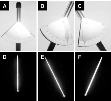
he measurement of the SVV was performed in PWMS and HC using a 24 cm long luminous portable rod. he rod
was positioned 1.5 meters in front of the subject who was sitting upright and wearing glasses with dark lenses that made use of any other visual reference impossible (Figure 1). Patients remained sitting and the head was aligned with trunk position. Measurements were stopped if the head tilted to either side. Starting the rod from 30 degree oblique posi-tion, to clockwise (positive) and counterclockwise (negative) initial positions, the subject verbally instructed the examiner to set the rod into a vertical position. Both, HC and PWMS made ten adjustments, 5 from positive and 5 from negative initial positions. A previous study published by our group used the same methodology to evaluate perception of verti-cality in patients with Parkinson’s disease22.
Subjective visual vertical calculation
Two diferent SVV-analyses were performed. In the irst
analysis the objective was to detect otolithic tonus im-balance, since SVV tilts are known to be a sensitive sign of otolithic tone imbalance and a lesion of the
gravicep-tive pathways. hese SVV tilts were calculated as a mean
value and expressed as either clockwise or anticlockwise.
he values of SVV-deviations from true vertical to the right
intraindividual variability implies a compromised perception of verticality and a disturbed processing of the graviceptive
pathways, but not necessarily an otolithic tone imbalance. In
this analysis the absolute values of SVV-deviations were con-sidered, which means that we should not considered positive or negative values, since increased shift for either direction of rotation may be symmetrical and a normal mean value may not be representative of abnormal deviations.
Statistical analysis
Data were expressed as means (standard deviation) or median [range], as appropriate. Statistical analysis was
per-formed by the Student t-test to compare EDSS and DGI scores
between PWMS groups. Mann-Whitney test to compare the mean of relative and absolute SVV values between PWMS
and HC. To compare the diferences among each group, RF-, RF+ and control group, the Kruskal Wallis test was performed.
Spearman´s correlation test was performed to assess the
cor-relation between SVV values with DGI, considering all PWMS in the same group. he r-values were considered as follows:
r < 0.4 poor correlation; 0.4 < r < 0.6 moderate correlation;
r > 0.6 strong correlation. he level of signiicance was 5%.
RESULTS
Table shows the demographic and clinical characteristics of the 98 patients according to risk of fall. Disability degree
measured by EDSS scale was signiicantly worse in RF+ group than in RF- group (p < 0.001). Patients with risk of fall had a
higher EDSS score than the patients without.
Analysis of SVV mean values
In the irst analysis, mean SVV-values were considered in
order to detect otolithic tonus imbalance. Mean SVV-value
in HC was +0.5 (-0.12 - 0.7). In the RF- group the mean SVV-deviation was +0.3º (-0.75 - 1.72), while in the RF+ group it was +0.65º (-1.3 - 1.65). No signiicant diferences were
detected between PWMS with and without risk for falling
(2-tailed Mann-Whitney test, p = 0.54). We did not also ind diferences in mean SVV-values when we compared PWMS
with HC (2-tailed Mann-Whitney test, p = 0.97).
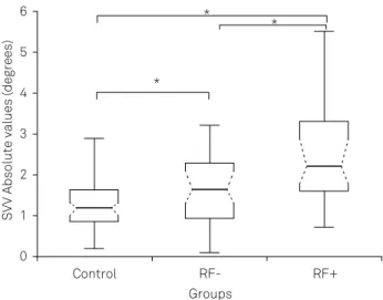
Analyses of SVV absolute values
In the second analysis which was done to detect intraindi
-vidual variability and the disturbed processing of graviceptive
pathways, the SVV absolute values were considered. he ab
-solute mean SVV deviation in HC was 1.2 (0.87 - 1.62), in
RF- group was 1.6º (0.97 - 2.3), and in the RF+ group was 2.2º
(1.62 - 3.25). Statistical comparison of absolute mean values of
SVV proved signiicant diference between HC, RF- and RF+
(2-tailed Mann-Whitney test, p < 0.001, Figure 2). According to our results, misperception of verticality was higher in pa-tients with risk of fall.
Increased misperception of verticality correlates to risk of fall in PWMS
he risk of fall was assessed by DGI scale. he median (range) of DGI score was 24 (21 - 24) in RF- group and 16 (13 - 16) in RF+ group. Since only SVV absolute values were
diferent between groups, we considered these absolute val
-ues to correlate with DGI scores. An oposite correlation was found between the SVV absolute values and the DGI-scores
(rs = -0.325, p < 0.001), suggesting that the larger the error for SVV judgment, the greater the risk for falling (Figure 3).
DISCUSSION
A lot of previous studies have classiied PWMS as fall -ers and no fall-ers according to their fall history. We chosen to use another method because some of our patients with balance disorders do not report falls since they restrict their
diary activity at home, because they feel fear of falling. hus,
Figure 1. The igure shows the Subjective visual vertical (SVV) test. SVV view of the examiner (A, B, C), and view of the subject (D,E,F). The SVV-deviations from true vertical to the left (anticlockwise) of the subject, were deemed negative (B,E), while deviations to the right (clockwise) of the subject were considered positive (C,F).
A
B
C
D
E
F
Table. Demographic and clinical differences between PWMS with risk of fall (RF+) and without risk of fall (RF-).
Clinical findings RF+ group n = 41
RF- group
n = 57 p-value
Gender, F/M 28 - 13 39 - 18 0.42
Age, years 37.4 (10) 32.5 (8.1) 0.009
EDSS score 2.5 [2.0 - 3.5] 1.0 [1.0 - 1.5] < 0.001
Duration disease, years 9.4 (7.1) 6.6 (4.3) 0.014
the number of fall could not be considered. Our study shows that PWMS with risk of fall have worse misperception of ver-ticality than patients without risk. Moreover, comparing to HC misperception of verticality was also found in patients without risk of fall, suggesting that the misperception of ver-ticality is present even before the clinical manifestation of
balance disorders. he misperception of verticality as shown
by higher absolute means of SVV deviation, suggesting a disturbed processing of graviceptive pathways, rather than otolithic tonus imbalance.
Only a few previous studies have evaluated the percep-tion of verticality in MS populapercep-tion, and none of them had considered the relationship between misperception of ver-ticality and both dynamic balance disorders or risk of fall. SVV is a test of otolithic function and is able to provide information about the integrity of the vestibular pathways, both peripheral and central10,11,23. his suggests that SVV test
may be a useful tool for evaluation of vestibular system and sensorial integration16,17. he wrong judgment of verticality
has been associated with acute unilateral brainstem le-sions and thalamic or cortical lele-sions12,17,24,25. In order to as
-sess if the misperception of SVV was related to an otolithic tonus imbalance or a disturbed processing of graviceptive pathways, our study considered two analyses. We found that
perception of verticality was diferent among HC and both PWMS groups, RF+ and RF-, when absolute values of SVV
deviation were considered, but not when the arithmetic
mean was considered. his inding means that MS patients
do not have a graviceptive tone imbalance, but they have an impaired precision of vertical judgment and a defective pro-cessing of graviceptive pathways.
he analysis of the SVV test may consider the tilt direc
-tion or the intraindividual variability. Intraindividual SVV variability relects the precision of the rod’s adjustment, and
is a measure of the precision of vertical perception26. So, an
increased intraindividual variability is considered to be a
decreased efectiveness of the otolithic organs, a disturbed
processing of graviceptive pathways with an impaired sen-sorial integration13. Our indings show that even patients
without risk of fall have misperception of verticality, proba-bly resulted from impaired central integration, since patients with visual or acute vestibular dysfunction were excluded. We also found that misperception of verticality in MS
pa-tients is correlated to the risk for falling evaluated by DGI
scale. Prior studies suggested that the incapacity to detect vi-sual verticality could be related to poor balance recover27,28.
Despite of these studies have evaluated patients that sufered
stroke, the indings highlight that some cortical areas are re
-lated to perception of verticality, and these sense is not just related to peripheral lesions.
he upright stability requires the determination of body
orientation through CNS information and depends on the in-tegration of visual, vestibular and somatosensory inputs, and
also an adequate integration of these aferences in the mul
-tissensorial cortex areas4,5. When the visual information is
excluded, the capacity of judgment of gravitational
vertical-ity relies on the vestibular system, speciically, the otolithic
organs inputs13,24.
Due to the widespread distribution of lesions in CNS,
per-ception of verticality in MS patients could be afected by the deicient integration of sensorial pathways. Several studies of
SVV deviation - Absolute values (degrees)
DGI Scores 6
4 5
3
2
1
6 8 10 12 14 16 18 20 22 24
Figure 3. A signiicant and negative correlation was proved between Subjective visual vertical (SVV) test absolute values and DGI scores in the MS patients group. The results showed that the more risk for falling is correlated with worse perception of verticality. The circles represent the individual SVV deviation in degrees (p < 0.001).
SVV Absolute values (degrees)
Groups 6
4 5
3
2
1
0
Control
RF-*
* *
RF+
balance in MS have suggested that impaired central integra-tion seems to be the main mechanism involved with balance
deicits14,15. Our results suggest that SVV tilt might be a
sen-sitive sign to evaluate impaired aferent pathways related to
balance control.
Consistent with our study, Crevits et al.16 found a positive
correlation between SVV tilt and EDSS total scores and con-cluded that misperception of gravity may interfere with
dis-ability. In addition, these authors also found highly signiicant
correlation between SVV deviation and subscores of EDSS for brainstem and cerebellum, associating with dysfunction of oculomotor or otolithic pathways and cerebellar dysfunc-tion. Other authors had also proposed that SVV could be an index for cerebellar dysfunction in MS16,17. Our data
comple-ment these previous studies because we also found the cor-relation between disability degree (EDSS) and misperception of SVV. However, we chose not to consider subscores of EDSS. SVV tilt has not been shown to be a cerebellar sign, and the correlation of SVV tilt and cerebellar dysfunction demon-strated by these authors could be probably explained by con-comitant brainstem and cerebellar lesions on MS patients.
In our study, we evaluated vestibular function using clini
-cal tests. We included PWMS with normal VOR and without nystagmus positional or evocade. When VOR is abnormal or
nystagmus is present, the lesion is characterized by
unilater-al in peripherunilater-al or centrunilater-al vestibular pathways. In these cases,
SVV test shows deviation from verticality for ipsiversive or contraversive side from lesions. Because our objective was to analyze the vertical perception which depends on cen-tral processing of the vestibular function and visual and proprioceptive functions as well, we excluded patients with
vestibular symptoms. hus, we considered the vestibular
clinical tests enough for our purpose. However, we did not use caloric test and this could be a limitation of this study.
Because balance impairment reported as a frequent and disabling consequence of MS, even at the initial stage of the disease, the need for reliable measures to identify subtle im-pairments that damage balance control is necessary14. SVV
evaluation proved to be a simple and easy method to de-tect impairments on central integration areas, even when
aferent signs seems normal. Further, the correlation of verti
-cality misperception and impaired DGI scores in MS patients
suggests that a disturbed processing of graviceptive path-ways may be involved into the pathophysiology of balance
disorders in these patients. his inding also suggests that
misperception of verticality should be taken into account in rehabilitation programs for prevention of risk of falls and im-prove balance strategies in MS patients.
References
1. Cattaneo D, Jonsdottir J. Sensory impairments in quiet standing in subjects with multiple sclerosis. Mult Scler. 2009;15(1):59-67. doi:10.1177/1352458508096874
2. Prosperini L, Kouleridou A, Petsas N, Leonardi L, Tona F, Pantano P et al. The relationship between infratentorial lesions, balance deicit and accidental falls in multiple sclerosis. J Neurol Sci.
2011;304(1-2):55-60. doi:10.1016/j.jns.2011.02.014
3. Cattaneo D, De Nuzzo C, Fascia T, Macalli M, Pisoni I, Cardini R. et al. Risks of falls in subjects with multiple sclerosis. Arch Phys Med Rehabil. 2002;83(6):864-7. doi:10.1053/apmr.2002.32825 4. Horak FB, Wrisley DM, Frank J. The Balance Evaluation Systems
Test (BESTest) to differentiate bal- ance deicits. Phys Ther. 2009;89(5):484-98. doi:10.2522/ptj.20080071
5. Horak FB. Postural orientation and equilibrium: what do we need to know about neural control of balance to prevent falls? Age Ageing. 2006;35(Suppl 2): ii7-11. doi:10.1093/ageing/al077
6. Bonan I V, Guettard E, Leman M C, Colle FM, Yelnik AP. Subjective visual vertical perception relates to balance in acute stroke. Arch Phys Med Rehabil. 2006;87(5):642-6. doi:10.1016/j.apmr.2006.01.019
7. Frzovic D, Morris ME, Vowels L. Clinical tests of standing balance: performance of persons with multiple sclerosis. Arch Phys Med Rehabil. 2000;81(2):215-21. doi:10.1016/S0003-9993(00)90144-8
8. Massion J, Woollacoot MH. Posture and equilibrium: clinical disorders of balance, posture and gait. London: Arnold; 2004. 9. Dyde RT, Jenkin MR, Harris LR. The subjective visual vertical
and the perceptual upright. Exp Brain Res. 2006;173(4):612-22. doi:10.1007/s00221-006-0405-y
10. Bisdorff AR, Wolsley CJ, Anastasopoulos D, Bronstein AM, Gresty MA The perception of body verticality (subjective postural
vertical) in peripheral and central vestibular disorders. Brain. 1996;119(5):1523-34. doi:10.1093/brain/119.5.1523
11. Böhmer A. The subjective visual vertical as a clinical parameter for acute and chronic vestibualr (otolith) disorders. Acta Otolaryngol. 1999;119(2):126-7. doi:10.1080/00016489950181495
12. Baier B, Suchan J, Karnath HO, Dieterich, M. Neural correlates of disturbed perception of verticality. Neurology. 2012;78(10):728-35. doi:10.1212/WNL.0b013e318248e544
13. Tarnutzer AA, Bockisch C, Straumann D, et al. Gravity dependence of subjective visual vertical variability. J Neurophysiol.
2009;102(3):1657-71. doi:10.1152/jn.00007.2008
14. Cameron MH, Lord S. Postural control in multiple sclerosis: implications for fall prevention. Curr Neurol Neurosci Rep. 2010;10(5):407-12. doi:10.1007/s11910-010-0128-0 15. Jackson RT, Epstein CM, De l’Aune WR. Abnormalities in
posturography and estimations of visual vertical and horizontal in multiple sclerosis. Am J Otol. 1995;16(1):88-93.
16. Crevits L, Venhovens J, Vanoutrive J, Debruyne J. False
perception of visual verticality in multiple sclerosis. Eur J Neurol. 2007;14(2):228-32. doi:10.1111/j.1468-1331.2006.01636.x 17. Serra A, Derwenskus J, Downey DL, Leigh RJ. Role of eye
movement examination and subjective visual vertical in clinical evaluation of multiple sclerosis. J Neurol. 2003;250(5):569-75. doi:10.1007/s00415-003-1038-8
18. McDonald WI, Compston A, Edan G, Goodkin D, Hartung HP, Lublin FD et al. Recommended diagnostic criteria for multiple sclerosis: guidelines from the International Panel on the diagnosis of multiple sclerosis. Ann Neurol. 2001;50(1):121-7. doi:10.1002/ana.1032 19. Kurtzke JF. Rating neurologic impairment in multiple sclerosis:
20. Shumway-Cook A, Baldwin M, Polissar N, Gruber W. Predicting the probability for falls in community-dwelling older adults. Phys Ther. 1997;77(8):812-9.
21. Forsberg A, Andreasson M, Nilsagård YE. Validity of the Dynamic Gait Index in people with multiple sclerosis. Phys Ther. 2013;93(10):1369-76. doi:10.2522/ptj.20120284
22. Pereira CB, Kanashiro AK, Maia FM, Barbosa ER. Correlation of impaired su bjective visual vertical and postural instability in Parkinson’s disease. J Neurol Sci. 2014;346(1-2):60-5. doi:10.1016/j.jns.2014.07.057
23. McConvey J, Bennett SE. Reliability of the Dynamic Gait Index in individuals with multiple sclerosis. Arch Phys Med Rehabil. 2005;86(1):130-3. doi:10.1016/j.apmr.2003.11.033
24. Brandt T, Dieterich M. Vestibular syndromes in the roll plane: topographic diagnosis from brainstem to cortex. Ann Neurol. 1994;36(3):337-47. doi:10.1002/ana.410360304
25. Lopez C, Lacour M, Ballester M, Dumitrescu M, Anton J, Nazarian B et al. Brain activation during subjective visual vertical judgement: a functional magnetic resonance imaging study. Gait Posture. 2005;21(1):S49-50. doi:10.1016/S0966-6362(05)80164-X 26. Tarnutzer AA, Shaikh AG, Palla A, Straumann D, Marti
S. Vestibulo-cerebellar disease impairs the central representation of self-orientation. Front.Neur. 2011;2:11. doi:10.3389/fneur.2011.00011
27. Bonan IV, Leman MC, Legargasson JF, Guichard JP, Yelnik AP. Evolution of subjective visual vertical perturbation after stroke. Neurorehabil Neural Repair. 2006;20(4):484-91. doi:10.1177/1545968306289295