Auditory evoked potentials
and multiple sclerosis
Carla Gentile Matas1, Sandro Luiz de Andrade Matas2,
Caroline Rondina Salzano de Oliveira3, Isabela Crivellaro Gonçalves4
ABSTRACT
Multiple sclerosis (MS) is an inflammatory, demyelinating disease that can affect several areas of the central nervous system. Damage along the auditory pathway can alter its integrity significantly. Therefore, it is important to investigate the auditory pathway, from the brainstem to the cortex, in individuals with MS. Objective: The aim of this study was to characterize auditory evoked potentials in adults with MS of the remittent-recurrent type. Method: The study comprised 25 individuals with MS, between 25 and 55 years, and 25 age- and gender-matched healthy controls (research and control groups). Subjects underwent audiological and electrophysiological evaluations. Results: Statistically significant differences were observed between the groups regarding the results of the auditory brainstem response and the latency of the Na and P300 waves. Conclusion: Individuals with MS present abnormalities in auditory evoked potentials indicating dysfunction of different regions of the central auditory nervous system.
Key words: auditory evoked potentials, auditory brain stem evoked potentials, P300 event-related potentials, multiple sclerosis.
Potenciais evocados auditivos e esclerose múltipla
RESUMO
A esclerose múltipla é uma doença inflamatória desmielinizante que pode se desenvolver em diversas regiões do sistema nervoso central. O comprometimento da via auditiva central pode alterar significativamente a integridade desta e, portanto, a investigação desta região em indivíduos com esclerose múltipla, desde o tronco encefálico até o córtex, torna-se importante. Objetivo: Caracterizar os resultados dos potenciais evocados auditivos em adultos com esclerose múltipla do tipo remitente-recorrente. Método: Foram submetidos às avaliações audiológica e eletrofisiológica 25 indivíduos pertencentes ao grupo controle e 25 ao grupo pesquisa, com idades entre 25 e 55 anos. Resultados: Verificou-se diferença estatisticamente significante entre os grupos quanto à ocorrência de resultados normais e alterados no potencial evocado auditivo de tronco encefálico e latências das ondas Na e P300. Conclusão: Indivíduos com esclerose múltipla apresentam alterações nos potenciais evocados auditivos, indicando comprometimento de diferentes regiões do sistema nervoso auditivo central.
Palavras-chave: potenciais evocados auditivos, potenciais evocados auditivos do tronco encefálico, potencial evocado P300, esclerose múltipla.
Correspondence
Carla Gentile Matas Rua Princesa Isabel 17 / 204-A 04601-000 São Paulo SP - Brasil E-mail: [email protected]
Received 14 July 2009
Received in final form 9 February 2010 Accepted 18 February 2010
Auditory Evoked Potentials Laboratory from the Speech Pathology and Audiology Course, School of Medicine, University of São Paulo (FMUSP), São Paulo SP, Brazil: 1Audiologist, PhD, Professor at Speech Pathology and Audiology Course, FMUSP; 2Neurologist, PhD, Physician at São Paulo Hospital, Federal University of São Paulo, São Paulo SP, Brazil; 3Audiologist, MD; 4Audiologist, MD, PhD Student at Rehabilitation Sciences Program, FMUSP.
Multiple sclerosis (MS) is a progressive, inlammatory, demyelinating disease that was irst identiied by Jean Charcot in 1860. MS is caused by the destruction of the my-elin sheath by autoantibodies and the
speeds within the brain parenchyma. herefore, chang-es in brain functions observed in individuals with MS are a result of impairment in the transmission of informa-tion caused by the destrucinforma-tion of the myelin nerve wrap1.
MS can afect any region of the central nervous sys-tem. Therefore, special attention must be given to the central auditory nervous system because hearing func-tion is dependent on nervous system’s integrity. hus, au-diological diagnoses based only on conventional audio-logical evaluation do not consider the many changes that occur along the central auditory pathway, some of which are not clinically manifested2.
Auditory evoked potentials (AEP) relect the neuro-electric activity within the auditory pathway, from the auditory nerve to the cerebral cortex, in response to an acoustic stimulus or event. he most studied AEP are the brainstem auditory evoked potential (BAEP), the audi-tory middle latency response (AMLR) and the cognitive potential (P300).
BAEP, which consists of seven waves generated by one or more structures along the auditory pathway, assesses the integrity of the auditory pathway from the auditory nerve to the brainstem. AMLR, consisting of a series of waves that appear after the BAEP, relects the activation of several subcortical structures. P300 is an endogenous, event-related potential and its generation involves skills such as attention, auditory discrimination, memory and semantic perspective. It may be more correlated to the degree of global auditory dysfunction than any speciic diagnosis because its results are afected by a variety of disorders that alter cognition3.
he appearance of sclerotic plaques along the audi-tory pathway signiicantly alters its integrity. herefore, it is important to investigate the functioning of the cen-tral auditory nervous system in individuals with MS. Sev-eral studies have been performed to investigate the AEP in subjects with MS. he results of these studies varied but have mainly indicated BAEP abnormalities in MS pa-tients4-7. Furthermore, researchers have emphasized the
use of P300 as a clinical diferential in patients with MS as P300 is efective in detecting cognitive dysfunctions thus increasing the chances of neuropsychological interven-tion with an emphasis on cognitive rehabilitainterven-tion8-11.
AEP, besides its usefulness in identifying lesions in the auditory pathway, is useful in monitoring changes in the auditory pathway. Schochat et al.7 state that although MRI
has advanced the diagnosis of MS, it is feasible to use the AEP resources with precision in the follow-up treatment of pre-established proiles and in the diagnosis of new le-sions that might develop, including those lele-sions that are clinically silent.
Due to the considerations presented, this study aimed to characterize AEP of short (BAEP) and middle (AMLR)
latencies and P300 in adults with MS of the remittent-recurrent type and normal hearing. hese results were compared to the results obtained in individuals with nor-mal hearing and no history of neurological alteration.
METHOD
Institutional review board approval for this study was obtained from CAPPesq - HC/FMUSP and was registered under protocol number 274/06.
The inclusion criteria for the control group (CG) were history of normal neurological development, nor-mal hearing thresholds, absence of psychiatric diagnoses, no complaints of tinnitus, and no auditory processing dis-orders. For the research group (RG), the inclusion criteria were medical diagnosis of MS of the remittent-recurrent type based on the criteria proposed by Poser et al.9,
nor-mal hearing thresholds, and no outbreak for at least six months before the recording of auditory evoked poten-tials. All the participants signed an inform consent.
he two groups were composed of 25 individuals each with 19 females and six males aged between 25 and 55 years (mean age of 35.16 years for the CG and of 34.88 years for the RG). Participants in the RG were referred by a neurologist. At the time of data collection the RG partici-pants were undergoing medical follow-ups. Participartici-pants in the CG were age- and gender-matched to RG participants.
Initially, an anamnesis was performed with each par-ticipant to obtain personal data related to the history of the disease (MS), use of medications and hearing devel-opment. Information gathered during the anamnesis was conirmed by neurologists. All participants of the RG un-derwent MRI and received interferon beta 1a drug treat-ment. Of the 25 subjects evaluated, besides the character-istic lesions of MS in the central semi-oval center, two had sclerotic plaques in the regions that generate the brainstem auditory evoked potentials. It was determined from the subjects’ medical histories that the average duration of dis-ease was four years and three months. Patients were evalu-ated for MS12 outbreaks up until the time of data collection
and the results are as follows: two had one outbreak (8%), ive had two outbreaks (20%), four had three outbreaks (16%), eight had four outbreaks (32%), four had ive out-breaks (16%), one had six outout-breaks (4%) and one had sev-en outbreaks (4%). According to the EDSS scale, eight pa-tients (32%) were in the range zero, six (24%) in range one, eight in range two (32%) and three (12%) in range three.
(tym-panometry and acoustic relex research, at frequencies of 500, 1000, 2000 and 4000 Hz) performed with the middle ear analyzer GSI-33.
Electrophysiological evaluation consisted of record-ing BAEP, AMLR and P300. hese evaluations were con-ducted on Masbe equipment - Contronic® and the ATC-plus 2.1® software installed on a computer.
Initially, the skin of each individual was cleansed with abrasive paste and the electrodes were attached with elec-trolytic paste and adhesive tape and positioned according to the International Electrode System (IES) 10-2013. he
value of the electrode impedance was checked prior to use and maintained below 5 kOhms. he acoustic stimu-lus was presented through a pair of TDH-39® headphones.
he selection of parameters for the acoustic stimulus that was used in these experiments to record the auditory evoked potentials was based on the most frequently uti-lized parameters in the speciauti-lized literature.
For BAEP, the acoustic stimulus used was the click, rarefaction polarity, presented monaurally at 80 dBnHL, at a rate of 19.9 clicks per second (total of 2000 stimuli) with low-pass ilter of 3000 Hz and high-pass ilter of 100 Hz. Two recordings were obtained at the same intensity to ensure the trace reproducibility. he latencies of waves I, III, V and interpeaks I-III, III-V, I-V were marked using the standard values proposed by Hall 3.
he click stimulus was used to obtain the AMLR. he stimulus was presented monaurally at 70 dBnHL, at a rate of 9.9 clicks per second (total of 1000 stimuli) with a low-pass ilter of 150 Hz and a high-low-pass ilter of 10 Hz. he values of Na-Pa amplitude at the derivations C3/A1, C4/ A2, C3/A2 and C4/A1 were analyzed. The Na-Pa am-plitude values were analyzed using a two-by-two design comparing the ipsilateral and contralateral values14. he
Pa and Na wave latency values were also analyzed. he values proposed by McGee et al.15 were used as
normal-ity criteria.
he acoustic stimulus tone burst was used to obtain the P300. he 75 dBnHL stimuli were randomly present-ed by the computer at the frequencies of 1000 Hz (fre-quent stimulus) and 1500 Hz (rare stimulus) at a rate of 1.1 stimuli per second with low-pass ilters of 30 Hz and high-pass ilters of 1 Hz. he rare stimulus consisted of 15% to 20% of the total of the 300 stimuli. he individ-ual was instructed to identify the rare stimulus by rais-ing his/her hand every time it was heard.16 he presence
and absence of this potential and the latency value of the P300 wave were marked according to the normal values proposed by McPherson17.
Results that were not in accordance with the criteria previously described were considered abnormal. he in-dividual was considered abnormal when at least one of the ears and/or sides presented abnormality. he
abnor-malities were classiied according to each auditory evoked potential as follows:
For BAEP, the abnormalities were divided according to their location as follows: lower brainstem (LB) - in-creased latencies of waves III and V and interpeaks I-III e I-V; higher brainstem (HB) - increased latencies of wave V and interpeaks I-V e III-V with normal values of laten-cies of waves I and III; and both - LB and HB types oc-curring concurrently in the same individual.
For AMLR, with respect to the Na-Pa amplitude, ab-normalities were classiied as electrode efect, ear efect or both (the latter used when the individual presented both types of abnormalities, electrode efect and ear ef-fect, occurring concurrently). he electrode efect is char-acterized by the presence of a diference greater than 50% when comparing the magnitude of measures obtained on C3/A1 and C4/A1 with the measures obtained on C3/ A2 and C4/A2. he electrode efect is, therefore, an ab-normality that can be detected in the AMLR and is not linked to poor contact of the electrode. he ear efect is characterized by the presence of a diference greater than 50% when comparing the magnitude of the measures ob-tained on C3/A1 and C3/A2 with the measures obob-tained on C4/A1 and C4/A2. he Pa and Na wave latency val-ues were considered abnormal when the valval-ues exceed-ed the adoptexceed-ed normal value. his abnormality was clas-siied as latency delay.
he abnormalities observed on P300 were classiied as latency delay, absence or both (the latter was used when the individual presented both types of abnormalities, la-tency delay and absence, occurring concurrently with one type of abnormality in each ear). Analyses of quantita-tive and qualitaquantita-tive data were performed. In the quanti-tative data analysis, the mean, median and standard de-viation for each parameter analyzed were calculated and the between groups comparison of means was conduct-ed. he standard tests, Anderson-Darling and the Con-idence Interval for the Mean, were used for the statisti-cal analysis of quantitative data. Regarding the qualitative data, the results were classiied into normal and abnor-mal and further classiied into types of abnorabnor-malities. he Fisher Exact Test and the Conidence Interval for Propor-tion were used for the statistical analysis of the qualita-tive data with a signiicance level of 0.05 (5%). he con-idence intervals of the study were calculated with a 95% statistical conidence.
RESULTS
vari-ables studied were observed in the comparison between ears in either group.
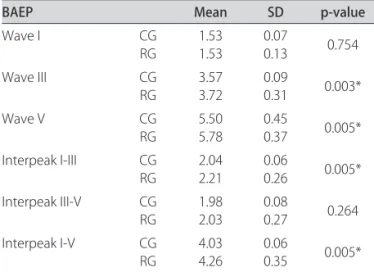
Statistically signiicant diferences were observed be-tween the control and research groups for latencies of waves III and V and interpeaks I-III and I-V, with high-er values for the RG (Table 1). In the analysis of the qual-itative data, the RG presented signiicantly higher occur-rence of abnormal results than the CG (Table 2). Higher occurrence of abnormalities of LB type (41%), HB type and both abnormality types (29.5%) was observed.
Regarding the AMLR quantitative data analysis, no statistically signiicant diferences between groups were observed for either latency of wave Pa or Na-Pa ampli-tude. However, there were statistically signiicant difer-ences for the Na wave latency in the derivations C3/A1, C4/A1 and C4/A2 (Table 3). he qualitative analysis of the Na - Pa amplitude (Table 4) demonstrated a greater per-centage of abnormal results in the RG with no statistical-ly signiicant diference between groups. he abnormali-ties most frequently observed in the CG were both (43%) and, in the RG, the most frequently observed abnormali-ties were ear efect (36%) and electrode efect (36%). he RG also showed a higher occurrence of abnormalities in latencies of Na and Pa waves and latency delay was the most frequently observed abnormality.
For the P300 quantitative analysis, CG presented a mean latency of 313.27 ms and the RG presented a mean latency of 312.24 ms with no statistically signiicant difer-ence between groups for this parameter (p-value=0.853). here was no statistically signiicant diference be-tween groups when comparing the occurrence of nor-mal and abnornor-mal results (Table 5), however, it was ob-served a higher percentage of abnormal results for the
Table 1. Between groups comparison of BAEP mean values of absolute latencies of waves I, III and V and interpeaks I-III, III-V and I-V.
BAEP Mean SD p-value
Wave I CG
RG
1.53 1.53
0.07
0.13 0.754
Wave III CG
RG
3.57 3.72
0.09
0.31 0.003*
Wave V CG
RG
5.50 5.78
0.45
0.37 0.005* Interpeak I-III CG
RG
2.04 2.21
0.06
0.26 0.005* Interpeak III-V CG
RG
1.98 2.03
0.08
0.27 0.264 Interpeak I-V CG
RG
4.03 4.26
0.06
0.35 0.005*
BAEP: brainstem auditory evoked potential; CG: control group; RG: research group; SD: standard deviation; *p-value statistically signiicant.
Table 2. Distribution of the occurrence of normal and abnormal BAEP results in control and research groups.
AEP
CG RG
p-value
N % N %
BAEP Normal Abnormal 25 0 100 0 8 17 32 68 <0.01
BAEP: brainstem auditory evoked potential; AEP: auditory evoked potential; CG: control group; RG: research group.
Table 3. Between groups comparison of AMLR mean latency values of waves Na and Pa (in ms) and Na-Pa amplitude (in microvolts) in the derivations C3/A1, C3/A2, C4/A1, C4/A2.
AMLR
C3/A1 C3/A2 C4/A1 C4/A2
CG RG CG RG CG RG CG RG
Na Latency Mean Median SD 18.13 17.43 2.6 21.84 20.86 4.2 18.53 17.19 2.4 21.92 21.36 5.2 18.09 17.76 2.77 22.35 21.02 4.8 17.52 16.99 2.05 22.57 17.72 5.3
p-value 0.036* 0.72 0.045* 0.025*
Pa Latency Mean Median SD 31.95 31.90 2.34 28.53 29.43 4.5 30.90 30.34 3.31 28.42 29.34 4.8 31.09 32.02 4.04 27.56 27.79 4.3 30.31 30.71 3.21 28.58 24.03 4.7
p-value 0.84 0.66 0.75 0.44
Na-Pa Amplitude Mean Median 2.57 1.61 1.99 1.45 4.23 1.48 3.33 1.62 3.43 1.76 1.96 1.84 2.00 1.63 1.51 1.45
p-value 0.561 0.759 0.451 0.331
AMLR: auditory middle latency response; CG: control group; RG: research group; SD: standard deviation; *p-value statistically signiicant.
RG. he type of abnormality most frequently found was the latency delay of the P300 wave (75%).
DISCUSSION
structural integrity of the neural components of the au-ditory pathway. In the present study, electrophysiological assessment of hearing was performed through auditory evoked potential measurements in individuals with MS. he AEP can provide an objective measurement on the integrity of the auditory system as a whole18, thus
reaf-irming the importance of such measures in neuroscience. Authors report that the use of evoked responses can provide a sensitive index of MS when compared to CT or even MRI19,20. Abnormalities in AEP can be
identi-ied in patients with normal MRI, especially in the case of the brain stem. Moreover, it is important to consider that such diagnostic methods assess diferent features of the CNS (functional versus structural; sensory pathways versus CNS as a whole), therefore, they are complemen-tary. he relatively high cost of MRI, when compared to AEP, is an additional issue to consider regarding the clin-ical application of these two neurodiagnostic approach-es in MS patients.
Research indicates that improvement of MS symptoms has been related to changes in sensory evoked respons-es9-11. his suggests that such procedures may be used
to evaluate the efectiveness of treatment strategy, per-haps with more sensitivity and objectivity than neurolog-ical examination21. Additionally, MRI only evaluates the
morphological data regarding the evolution of the disease in relation to the temporal and spatial aspects but does not ofer information about the impact of therapy in MS. he BAEP allows the identiication of possible brain-stem abnormalities. hese measures are important in aid-ing neurological diagnosis once they have well-established
criteria and a generator set. In the analysis of results ob-tained on the BAEP, we observed that the individuals with MS showed signiicantly.
longer latency values and signiicantly higher occur-rence of abnormal results (68%) when compared to the CG. Similar results were obtained by Celebisoy et al.4,
who observed BAEP abnormalities in 60% of assessed in-dividuals with MS. hese analyses suggest the presence of abnormalities on the lower brainstem auditory pathway of individuals with MS. his is consistent with the study by Bergamaschi et al.5 who described the presence of
ab-normalities on the distal portion of the auditory nerve in patients with MS using MRI. Such results corrobo-rate the indings of Hall3 regarding the
electrophysiolog-ical abnormalities that may be observed in BAEP in cas-es of MS. hcas-ese abnormaliticas-es may include increased la-tencies of waves III and V, increased I-III, III-V and I-V interpeaks, the absence of one or more components, and poor reproducibility and morphology of the later compo-nents suggesting dyssynchrony.
BAEP abnormalities were also reported by Santos et al.6. he authors observed BAEP abnormality in 58.62%
of the individuals with MS with no signs of brainstem in-volvement according to the MRI. he types of abnormali-ties observed were poor wave morphology, increased I-V interpeak, the presence of only wave I, or the absence of the latest waves with normal irst absolute latencies. Lima et al.22 also reported BAEP abnormalities in 36% of
indi-viduals with MS.
herefore, data found in this and in other studies4-7
reinforce the importance of recording the BAEP in cas-es of clinical suspicion of demyelinating diseascas-es and cas- es-pecially with proven diagnosis of MS, because BAEP re-cording aids in the diagnosis and the deinition of the type of brain impairment presented. Moreover, the record can provide information about the functional aspect of im-provement after treatment. As BAEP is an objective, easy and inexpensive method, it can be performed serially to evaluate the efectiveness of treatment. his is diicult to accomplish with MRI given its high cost.
Table 4. Distribution of the occurrence of normal and abnormal AMLR results in control and research groups.
AMLR
CG RG
p-value
N % N %
Na Latency Normal
Abnormal 214 8416 232 928 <0.01* Pa Latency Normal
Abnormal 205 8020 232 928 0.42
Na-Pa Amplitude Normal Abnormal
18 7
72 28
11 14
44
56 0.08
AMLR: auditory middle latency response; CG: control group; RG: research group; *p-value statistically signiicant.
Table 5. Distribution of the occurrence of normal and abnormal P300 results in control and research groups.
AEP
CG RG
p-value
N % N %
P300 Normal 25 100 21 84 0.11 Abnormal 0 0 4 16
he AMLR is considered one of the best tests to eval-uate the central auditory nervous system, and it is also a useful tool in the design and monitoring of the therapeu-tic process. here are few studies in the literature that use the AMLR as an electrophysiological measure for the in-vestigation of the auditory pathway in patients with MS. In the present study, RG presented higher incidence of abnormal results than the CG with respect to the Na - Pa amplitude and the latencies of Pa and Na waves. Howev-er, this diference was not statistically signiicant. he ab-normalities of higher occurrence in the RG were ear ef-fect, electrode eef-fect, and delay in latencies of Pa and Na waves. he presence of AMLR abnormalities in individ-uals with MS was also evidenced by Celebisoy et al.4 who
reported the occurrence of abnormalities in 73.4% of indi-viduals assessed. AMLR abnormalities were also observed in the MS case study presented by Schochat et al.7, thus
demonstrating the possibility of involvement of the audi-tory pathway at the subcortical level.
he latency of the P300 wave is the most reliable in-dex for analysis of this potential17. Statistically signiicant
diferences between groups were not observed in either the qualitative or quantitative analysis. However, regard-ing the distribution of normal and abnormal results, it was observed that the RG showed higher percentage of abnormal results than the CG. he latency delay was the type of abnormality most frequently observed.
he indings of this study regarding the presence of P300 abnormalities in individuals with MS and the types of abnormalities often encountered, corroborate those described by Schochat et al.7, Magnano et al.8, Giesser
et al.23, Gonzáles-Rosa et al.24, Newton et al.25, Gil et al.26,
and Dijk et al.27.
Several studies8-11,18,23,24,28 describe the presence of
cog-nitive changes in patients with MS demonstrating that they may present dysfunctions in memory, attention, ver-bal luency, task performance, and visual perception, all of which are important skills related to P300. hese data justify the use of this potential, in association with oth-er tests, in evaluation and follow-up of MS as reported by Magnano et al.8, Aminof and Goodin9, Magnié et al.10
and Kurokawa et al.29. In MS, the CNS
electrophysiolog-ical evaluations performed through visual evoked poten-tials, auditory evoked potentials of short latency, and so-matosensory potentials are well-established.
Although the visual and somatosensory evoked po-tentials have greater sensitivity than the AEP, the use of a multimodal battery promotes increased sensitivity when compared to using a single modality alone. hom-as30 states that when used in combination with
structur-al and functionstructur-al MRIs, the electrophysiologicstructur-al mea-sures can provide additional information regarding the temporal dynamics of brain activity. Moreover,
accord-ing to Aminof and Goodin9 and Stockard and Rossiter21,
there is a relationship between improvement of symp-toms and reversal of abnormalities in somatosensory po-tential, suggesting that this potential could be used in as-sessing the efectiveness of medical treatment. Further-more, the AMLR and event-related potentials have been widely studied because of their correlation with cognitive aspects. he abnormalities observed in these potentials are deined by the increase of the latency waves or by the absence of characteristic peaks6.
hus, considering that individuals with MS may pres-ent abnormalities in AEP and this procedure is both in-expensive and easy to implement, we would like to em-phasize that AEP can be considered by neurologists as an additional useful tool in assessing and monitoring of in-dividuals with MS.
In conclusion, individuals with MS of the remittent-recurrent type present abnormalities in BAEP. his sug-gests that the brainstem auditory pathway, in the regions from the cochlear nucleus to the lateral lemniscus, may be disrupted due to structural and/or functional chang-es in the transmission of the acoustic stimulus along the auditory pathway. Abnormalities in AMLR suggest im-pairment in subcortical regions of the auditory pathway and/or central auditory processing disorder. he presence of abnormalities in cognitive potential (P300) suggests impairment in cortical regions of the auditory pathway and deicits in cognitive processing, memory, attention and auditory discrimination. Taking into consideration these aspects and the fact these individuals have hearing thresholds within normal limits, the combination of dif-ferent objective methods of electrophysiological assess-ment of hearing (BAEP, AMLR and P300) contributes to the detection of changes in central auditory pathway in individuals with MS.
REfERENCES
Ropper AH, Brown RH. Multiple sclerosis and allied demyelinative diseases. 1.
In: Ropper AH, Brown RH (Eds). Adams and Victors Principles of neurology. 8th Ed. New York: McGraw-Hill, 2005:771-796.
Mustillo P. Auditory deicits in multiple sclerosis: a review. Audiology 1984; 2.
23:145-164.
Hall III JW. Handbook of auditory evoked responses. Boston: Allyn and 3.
Bacon, 2007.
Celebisoy N, Aydogdu I, Ekmekci O, Akurekli O. Middle latency auditory 4.
evoked potentials (MLAEPs) in MS. Acta Neurol Scand 1996;93:318-321. Bergamaschi R, Romani A, Zappoli F, Versino M, Cosi V. MRI and brainstem 5.
auditory evoked potential evidence of eighth cranial nerve involvement in multiple sclerosis. Neurology 1997;48:270-272.
Santos MAR, Lana-Peixoto MA, Munhoz MSL, Almeida AV. Avaliação dos po-6.
tenciais evocados auditivos do tronco encefálico na esclerose múltipla. Arq Neuropsiquiatr 2003;61:392-397.
Schochat E, Matas CG, Sanches SGG, Carvallo RMM, Matas S. Central auditory 7.
evaluation in multiple sclerosis Arq Neuropsiquiatr 2006;64:872-876. Magnano I, Aiello I, Piras, MR. Cognitive impairment and neurophysiological 8.
correlates in MS. J Neurol Sci 2006;245:117-122.
Aminof JC, Goodin DS. Long-latency cerebral event-related potentials in 9.
Magnié MN, Bensa C, Laloux L, Bertogliati C, Faure S, Lebrun C. Contribution 10.
of cognitive evoked potentials for detecting early cognitive disorders in mul-tiple sclerosis. Rev Neurol 2007;163:1065-1074.
Fletcher S, Vardi J, Finkelstein Y, Pollak L. Cognitive dysfunction evaluation 11.
in multiple sclerosis patients treated with interferon beta 1-b: an open-label prospective 1 year study. Isr Med Assoc J 2007;9:457-459.
Poser CM, Paty DW, Scheinberg L, et al. New diagnostic criteria for multiple 12.
sclerosis: guidelines for research protocols. Ann Neurol 1983;13:227-231. Jasper HA. The ten–twenty system of the International Federation. Electro-13.
encephalogr Clin Neurophysiol 1958,10:371-375.
Musiek FE, Lee WW. Potenciais auditivos de média e longa latência. In: Mus-14.
iek FE, Rintelmann WF (Eds). Perspectivas atuais em avaliação auditiva. Ba-rueri: Manole, 2001:239-256.
McGee T, Kraus N, Manfredi C. Toward a strategy for analyzing the auditory 15.
middle-latency response waveform. Audiology 1988;27:119-130. Picton TW. The P300 wave of the human event-related potential. J Clin Neu-16.
rophysiol 1992;9:456-479.
McPherson DL. Late potentials of the auditory system. San Diego: Singu-17.
lar Pub. Group, 1996.
Rao SM, Leo GJ, Bernardin L, Unverzagt F. Cognitive dysfunction in multiple 18.
sclerosis. I. Frequency, patterns, and prediction. Neurology 1991;41:685-691. Giesser BS, Kurtzberg D, Vaughan Jr HG, et al. Trimodal evoked potentials 19.
compared with magnetic resonance imaging in the diagnosis of multiple sclerosis. Arch Neurol 1987;44:281-284.
Tramo MJ, Schneck MJ, Lee BCP, Rapoport S. Evoked potentials and mag-20.
netic resonance imaging in the diagnosis of multiple sclerosis. Neurology 1985;35(Suppl 1):S105.
Stockard JJ, Rossiter VS. Clinical and pathologic correlates of brainstem au-21.
ditory response abnormalities. Neurology 1977;27:316-325.
Lima TMA, Crato AN, Mancini PC, Simões LC, Gonçalves DU. Alterações dos 22.
potenciais evocados auditivos do tronco encefálico em pacientes com es-clerose múltipla. Braz J Otorhinolaryngol 2009;75:177-181.
Giesser BS, Schroeder MM, LaRocca NG, Kurtzberg D Ritter W, Vaughan HG, 23.
Scheinberg LC. Endogenous event-related potentials in multiple sclerosis pa-tients. Eletroenceph Clin Neurophisiol 1992;82:320-329.
Gonzales-Rosa JJ, Vazquez-Marrufo M, Vaquero E, et al. Diferential cognitive 24.
impairment for diverse forms of multiple sclerosis. BMC Neurosci 2006;7:39. Newton MR, Barret G, Callanan MM, Towell AD. Cognitive event-related po-25.
tentials in multiple sclerosis. Brain 1989;112:1637-1660.
Gil R, Zai L, Neau JP, Jonveaux T, et al. Event-related auditory evoked poten-26.
tials and multiple sclerosis. Eletroenceph Clin Neurophysiol 1993;88:182-187. Dijk JG, Jennekens-Schikel A, Caekebeke JFV, Singh A, Zwinderman AH. What 27.
is the validity of an “abnormal” evoked or event-related potential in MS. J Neurol Sci 1992;109:11-17.
Fischer JS. Cognitive impairment in multiple sclerosis. In: Cook SD (Ed). Hand-28.
book of multiple sclerosis. New York: Marcel Dekker, 2001:233-255. Kurokawa T, Kira J, Tobimatsu S. Electrophysiolgical diagnosis for multiple 29.
sclerosis. Nippon Rinsho 2003;61:1347-1354.
Thomas KM. Assessing brain development using neurophysiologic and be-30.