Neuropsychiatric symptoms in
Vascular Cognitive Impairment
A systematic review
Chan Tiel1, Felipe Kenji Sudo2, Gilberto Sousa Alves2,3, Letice Ericeira-Valente2, Denise Madeira Moreira4, Jerson Laks2,5, Eliasz Engelhardt1
ABSTRACT. Neuropsychiatric symptoms or Behavioral and Psychological Symptoms of Dementia (BPSD) are common and invariably appear at some point during the course of the disease, mediated both by cerebrovascular disease and neurodegenerative processes. Few studies have compared the profiles of BPSD in Vascular Cognitive Impairment (VCI) of different subtypes (subcortical or cortical) and clinical stages (Vascular Cognitive Impairment No Dementia [VaCIND] and Vascular Dementia [VaD]). Objective: To review the BPSD associated with different subtypes and stages of VCI using the Neuropsychiatric Inventory (NPI). Methods: Medline, Scielo and Lilacs databases were searched for the period January 2000 to December 2014, with the key words: “BPSD AND Vascular Dementia, “NPI AND Vascular Dementia” and “NPI AND VCI. Qualitative analysis was performed on studies evaluating BPSD in VCI, using the Neuropsychiatric Inventory (NPI). Results: A total of 82 studies were retrieved of which 13 were eligible and thus included. Among the articles selected, 4 compared BPSD in Subcortical Vascular Dementia (SVaD) versus Cortical-Subcortical Vascular Dementia (CSVaD), 3 involved comparisons between SVaD and VaCIND, 1 study analyzed differences between CSVaD and VaCIND, while 5 studies assessed BPSD in CSVaD. Subcortical and Cortical-Subcortical VaD were associated predominantly with Apathy and Depression. VaCIND may present fewer behavioral symptoms than VaD. Conclusion: The profile of BPSD differs for different stages of VCI. Determining the most prevalent BPSD in VCI subtypes might be helpful for improving early diagnosis and management of these symptoms.
Key words: neuropsychiatric symptoms, BPSD, NPI, Vascular Cognitive Impairment, Vascular Dementia, Alzheimer’s disease.
SINTOMAS NEUROPSIQUIÁTRICOS NO COMPROMETIMENTO COGNITIVO VASCULAR: REVISÃO SISTEMÁTICA
RESUMO. Sintomas Neuropsiquiátricos ou Sintomas de Comportamento e Psicológicos da Demência (SCPD) são comuns e aparecem em algum ponto no curso da enfermidade, mediado por doenças cerebrovasculares e processos neurodegenerativos. Poucos estudos compararam os perfis dos SCPD no Comprometimento Cognitivo Vascular (CCV) de diferentes subtipos (subcortical ou córtico-subcortical) e diferentes estágios clínicos (Comprometimento Cognitivo Vascular Não Demência [CCV-ND] e Demência Vascular [DV]). Objetivo: Revisar os SCPD associados aos diferentes subtipos e estágios do CCV, com o Inventário Neuropsiquiátrico (INP). Métodos: Medline, Scielo e Lilacs foram pesquisados de janeiro de 2.000 até dezembro de 2.014, com as palavras chaves: “BPSD AND Vascular Dementia”, “NPI AND Vascular Dementia” e “NPI AND VCI”. Uma análise quantitativa foi feita nos estudos que avaliaram sintomas comportamentais no CCV através do INP. Resultados: Um total de 82 estudos foram identificados e 13 estudos foram considerados elegíveis e incluídos. Destes artigos, 4 compararam SCPD entre DV Córtico-Subcortical (DVCS) e DV Subcortical (DVS), 3 artigos compararam DVS e CCVND, 1 artigo analisou diferenças entre DVCS e CCVND, e 5 estudos avaliaram os SCPD em DVCS. Tanto a DVCS quanto a DVS associaram-se principalmente a Apatia e Depressão. O CCVND pode apresentar menos sintomas comportamentais que a DV.Conclusão: O perfil dos SCPD difere entre diferentes estágios do CCV. O entendimento dos SCPD mais prevalentes nos subtipos do CCV poderia ser útil para facilitar o diagnóstico precoce e o manejo desses sintomas.
Palavras-chave: sintomas neuropsiquiátricos, SCPD, INP, Comprometimento Cognitivo Vascular, Demência Vascular, doença de Alzheimer.
The study was conducted at the Institute of Psychiatry and Institute of Neurology – UFRJ, Rio de Janeiro RJ – Brazil.
1Instituto de Neurologia Deolindo Couto, Setor de Neurologia Cognitiva e do Comportamento-INDC-CDA/IPUB, UFRJ, Rio de Janeiro RJ, Brazil. 2Instituto de
Psiquia-tria, Universidade Federal do Rio de Janeiro (UFRJ), Rio de Janeiro RJ, Brazil. 3Departamento de Medicina Clínica, Universidade Federal do Ceará, Fortaleza CE,
Brazil. 4Serviço de Radiologia, Instituto de Neurologia Deolindo Couto (UFRJ); Hospital Pró-Cardíaco, Rio de Janeiro RJ, Brazil. 5Universidade do Estado do Rio de
Janeiro, Rio de Janeiro RJ, Brazil.
Chan Tiel. Rua General Goes Monteiro, 184/904 – 22290-080 Rio de Janeiro RJ – Brazil. E-mail: [email protected]
Dement Neuropsychol 2015 September;9(3):230-236
231
Tiel C, et al. BPSD in Vascular Cognitive Impairment
INTRODUCTION
V
ascular Dementia (VaD) is considered thesec-ond-most-common type of dementing illness, accounting for a signiicant proportion of total demen-tia cases.1,2 Vascular Cognitive Impairment (VCI) is a
broader dimensional term,3 encompassing subjects with
mild cognitive impairment and no incapacity on activi-ties of daily living, referred to as Vascular Cognitive Impairment No Dementia (VaCIND), as well as condi-tions associated with signiicant cognitive impairment and decline in functional status (VaD).4,5 Clinical
fea-tures can vary considerably depending on the extension and location of vascular lesions. Moreover, VCI may be associated with large-vessel disease, which often leads to extensive cortical-subcortical damage, or with insidi-ous occlusion/semi-occlusion of small penetrating arte-rioles, resulting in subcortical vascular disease.6,7
Comparisons among signs and symptoms of difer-ent subtypes of VCI may help understand how diferdifer-ent mechanisms of brain damage can produce convergent or divergent clinical features, which in turn may lead to a better comprehension of the pathophysiology of the disorder. Among these manifestations, Neuropsychi-atric Symptoms are of great importance, considering that they almost invariably appear at some point of the natural course of VCI in addition to cognitive impair-ment.8-12 Also referred as Behavioral and Psychological
Symptoms of Dementia (BPSD),13 these features are
fre-quently associated with growing levels of distress both in persons with dementia and in their caregivers, as well as with higher risk for adverse outcomes and increased use of health care resources.14 Clinically, BPSD have a major
impact on the patient’s functional and cognitive status.15
Despite considerable advances in the detection of brain vascular-related syndromes in recent years, the association between vascular lesions and both cognitive symptoms and BPSD in VCI remains controversial. For instance, data in the literature suggests that BPSD may occur in VCI, regardless of the development of demen-tia.16,17 BPSD may also appear at any stage, induced by
cerebrovascular lesions disrupting the cortical-subcor-tical circuits between prefrontal cortex connections to limbic nuclei of the basal ganglia and thalamus, and other limbic system structures.18-20 his suggests that
better characterization of vascular-related BPSD, and the underlying mechanisms of brain injury associated with these features, is still needed so as to allow the adoption of efective evidence-based prophylactic and therapeutic measures.
Among methods for assessing BPSD, the Neuropsy-chiatric Inventory (NPI)21 is a valid and reliable
instru-ment, originally developed to assess 10 neuropsychiatric disturbances commonly seen in dementia. Subsequently, it has been modiied to evaluate 12 disturbances includ-ing agitation, aberrant motor behavior, anxiety, elation, irritability, depression, apathy, disinhibition, delusions, hallucinations, sleep and appetite changes.22 Currently,
the NPI is the most frequently used instrument for eval-uating these symptoms in research studies and clinical practice. It is a semistructured interview which rates the frequency and severity of BPSD for the month preced-ing assessment. he NPI has been translated and vali-dated to Brazilian Portuguese.23
To date, few studies have compared neuropsychiatric disturbances among VCI subgroups (cortical or subcor-tical, “pure” or mixed etiology, VaD or VaCIND). hus, the aim of the present review was to examine BPSD pro-iles in VCI subgroups, primarily addressing two issues: [1] which are the most prevalent BPSD in VCI, and [2] whether diferent clinical presentations of VCI present diferent BPSD proiles. It can be hypothesized that the BPSD in VCI have a heterogeneous pattern, depending on the clinical stage and mechanism of brain lesion.
METHODS
Search strategy and criteria. To identify articles, a
system-atic search was performed in PUBMED/MedLine, Scielo and Lilacs databases for English-language articles published between January 2000 and December 2014, followed by a manual search of the reference lists of relevant articles retrieved. he results of three searches, which employed the following key words: “BPSD AND Vascular Dementia”, “NPI AND Vascular Dementia” and “NPI AND VCI” were combined.
Studies were selected for inclusion in the present systematic review if they met all of the following crite-ria: [a] studies that assessed patients diagnosed with VaD according to the 4th edition of the Diagnostic and
Statistical Manual of Mental Disorders (DSM-IV)24
cri-teria or the NINDS-AIREN cricri-teria1 and/or studies that
assessed Va-CIND subjects, diagnosed according to well-recognized criteria;24,26 [b] studies that assessed BPSD
in VCI; [c] studies that evaluated BPSD using the NPI; [d] studies that presented data on the prevalence and/or incidence rates of BPSD. Studies involving Alzheimer’s disease (AD) or mixed dementia (MD) subjects were included if within a comparison group for VCI subjects.
Data extraction. Data from those studies meeting the inclusion criteria were extracted by one of the authors (CT) and reviewed by other researchers from the group (JL, FKS). In cases of disagreement among the two, discussion included the whole group to reach a consensus. Information about the clinical diagnosis, sample size, assessment instrument for BPSD, and the prevalence of BPSD categories were drawn from the selected studies.
18 studies selected
63 studies excluded
(RCT with medication, studies assessing only AD patients, studies
not applying NPI, and Reviews)
13 elegible articles
5 articles excluded
(studies assessing DLB and FTD)
7 articles assessing VCI
6 articles assessing VaD
82 articles retrieved on PUBMED
RCT: Randomized clinical trial; AD: Alzheimer’s Disease; NPI: Neuropsychiatric Inventory; DLB: Dementia with Lewy Bodies; FTD: Frontotemporal dementia; VCI: Vascular Cognitive Impair-ment; VaD: Vascular Dementia.
Figure 1. Flowchart of search strategy and article selection.
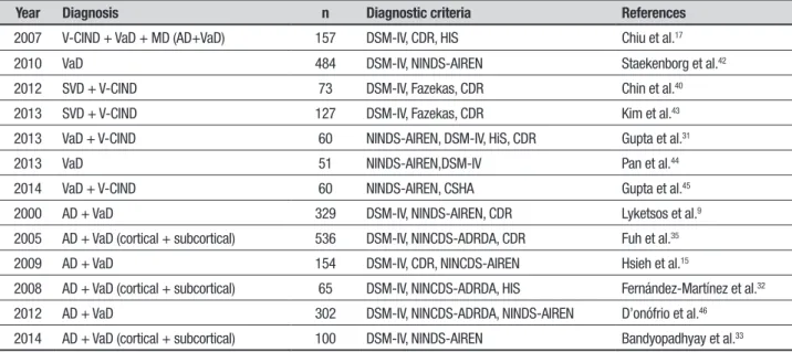
Table 1. Characteristics of samples, diagnostic groups and operational criteria in the studies selected.
Year Diagnosis n Diagnostic criteria References
2007 V-CIND + VaD + MD (AD+VaD) 157 DSM-IV, CDR, HIS Chiu et al.17
2010 VaD 484 DSM-IV, NINDS-AIREN Staekenborg et al.42
2012 SVD + V-CIND 73 DSM-IV, Fazekas, CDR Chin et al.40
2013 SVD + V-CIND 127 DSM-IV, Fazekas, CDR Kim et al.43
2013 VaD + V-CIND 60 NINDS-AIREN, DSM-IV, HiS, CDR Gupta et al.31
2013 VaD 51 NINDS-AIREN,DSM-IV Pan et al.44
2014 VaD + V-CIND 60 NINDS-AIREN, CSHA Gupta et al.45
2000 AD + VaD 329 DSM-IV, NINDS-AIREN, CDR Lyketsos et al.9
2005 AD + VaD (cortical + subcortical) 536 DSM-IV, NINCDS-ADRDA, CDR Fuh et al.35
2009 AD + VaD 154 DSM-IV, CDR, NINCDS-AIREN Hsieh et al.15
2008 AD + VaD (cortical + subcortical) 65 DSM-IV, NINCDS-ADRDA, HIS Fernández-Martínez et al.32
2012 AD + VaD 302 DSM-IV, NINCDS-ADRDA, NINDS-AIREN D’onófrio et al.46
2014 AD + VaD (cortical + subcortical) 100 DSM-IV, NINDS-AIREN Bandyopadhyay et al.33
AD: Alzheimer Dementia; VaD: Vascular Dementia; MD: Mixed Dementia; SVD: Subcortical Vascular Dementia; VCIND: Vascular cognitive Impairment-No Dementia; DSM-IV: Diagnostic and Statistical Manual of Mental Disorders 4th edition; CDR: Clinical Dementia Rating; HIS: Hachinski ischaemic Score; Fazekas: Fazekas scale; CSHA: Canadian Study of Health and Ageing; NINDS-AIREN: National
Institute of Neurological Disorders and Stroke and Association - Internationale pour la Recherché et lÉnseignement en Neurosciences; NINCDS-ADRDA: National Institute of Neurological and Communicative Disorders and Stroke and the Alzheimer’s Disease and Related Disorders Association.
Statistical analysis. Data were analyzed using the IBM
Statistical Package for Social Sciences version 20 for Windows. Initially, the Shapiro-Wilk test was performed to verify the normality of data (prevalence of BPSD). Since signiicance exceeded .05 for all variables, One-Way ANOVA was performed to compare statistically signii-cant diferences between BPSD prevalence in the SVaD (Subcortical Vascular Dementia), VaCIND (Vascular CIND) and CSVaD (Cortical-Subcortical Vascular Dementia) groups. Tukey’s Post-hoc test was performed to identify groups with signiicantly diferent preva-lences of BPSD. he level of signiicance was set at .05.
RESULTS
Of the total 82 articles initially retrieved, 13 fulilled the inclusion criteria and were included in this review. No studies were identiied on the Lilacs and Scielo data-bases using the previously described search strategy. Figure 1 shows the stages of selection of papers included in this article.
he number of participants in the studies ranged from 51 to 938 patients. Diagnostic groups included diferent stages of VCI (VaCIND and VaD), etiologies (VCI, AD and MD) and subtypes of VaD (cortical and cortical-subcortical). Operational criteria for VCI were the DSM-IV,24 NINDS-AIREN,1 Hachinski’s Ischemic
Score,27 Clinical Dementia Rating,28 Fazekas scale,29 and
Dement Neuropsychol 2015 September;9(3):230-236
233
Tiel C, et al. BPSD in Vascular Cognitive Impairment
BPSD were identiied in all diagnostic groups at a high prevalence: 83.8 [96.4%] in SVaD, 59.7 [100%] in CSVaD and 47.5 [89%] in VaCIND. Apathy and Depres-sion were the most frequent BPSD, followed by Irrita-bility, Anxiety and Agitation. In another study, a higher prevalence of Euphoria was identiied in VaCIND than in VaD (7.14% vs 3.13%).31 Agitation/Aggression
symp-toms appeared to be equally prevalent in CSVaD (21.4 [62.9%]) and in SVaD (22.7 [47.62%]). Also, patients with SVaD presented comparable Aberrant Motor Behavior (up to 38.10%) and Hallucinations (up to 28%) as patients with CSVaD (5-61.5% and 1-30.8%, respec-tively). Table 2 depicts the prevalences of BPSD and the NPI scores in the studies.
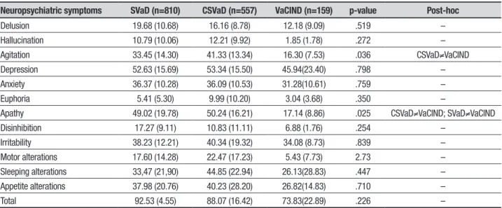
On comparison of mean prevalence of BPSD among subtypes of VCI, Apathy and Agitation were found to difer signiicantly in the assessed groups. Agitation was signiicantly more prevalent in CSVaD than in VaCIND (p=0.036), whereas the VaCIND group showed signii-cantly less Apathy than both SVaD and CSVaD groups (p=0.025). Table 3 shows these results.
DISCUSSION
he present study reviewed the characteristics of BPSD in VCI of diferent subtypes and clinical stages. he arti-cles retrieved in this search showed that at least half of those individuals with VCI presented BPSD. Moreover, comparison between BPSD in SVaD and CSVaD revealed no diferences in prevalence and proile of BPSD domains. herefore, both subtypes of VaD are likely to present Depression and Apathy during the course of the disease. Other symptoms varied widely in prevalence among groups. As expected, VaCIND subjects had a lower prevalence of BPSD than VaD patients, a inding consistent with larger areas of spared brain tissue. Studies showed higher frequency of Delusions, Aber-rant Motor Behavior and Sleep disorders in AD than in VaD subjects.9,15,32,35
Although AD was not part of the present search, available data on the diferences between BPSD in vas-cular and neurodegenerative cognitive impairments were also examined. Studies that assessed AD also showed high prevalence of BPSD (up to 53.3%). More than half of the patients had at least one symptom, both in VaD and AD (AD 53.3% vs VaD 59.7%).9 In three
studies, almost all patients with AD or VaD had some
type of BPSD (AD=100% vs VaD=100%; AD=94.6% vs
VaD=96.4%).15,29,30 Comparisons of BPSD prevalence
in VaD and AD showed heterogeneous results. Depres-sion and Apathy were more prevalent in VaD, whereas prevalence of the other symptoms varied widely across
the studies. In another study, the prevalence for Depres-sion was also higher in VCI (VCI=30% vs AD=15%).31
Several studies reported a higher prevalence of speciic BPSD domains in AD than in VaD, as follows: Delusions (AD=22.4% vs VaD=8.1%),9 Aberrant Motor Behavior
(AD=24.3% vs VaD=7.1%),32 Nocturnal Behavior/Sleep
(AD=96.0% vs VaD=73.1%/AD=35.1% vs VaD=3.6%).15,32
hese results indicate diferent patterns of behavioral symptoms between VCI and AD. However, since AD was not part of the search strategy for this review, further studies evaluating BPSD diferences between VCI and AD are needed to allow more robust conclusions.
A number of issues in assessing BPSD may account for the disparity in results among studies. Neuropsychi-atric Symptoms in dementia tend to luctuate over time. herefore, estimating prevalence of BPSD using a cross-sectional approach may not be appropriate. Moreover, since assessment of individual BPSD domains resulted in great variability across studies, identifying clusters of symptoms with similar underlying neurobiological cor-relates may allow better characterization of the behav-ioral proiles of VCI subtypes. In this regard, Frisoni et al. (1999)36 described a three-factor model of BPSD in
AD: Mood (indicated by Anxiety, Apathy and Depres-sion), Psychotic (Irritability/Lability, Delusions, Hallu-cinations and Agitation/Aggression), and Frontal Symp-toms Domain (Euphoria and Disinhibition). Aalten et al. (2008)37 identiied four neuropsychiatric subsyndromes:
hyperactivity, psychosis, afective syndrome and apathy. Truzzi et al. (2013)38 compared BPSD clusters in
sub-jects with dementia evaluated in two diferent countries: the Brazilian sample showed factors, such as Psychosis (Delusion, Hallucination, Euphoria, and Disinhibition), Mood (Depression, Anxiety, Agitation/Aggression, and Irritability), and Psychomotor (Apathy and Aberrant Motor Behavior), whereas factors drawn from a Norwe-gian sample were Psychosis (Delusion, Hallucination, and Aberrant Motor Behavior), Mood (Depression, Anxi-ety, Agitation/Aggression, Apathy and Irritability), and Frontal (Euphoria, Disinhibition, and Irritability).
Applying the cluster-model to the results of this review allows several conclusions to be drawn. Mood and psychomotor symptoms were more prevalent in patients with VCI whereas patients with AD may have more psychotic symptoms. Mood symptoms appeared to be equally prevalent in both SVaD and CSVaD.
Dement Neuropsyc
hol 2015 September;9(3):230-236
BPSD in
Vascular Cognitive Impair
ment Tie l C , e t a l. D O I: 1 0.1 59 0/1 98 0-5 76 42 01 5D N 93 00 00 04
References (n) Del Hal Agi Dep Anx Eup Apa Dis Irr Ama Sle App Total
Subcortical Vascular Dementia (SVaD) Fuh et al., 200535
(n=161) (--) [31.1%] (--) [21.0%] (--) [44.1%] (--) [45.0%] (--) [33.8%] (--) [14.9%] (--)
[47.2%] [24.0%](--)
(--) [41.6%] (--) [27.9%] (--) [49.4%] (--) [40.3%] (--) [--] Chiu et al., 200717
(n=95) 1.7 (3.6) [26%] 1.6 (3.5) [28%] 2.1 (3.8) [32%] 2.8 (4.0) [49%] 1.9 (3.6) [32%] 0.4 (1.5) [6%] 4.6 (5.3) [48%] 0.9 (2.5) [16%] 2.9 (4.3) [41%] 2.3 (4.4) [26%] 5.2 (5.1) [60%] 2.5 (4.1) [31%] 27.7 (22.2) [92%] Fernández-Martínes et
al., 200832 (n=22)
(--) [9.1%] (--) [4.5%] (--) [22.7%] (--) [31.8%] (--) [27.3%] (--) [9.1%] (--) [54.5%] (--) [22.7%] (--) 31.8%] (--) [0.0] (--)
[4.5%] [13.6%](--) 11.82 (9.75) [96.4%] Staekenborg et al.,
201041 (n=401)
0.4 (1.4) [12%] 0.1 (1.3) [7%] 1.1 (3.0) [38%] 1.4 (2.5) [45%] 1.1 (3.1) [34%] 0.1 (1.0) [5%] 3.8 (3.4) [67%] 0.5 (1.6) [16%] 1.2 (2.1) [41%] 0.6 (1.6) [15%] 1.1 (2.8) [33%] 1.4 (2.3) [29%] --(--) [93%] Chin et al., 201242
(n=42)
0.31 (1.14) 0.33 (1.32) 1.40 (1.96) 1.33 (1.78) 1.43 (1.48) 0.31 (1.32) 3.86 (4.00) 1.00 (2.48) 2.10 (2.62) 2.26 (4.20) 2.21 (3.73) 2.40 (3.68) 18.95 (16.18)
Kim et al., 201343
(n=68) (--) [19.1%] (--) [10.3%] (--) [42.6%] (--) [50.0%] (--) [41.8%] (--) [2.9%] (--) [57.4%] (--) [27.9%] (--) [45.6%] (--) [16.2%] (--) [27.9%] (--) [35.3%] (--) [83.8%] Gupta et al., 201445
(n=21) 1.7 [33.33%] 0.1 [4.76%] 3.9 [47.62%] 6.0 [76.19%] 4.0 [57.14%] 0.0 [0.0%] 5.9 [61.90%] 0.4 [14.29%] 4.2 [52.38%] 2.8 [38.10%] 3.7 [52.38%] 4.3 [80.95%] (--) [95%] Cortical-Subcortical Vascular Dementia (CSVaD)
Lyketsos et al., 20009*
(n=62) 0.24 (0.98) [8.1%] 0.29 (0.96) [12.9%] 1.10 (2.03) [32.2%] 2.78 (10.80) [32.3%] 0.57 (1.70) [17.7%] 0.10 (0.79) [1.6%] 0.43 (3.30) [22.2%] 0.52 (1.80) [11.3%] 0.41 (0.94) [17.7%] 0.19 (0.76) [8.1%] --(--) [--] --(--) [--] 7.70 (13.50) [59.7%] Fuh et al., 200535
(n=35) (--) [31.4%] (--) [25.7%] (--) [62.9%] (--) [51.0%] (--) [51.0%] (--) [11.0%] (--) [62.9%] (--) [34.3%] (--) [51.4%] (--) [26.5%] (--) [65.7%] (--) [45.7%] (--) [--] Fernández-Martínes et
al., 200832 * (n=6)
0.75 (2.44) [14.3%] 0.43 (2.27) [3.6%] 0.96 (2.27) [21.4%] 0.96 (1.86) [35.7%] 0.96 (1.64) [28.6%] 0.14 (0.59) [7.1%] 4.11 (3.85) [64.3%] 0.54 (1.29) [17.9%] 1.75 (2.95) [35.7%] 0.29 (1.05) [7.1%] 0.21 (1.13) [3.6%] 0.71 (1.90) [14.3%] 11.82 (9.75) [96.4%] Hsieh et al, 200815*
(n=77) 2.46 (4.34) [26.9%] 1.46 (2.77) [30.8%] 1.73 (3.19) [30.8%] 3.30 (2.81) [65.4%] 1.23 (2.05) [34.6%] 2.00 (3.09) [34.6%] 5.04 (2.90) [65.4%] 0.00 (0.00) [0.0%] 0.92 (1.89) [23.1%] 3.31 (3.08) [61.5%] 5.23 (4.07) [73.1%] 0.00 (0.00) [0.0%] 26.69 (9.92) [100%] Staekenborg et al.,
201042 (n=83)
0.3 (1.5) [7%] 0.0 (0.1) [1%] 1.5 (2.3) [48%] 1.1 (1.6) [43%] 1.5 (2.3) [39%] 0.2 (0.7) [13%] 2.1 (2.9) [54%] 0.4 (1.1) [16%] 1.7 (2.5) [49%] 0.2 (0.9) [5%] 1.0 (2.8) [31%] 1.5 (2.4) [27%] (--) [--] D’onófrio et al. 201246*
(n=136) (--) [11.9%] (--) [9.7%] (--) [33.6%] (--) [59.7%] (--) [49.3%] (--) [1.5%] (--) [56.0%] (--) [0.0%] (--) [38.8%] (--) [17.9%] (--) [63.4%] (--) [54.5%] (--) [69.4%] Gupta et al., 201331*
(n=32) 0.96 [21.88%] 0.09 [6.25%] 3.81 [53.13%] 6.53 [75.0%] 3.03 [40.63%] 0.28 [3.13%] 5.37 [59.38%] 0.5 [9.38] 4.18 [59.38%] 1.78 [28.13%] 3.28 [50.0%] 4.43 [84.38%] (--) [100%] Pan et al., 201344*
(n=51)
1.55 (0.69) 1.30 (0.51) 5.55 (0.90) 4.21 (0.81) 2.32 (0.81) 3.75 (0.65) 3.33 (0.71) 2.75 (0.68) 3.2 (0.92) 4.25 (0.69) 5.32 (0.83) 4.08 (0.57) -- (--)
Gupta et al., 201445
(n=25) 0.5 [16%] 0.2 [8%] 3.8 [52%] 6.1 [72%] 2.3 [36%] 0.7 [12%] 1.9 [28%] 0.6 [8%] 4.8 [72%] 0.9 [20%] 1.8 [32%] 3.5 [68%] (--) [95%] Bandyopadhyay et al.,
201433 * n=50)
(--) [8%] (--) [12%] (--) [38%] (--) [46%] (--) [28%] (--) [6%] (--) [40%] (--) [10%] (--) [16%] (--) [28%] (--) [40%] (--) [28%] (--) [96%] Vascular CIND (VaCIND)
Chin et al., 201240
(n=31)
0.00 (0.00) 0.00 (0.00) 0.55 (1.73) 0.26 (0.58) 0.39 (0.76) 0.03 (0.18) 1.00 (2.14) 0.23 (1.09) 0.52 (1.29) 0.52 (1.29) 0.19 (0.65) 0.10 (0.54) 3.26 (5.64)
Kim et al., 201343
(n=59) (--) [1.7%] (--) [0.0%] (--) [11.9%] (--) [25.4%] (--) [22.0%] (--) [0.0%] (--) [20.3%] (--) [8.5%] (--) [25.4%] (--) [0.0%] (--) [5.1%] (--) [13.6%] (--) [47.5%] Chiu et al., 200717
(n=41) 0.7 (1.8) [17%] 0.0 (0.2) [2%] 0.5 (1.6) [12%] 1.4 (2.5) [41%] 0.9 (1.9) [29%] 0.1 (0.3) [2%] 1.7 (3.7) [24%] 0.3 (1.9) [5%] 1.3 (2.6) [34%] 0.2 (1.3) [2%] 5.1 (5.4) [59%] 1.7 (3.4) [24%] 13.5 (11.6) [85%] Gupta et al., 201331
(n=28) 0.6 [17.86%] 0.07 [3.57%] 2.25 [25%] 4.6 [71.43%] 2.5 [42.86%] 0.5 [7.14%] 0.46 [7.14%] 0.25 [7.14%] 3.14 [42.86%] 0.96 [14.29%] 0.82 [14.29%] 2.1 [42.86%] -- (--) [89%]
Dement Neuropsychol 2015 September;9(3):230-236
235
Tiel C, et al. BPSD in Vascular Cognitive Impairment
found in relation to SVaD. his inding suggests a pos-sible role of cortical function in the pathophysiology of this Neuropsychiatric Symptom. In fact, neuroibrillary tangles involving orbitofrontal cortex bilaterally have been previously associated with agitation among AD patients.19 Furthermore, both SVaD and CSVaD
signii-cantly difered from VaCIND in the mean prevalence of Apathy, which might indicate the importance of subcor-tical changes for the genesis of these symptoms. Apathy may be induced by changes in the neural networks gen-erating and controlling goal-directed actions,39 which
predominantly involve prefrontal cortex connections to basal ganglia, thalamus and limbic system structures.20
hus, it seems plausible that disruption of the white-matter tracts between frontal cortex and basal ganglia by severe white-matter hyperintensities may result in Apathy. In addition, the severity of vascular load might also contribute to the occurrence of Apathy, as sug-gested by Chin et al. (2012),40 who found that patients
with SVaD had higher scores for most individual items of the NPI than patients with SVaMCI, especially Apathy. he present review has some limitations. Studies were included in which NPI was the only instrument evaluating BPSD. he inclusion of studies using other behavioral measurements, such as the Behave-AD, may have precluded direct comparisons among studies due to diferences in the evaluated BPSD items evaluated. How-ever, additional instruments would have provided a more overarching characterization of BPSD proiles in demen-tia subtypes. Moreover, since the NPI was not designed to assess BPSD in subjects with cognitive impairment below the dementia threshold, behavioral disturbances
in VaCIND might have been underestimated. Secondly, some studies did not report the prevalence of each NPI item, which may have afected the results in this review. Finally, scoring on the NPI may be inluenced by vari-ables associated with the caregiver, such as burden. A version of the NPI which includes the clinician’s impres-sion of the patient’s behavioral features (Neuropsychi-atric Inventory-Clinician rating scale [NPI-C]) has been proposed and data has suggested that it may reduce bias associated with caregivers’ imprecise information.41
Given the high prevalence, particularly of mood dis-orders, it is clear that a rigorous assessment of psychiat-ric features in VCI should be part of the routine exami-nation of this patient group. Characterization of the behavioral proile of these subjects may allow a better comprehension of the disorder’s pathophysiology and enable the development of more efective treatments for these conditions, positively impacting patient qual-ity of life.
Author contribution. Design of the study: Eliasz Engelhardt,
Jerson Laks. Analysis of the data: Chan Tiel, Felipe Kenji Sudo, Jerson Laks. Intellectual contribution to the writing of the manuscript: Chan Tiel, Felipe Kenji Sudo, Gilberto Sousa Alves, Letice Ericeira-Valente, Denise Madeira Moreira, Eliasz Engelhardt. Statistics: Felipe Kenji Sudo. Manuscript written by: Chan Tiel, Felipe Kenji Sudo. Interim and inal revision: Eliasz Engelhardt, Jerson Laks.
Support. Conselho Nacional de Pesquisa (CNPq) for the
support to Jerson Laks, who is a Researcher 2 of this council.
Table 3. Comparison of mean Neuropsychiatric Symptom prevalences among SVaD, CSVaD and VaCIND groups.
Neuropsychiatric symptoms SVaD (n=810) CSVaD (n=557) VaCIND (n=159) p-value Post-hoc
Delusion 19.68 (10.68) 16.16 (8.78) 12.18 (9.09) .519 – Hallucination 10.79 (10.06) 12.21 (9.92) 1.85 (1.78) .272 – Agitation 33.45 (14.30) 41.33 (13.34) 16.30 (7.53) .036 CSVaD≠VaCIND Depression 52.63 (15.69) 53.34 (15.50) 45.94(23.40) .798 – Anxiety 36.37 (10.28) 36.09 (10.53) 31.28(10.61) .759 – Euphoria 5.41 (5.30) 9.99 (10.20) 3.04 (3.68) .350 –
Apathy 49.02 (19.78) 50.24 (16.21) 17.14 (8.86) .025 CSVaD≠VaCIND; SVaD≠VaCIND Disinhibition 17.27 (9.11) 10.83 (11.11) 6.88 (1.76) .254 –
Irritability 38.23 (12.21) 40.34 (19.32) 34.08 (8.73) .839 – Motor alterations 17.60 (14.28) 22.47 (17.23) 5.43 (7.73) 2.73 – Sleeping alterations 33,47 (21,90) 44.85 (22.94) 26.13(28.83) .447 – Appetite alterations 37.98 (20.76) 40.23 (28.20) 26.82(14.83) .710 – Total 92.53 (4.55) 88.07 (16.42) 73.83(22.89) .226 –
REFERENCES
1. Román GC, Tatemichi TK, Erkinjunti T, et al. Vascular Dementia: Diag-nostic criteria for research studies. Report of the NINDS-AIREN interna-tional workshop. Neurology 1993;43:250-260.
2. Román GC. Vascular dementia may be the most common form of dementia in the elderly. J Neurol Sci 2002;203-204:7-10.
3. Hachinski V. Vascular Dementia: A radical redefinition. Dement Geriatr Cogn Disord 1994;5:130-132.
4. Rockwood K, Wentzel C, Hachinski V, Hogan DB, MacKnight C, McDowell I. Prevalence and outcomes of vascular cognitive impair-ment. Neurology 2000;54:447-451.
5. Rockwood K. Vascular cognitive impairment and vascular dementia. J Neurol Sci 2002;203-204:23-27.
6. Román GC, Erkinjuntti, Wallin A, Pantoni L, Chui HC. Subcortical isch-aemic vascular dementia. Lancet Neurol 2002;1:426-436.
7. Levine DA, Langa KM. Vascular cognitive impairment: disease mecha-nisms and therapeutic implications. Neurotherapeutics 2011;8:361-373.
8. O’Brien J. Behavioral symptoms in vascular cognitive impairment and vascular dementia. Int Psychogeriatr 2003;15:133-138.
9. Lyketsos CG, Steinberg M, Tschanz JT, Norton MC, Steffens DC, Breitner JC. Mental and Behavioral Disturbances in Dementia: Findings from the Cache County Study on memory in aging. Am J Psychiatry 2000;157:708-714.
10. Aalten P, Vugt ME, Jaspers N, Jolles J, Verhey FR. The course of neuro-psychiatric symptoms in dementia. Part 1: findings from the two year longitudinal Maasbed study. Int J Geriatr Psychiatry 2005;20:523-530. 11. Aalten P, Verhey FRJ, Boziki M, et al. Neuropsychiatric syndromes in
Dementia. Results from the European Alzheimer Disease Consortium part I. Dement Geriatr Cogn Disord 2007;24:457-463.
12. Caputo M, Monastero R, Mariani E, et al. Neuropsychiatric symptoms in 921 derly subject with dementia: a comparison between vascular and neurodegenerative types. Acta Psychiatr Scand 2008;117:455-464. 13. Finkel SI, Costa e Silva J, Cohen G, Miller S, Sartorius N. Behavioral and
psychological signs and symptoms of dementia: a consensus state-ment on current knowledge and implications for research and treat-ment. Int Psychogeriatr 1996;8:497-500.
14. Cerejeira J, Lagarto L, Mukaetova-Landinska EB. Behavioral and Psychological symptoms of dementia. Front Neurol 2012;3:1-21. 15. Hsieh CJ, Chang CC, Lin CC. Neuropsychiatric profiles of patients
with Alzheimer’s disease and Vascular Dementia in Taiwan. Int J Geriatr Psychiatry 2009;24:570-577.
16. Eriksson S. Vascular dementia and Alzheimer’s disease: Should we study both within the same study? Int Psychogeriatr 1996;8:443-445. 17. Chiu PY, Liu CH, Tsai CH. Neuropsychiatric manifestations in vascular
cognitive impairment patients with and without dementia. Acta Neurol Taiwan 2007;16:86-91.
18. Alexopoulos GS. Depression in the elderly. Lancet 2005;365(9475): 1961-1970.
19. Tekin S, Cummings JL. Frontal-subcortical neuronal circuits and clinical neuropsychiatry: An update. J Psychosom Res 2002;53:647-654. 20. Tullberg M, Fletcher E, DeCarli C, et al. White matter lesions impair frontal
lobe function regardless of their location. Neurology 2004;63:246-253. 21. Cummings JL, Mega M, Gray K, Rosenberg-Thompson S, Carusi DA,
Gornbein J. The Neuropsychiatric inventory: comprehensive assess-ment of psychopathology in deassess-mentia. Neurology 1994;44:2308-2314. 22. Cummings JL. The Neuropsychiatric Inventory: assessing psychopa-hotlogy in dementia patients. Neurology 1997;48(5 Suppl 6):S10-S16. 23. Camozzato AL, Kochhann R, Simeoni C, et al. Reliability of the Brazilian
Portuguese version of the Neuropsychiatric Inventory (NPI) for patients with Alzheimer’s disease and their caregivers. Int Psychogeriatr 2008; 20:383-393.
24. American Psychiatric Association, Diagnostic and statistical manual of mental disorders, 4th ed. Washington, DC: American Psychiatric
Asso-ciation; 2000.
25. Petersen RC. Mild cognitive impairment as a diagnostic entity. J Intern Med 2004;256:183-194.
26. Davis HS, Rockwood K. Conceptualization of mild cognitive impair-ment: a review. Int J Geriatr Psychiatry 2004;19:313-319.
27. Hachinski VC, LLiff LD, Zilhka E, et al. Cerebral blood flow in dementia Arch Neurol 1975;32:632-637.
28. Hughes CP, Berg L, Danziger WL, Coben LA, Martin RL. A new clinical scale for the staging of dementia. Brit J Psychiatry 1982;140:566-572. 29. Fazekas F, Chawluk JB, Alavi A, Hurtig HI, Zimmerman RA. MR signal
abnormalities at 1.5 T in Alzheimer’s dementia and normal aging. AJR Am J Roentgenol 1987;149:351-356.
30. Ingles JL, Wentzel C, Fisk JD, Rockwood K. Neuropsychological predic-tors of incident dementia in patients with vascular cognitive impairment, without dementia. Stroke 2002:33:1999-2002.
31. Gupta M, Dasgupta A, Khwaja A, et al. The profile of behavioral and psychological symptoms in vascular cognitive impairment with and without dementia. Ann Indian Acad Neurol 2013;16:599-602. 32. Fernández-Martínez M, Castro J, Molano A, Zarranz J, Rodrigo R,
Ortega R. Prevalence of neuropsychiaric symptoms in Alzheimer’s disease and vascular dementia. Curr Alzheimer Res 2008;5:61-69. 33. Bandyopadhyay TK, Biswas A, Roy A, et al. Neuropsychiatric profiles
in patients with Alzheimer’s disease and vascular dementia. Ann Indian Acad Neurol 2014;17:325-330
34. Rockwood K, Moorhouse PK, Song X, et al. Disease progression in vascular cognitive impairment: Cognitive, functional and behavioural outcomes in the Consortium to Investigate Vascular Impairment of Cognition (CIVIC) cohort study. J Neurol Sci 2007;252:106-112. 35. Fuh JL, Wang SJ, Cummings JL. Neuropsychiatric profiles in patients
with Alzheirmer’s disease and vascular dementia. J Neurol Neurosurg Psychiatry 2005;76:1337-1341.
36. Frisoni B, Rozzini L, Gozetti A, et al. Behavioral syndromes in Alzheim-er’s disease: description and correlates. Dement and Geriatr Cogn Disord 1999;10:130-138.
37. Aalten P, Verhey FR, Bozii M, et al. Consistency of neuropsychiatric syndromes across dementias: results from the European Alzheimer Disease Consortium. Part II. Dement Geriatr Cogn Disord 2008;25:1-8. 38. Truzzi A, Ulstein I, Valente L, et al. Patterns of neuropsychiatric sub-syndromes in Brazilian and Norwegian patients with dementia. Int Psychogeriatr 2013;25:228-235.
39. Levy R, Dubois B. Apahty and the functional anatomy of the prefrontal cortex-basal ganglia circuits. Cereb Cortex 2006;16:916-928. 40. Chin J, Seo SW, Kim SH, et al. Neurobehavioral dysfunction in patients
with subcortical vascular mild cognitive impairment and subcortical vascular dementia. Clin Neuropsychol 2012;26:224-238.
41. Stella F, Forlenza OV, Laks J, et al. The Brazilian version of the neuro-psychiatric inventory-clinician rating scale (NPI-C): reliability and validity in dementia. Int Psychogeriatr 2013;25:1503-1511.
42. Staekenborg SS, Su T, van Straaten ECW, et al. Behavioural and psychological symptoms in vascular dementia; differences between small- and large vessel disease. J Neurol Neurosurg Psychiatry 2010; 81:547-551.
43. Kim HJ, Kang SJ, Kim C, et al. The effects of small vessel disease and amyloid burden on neuropsychiatric symptoms: a study among patients with subcortical vascular cognitive impairments. Neurobiol Aging 2013;34:1913-1920.
44. Pan WD, Yoshida S, Liu Q, et al. Quantitative evaluation of severity of behavioral and psychological symptoms of dementia in patients with vascular dementia. Transl Neurodegener 2013;2:9.
45. Gupta M, Dasgupta A, Khwaja GA, et al. Behavioural and psychological symptoms in poststroke vascular cognitive impairment. Behav Neurol 2014;2014:430128.