379
Original Article
Endomyocardial Biopsy Foretells Ventricular Function
Recovery After Coronary Artery Bypass Grafting
Paulo L. Moreno, Orlando B. Wender, Marinez Barra, Lucia C. Pellanda, Luiz E. Rohde,
Nadine Clausell
Porto Alegre, RS - Brazil
Instituto de Cardiologia do RS / Fundação Universitária de Cardiologia, Fundação Federal de Ciências Médicas de Porto Alegre and Hospital de Clínicas de Porto Alegre
Mailing address: Paulo L. Moreno - Av. Princesa Isabel, 370 Cep 90620-001 - Porto Alegre, RS, Brazil
E-mail: [email protected] Received for publication: 09/29/2003 Accepted for publication: 03/24/2004 English version by Stela Maris Costalonga
Objective
Patients with ischemic heart failure may benefit from coronary artery bypass grafting. The histopathological variables associated with improvement in ejection fraction 6 months after surgery were assessed.
Methods
This study comprised 24 patients indicated for coronary ar-tery bypass grafting, ejection fraction < 35%, functional class II-IV heart failure, and mean age 59 ± 9 years. Endomyocardial biopsies were performed during and 6 months after surgery. Extension of the fibrosis, number of cells with myocytolysis, and hypertrophy of the muscle fiber were quantified by using a system of image analysis. Clinical and functional review was repeated within 6 months.
Results
A significant improvement in heart failure functional class was observed in 16 patients after 6 months of follow-up (from NYHA functional class 2.8±0.7 to 1.7±0.6; P <0.001), but the ejection fraction did not change (25±6 % vs. 26±10%). Hypertrophy of the muscle fiber was similar in the specimens biopsied in the pre- and postoperative periods (21±4 vs. 22± 4 µm), but the extension of fibrosis (8±8 vs. 21±15% area) and the number of cells with myocytolysis (9±11 vs. 21±15% cell) significantly increased. However, the composition of a his-tological score combining those 3 variables indicated a greater increase in the ventricular function of those with a lower degree of preoperative histopathological alterations.
Conclusion
Patients with ischemic cardiomyopathy undergoing coronary artery bypass grafting improved their ventricular function when the preoperative adverse histopathological alterations were of a lower degree.
Key words
heart failure, ischemic cardiomyopathy, endomyocardial biopsy, coronary artery bypass grafting
Coronary artery disease is the major cause of heart failure in countries worldwide. After acute myocardial infarction, a patholo-gical process, known as ventricular remodeling, installs and contri-butes to progression of the heart failure syndrome 1. Several factors
are associated with left ventricular remodeling, including neurohor-mone mediators, such as the activators of the renin-angiotensin-aldosterone system (RAAS) and the increase in the sympathetic tonus. These hormones induce ventricular alterations, such as ventri-cular dilation and changes in shape, leading to mechanical dys-function in the process of contraction and relaxation 2.
In this process, the following histopathological alterations are observed: increase in fibrosis; hypertrophy and distortion of the myocyte; myocytolysis; alteration in the type I/type III collagen ratio; and some degree of inflammatory activity associated with an increase in the break down of the extracellular matrix 3-5. This
process is believed to be continuous and to self-perpetuate due to the progressive mechanical stress and activation of the renin-angiotensin-aldosterone and sympathetic systems 2.
Despite the improvement in the clinical treatment of heart failure with the introduction of angiotensin-converting enzyme inhibi-tors and beta-blockers, reducing ventricular remodeling, the prog-nosis of the patients with advanced ischemic cardiomyopathy still continues to be poor 6,7. In past years, coronary artery bypass
graf-ting has been indicated to improve the ventricular function and prognosis of those patients 8-10. Although no definitive clinical assay
exists in this context, a recent meta-analysis has suggested that myocardial viability in this type of patient is associated with a better postoperative outcome 11. Therefore, the identification of the patient
who can benefit from that surgery continues to be a challenge. In this study, the changes in ejection fraction occurring in pa-tients with severe ventricular dysfunction secondary to ischemic heart disease undergoing coronary artery bypass grafting were as-sessed, and the histopathological alterations in endomyocardial biopsies acquired in the pre- and postoperative periods were analyzed.
Methods
380
hypertrophic cardiomyopathy. Surgical indication was always dis-cussed with the attending physician based on clinical data, presence or absence of viable myocardium, and/or presence of angina or of anginal equivalent. The study was performed at the Hospital de Clínicas de Porto Alegre and the Instituto de Cardiologia of the Fundação Universitária de Cardiologia do Rio Grande do Sul. The study protocol was approved by the committees on ethics of both institutions, and all patients signed the written informed consent to participate in the study.
The left ventricular ejection fraction was assessed by use of radionuclide ventriculography in the preoperative period and 6 months after surgery. The procedure was performed by labeling the red blood cells of the patient with 99m Technetium
pertech-netate. The images were acquired in 3 planar angles (left anterior oblique, anterior, and lateral) using a camera equipped with a collimator. The ejection fraction was automatically determined using a specific computing program.
All patients underwent coronary artery bypass grafting according to the standardized techniques 12. With the patient under total
extracorporeal circulation and moderate hypothermia, myocardial protection was provided by anterograde infusion of a crystalloid cardioplegic solution (St. Thomas II) between 4 and 10°, repeated every 20 minutes, and cold topic saline solution. After finishing all anastomoses, rewarming was initiated. After a reperfusion of at least 10 minutes and the patient’s temperature reaching 37°, the extracorporeal circulation was progressively discontinued. All pa-tients were operated upon by the same surgeons.
The endomyocardial biopsies were performed according to the standard technique 13. The first biopsy was performed during
surgery with an incision in the right atrium and, through the tricuspid valve, the middle portion of the right ventricular septum was accessed, and 4 to 5 specimens were obtained before starting revascularization. The second biopsy was performed 6 months after surgery, through puncture of the internal jugular vein, using a 9 French sheath and a Stanford bioptome, under fluoroscopy, and 4 to 5 specimens were obtained in the same septal site previously biopsied. After the second procedure, the patients re-mained under observation for 2 hours before being discharged home. The specimens were immediately fixed in 10% formalin for subsequent histological study. All biopsies were performed by the same surgeon.
The specimens, fixed in 10% formalin immediately after ac-quisition, were then embedded in paraffin, and histological sections between 5 and 7µm were performed. To define the presence of hypertrophy and myocytolysis, the slides were stained with he-matoxylin-eosin. Fibrosis was assessed by using the Masson’s tri-chrome. All histopathological analyses were performed with an Olympus Bx40 microscope coupled to the digital image analysis system (Leica Q 500MC, Image Analysis System).
For determining the degree of hypertrophy, the diameter of the myocardial cells that had a central nucleus was measured, taking their shorter axis as a reference. By using the optical mi-croscope and a 400X magnification, a minimum of 4 and a maxi-mum of 24 fields were analyzed. For determining the degree of myocytolysis per field, between 3 and 18 fields were examined using a 400X magnification. The degree of fibrosis, identified by the blue color of the Masson’s trichrome, was delimitated and measured in 10 fields of 10.500 sqµ, with a 100X magnification.
The results are expressed as mean ± standard deviation (SD) or standard error of the mean (SEM) for continuous variables, and as proportions for categorical variables. The Student t test was performed for comparison involving continuous variables, and the Fisher exact test was used for categorical variables. A score was built encompassing histological findings for studying the association with ejection fraction in the pre- and postoperative periods. The score of fibrosis was as follows: 0 (zero) point, for fibrosis =1% of the area; 1 point, for fibrosis between 1 and 5% of the area; 2 points, between 5 and 20% of the area; and 3 points, for fibrosis > 20% of the area. The score of myocytolysis was as follows: 0 (zero) point, for myocytolysis =5 cells per field; 1 point, for myo-cytolysis between 5 and 10 cells per field; 2 points, between 10 and 20 cells per field; and 3 points, for myocytolysis >20 cells per field. For the score of hypertrophy, a cut-off point was esta-blished, defining as 0 (zero) point, for a diameter of the fiber =18 µm; and 1 point, for a diameter of the fiber >18 µm. The patients were divided according to the value of the score: whether =2 or >2. Differences were considered significant when P < 0.05.
Results
From January 1999 to August 2000, 24 patients were selected for this study. The mean ejection fraction was 24 ± 6 % (range from 9 to 34), the male sex predominated, and the mean age was 59.5 ± 9.8 years (range from 39 to 75). The New York Heart Association functional class for heart failure distribution was as follows: 5 patients in class II, 11 in class III, and 8 in class IV. All patients had angina or an anginal equivalent in the preope-rative period. The clinical characteristics are shown in table I.
Except for one patient, all the others were revascularized ex-clusively with the saphenous vein, and 96% received a graft to the anterior descending artery. The mean number of grafts was 2.9 ± 0.8 (range from 1 to 4). The mean time of extracorporeal circulation was 86 ± 19 minutes (range from 42 to 132 minutes), and the mean time of ischemia was 42 ± 10 minutes (range from 21 to 65 minutes). The intra-aortic balloon was used in 5 patients, only one in the preoperative period, and the mean time of use was 2.8 ± 0.8 days (range from 2 to 4).
Considering a period of 30 days, the operative mortality was 2 patients. One of them died in the operating room, right after
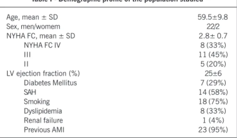
Table I - Demographic profile of the population studied
Age, mean ± SD 59.5±9.8
Sex, men/womem 22/2
NYHA FC, mean ± SD 2.8± 0.7
NYHA FC IV 8 (33%)
III 11 (45%)
II 5 (20%)
LV ejection fraction (%) 25±6
Diabetes Mellitus 7 (29%)
SAH 14 (58%)
Smoking 18 (75%)
Dyslipidemia 8 (33%)
Renal failure 1 (4%)
Previous AMI 23 (95%)
381
withdrawal of the extracorporeal circulation, and the other died due to respiratory failure in the early postoperative period, within the first 30 days. During surgery, 2 patients experienced stroke as follows: one had it in association with acute myocardial infarction, and partial recovery of the neurological deficit was observed; the other died on the 71st postoperative day. Other complications were as follows: one reintervention due to bleeding; one dehiscence of the sternum, which was managed with rotation of the greater pectoral; and one patient with arterial embolism to the right upper limb, who underwent embolectomy.
Five other patients died within the first 6 months of follow-up. One of them due to stroke and the others due to presumed sudden death, without confirmation on necropsy. The 17 surviving patients had a significant improvement in the NYHA functional class of heart failure from 2.8±0.7 to 1.7±0.9, P <0.0001. Although the left ventricular ejection fraction had no significant improvement (25±6 to 26±10%) in the group as a whole, the ventricular function had a significant increase in 8 patients, im-proving from 24±6 to 33±2%, P < 0.002. (fig. 1).
Initially, the histopathological variables of the surviving patients (n = 17) and of those who died (n = 7) were compared. In that analysis, the survivors had more myocytolysis (11±12 vs 4±3%; P = 0.05); however, fibrosis (25±5 vs 22±8%; P = 0.32), and hypertrophy (21.5±4 vs 21.4±5µm; P = 0.97) were similar in both groups.
Of the 17 patients alive 6 months after surgery, one refused to undergo the second biopsy; therefore, pre- and postoperative data were available for comparison in 16 patients. Comparing pre- and postoperative data, while the degree of hypertrophy in the speci-mens did not change (21±4 vs 22±4 µm), a significant increase was observed both in the number of cells with myocytolysis (9±11 vs 21±15% of cells, P < 0.001) and in the extension of fibrosis (8±8 vs 21±15%/area, P < 0.001). Stratifying the patients into a subgroup with maintained left ventricular ejection fraction (n=8) and another with patients who got worse (n=8), individually, none of the histological variables analyzed in the pre- and posto-perative period was different between the 2 groups (tab. II).
Ho-wever, when a score composed by the histological variables was used for analyzing the changes in the ejection fraction after surgery, lower scores in the preoperative period were associated with an improvement in the postoperative ventricular function (fig. 2). Microphotographs of the different histopathological characteristics are shown in figures 3 and 4.
Discussion
Patients with coronary artery disease and severe left ventricular dysfunction may benefit from coronary artery bypass grafting. The appropriate selection of patients who should undergo the procedure is still debatable. Our study showed that the individual analysis of 3 histopathological variables acquired through endomyocardial biopsies during surgery and in the late postoperative period of coronary artery bypass grafting of patients with severe ventricular dysfunction was insufficient to predict who would benefit with an increase in left ventricular ejection fraction after the procedure. However, the combination of histological alterations indicating a lower degree of remodeling was associated with an improvement in ventricular function in the postoperative period.
Our population is a typical cohort of patients with advanced cardiomyopathy, characterized by severe left ventricular dysfunction and significant functional limitation due to congestive heart failure. For these patients, despite the development of the medicamentous therapy, the prognosis continues to be poor 6,7. Thus, the
revas-cularization surgery has gained importance as a therapeutical option, due to the progress of the surgery and myocardial protection techniques 14. Luciani et al 15, in a study with 143 patients with
ejection fraction < 30%, compared the results according to the treatment performed (coronary artery bypass grafting, cardiac trans-plantation, or medicamentous treatment). Despite the relatively high surgical mortality (20%), the prognosis was significantly better in patients undergoing revascularization and cardiac transplantation as compared with those undergoing medicamentous treatment. In the present study, perioperative mortality, up to 30 days after surgery, was 8.3% (2 patients).
Our study showed no overall increase in the ejection fraction in the 17 patients who returned for reassessment after surgery. In fact, worsening of the ejection fraction > 1% occurred in 5 patients. However, 8 of the 17 patients showed a significant increase in their ejection fraction, which could be explained by the short time period for the recovery of contractility (6 months), because, according to some authors, in 2 to 6 months, only 35% of the expected recovery in ventricular function is noted 16. On the
other hand, independently of the success of myocardial revascu-larization, the impairment in contractility may lead to an overall progressive dilation, because the pathophysiological base, which may result in severely dilated ventricles, may depend not only on ischemia: the stress in the dilated ventricular wall perpetuates remodeling 17. Finally, variable quantities of viable myocardium
might exist in the patients, perhaps with areas of mixed fibrosis and muscle, which could justify the different degrees of ejection fraction improvement 10,18.
After a cardiac lesion, such as acute myocardial infarction, a series of events occur at the genomic, molecular, cellular, and interstitial levels leading to alterations in the cardiac size, form,
Ejection fraction (%)
Pre-op Postop
60
50
40
30
20
10
0
382
and function. At the histopathological level, the following may occur: hypertrophy of myocytes, necrosis, apoptosis, increase in fibrosis, fibrillary proliferation of collagen and fibroblasts 19. The
progression of the process of remodeling in several levels seems to be mediated by neurohormonal activation 20-22. On the other
hand, persistence of dilation leads to an increase in tension and overall stress, which lead to activation of several deleterious me-chanisms in a vicious cycle, perpetuating remodeling 23,24. This
process is the basis for understanding that a regional defect in contractility, after acute myocardial infarction, may later lead to the development of an overall cardiac dysfunction.
In our study, the 3 following important histological alterations
that characterize remodeling were analyzed: extension of the fi-brosis, degree of myocytic hypertrophy, and number of cells with myocytolysis. All 3 characteristics were clearly abnormal in the specimens examined in the preoperative period, because a mean amount of fibrosis of 8% was observed, as was the presence of cells with cytoplasmic vacuolization (myocytolysis) and hypertrophy. These findings are comparable to those reported by other investi-gators, who assessed the regional contractile function and histo-logical findings in patients with ischemic cardiomyopathy 25,26. It
is worth noting that, these 2 studies that correlate histopatholo-gical characteristics and postoperative functional reversibility were very careful to perform the biopsies in the myocardial segment with an alteration in regional motility, which had been previously detected through imaging techniques. Therefore, despite the fact that in the present study the biopsies were performed only in the right ventricle, our findings suggest that, independently of the area initially injured, the process of overall remodeling may lead to alterations distant from the myocardial area. Our results may suggest a different and safer approach for studying the histological alterations in patients with advanced cardiomyopathy.
If on the one hand, the histopathological alterations observed in the preoperative period of this study characterize cardiac re-modeling, on the other, the histological findings after myocardial revascularization are at least intriguing. It is worth noting that, to our knowledge, this is the first study that tried to perform histo-pathological comparisons in patients with severe ischemic left ventricular dysfunction before and after coronary artery bypass graf-ting. Despite the notion that restoration of blood flow necessarily leads to a benefit, we found no histological evidence indicating
∆∆∆∆∆
L
VEF (%)
p=0.01
Score ≤≤≤≤≤ 2
n=6
Score > 2 n=10
12
10
8
6
4
2
0
-2
-4
Fig. 2 - Graph representing the variation in ejection fraction between the pre- and postoperative periods of coronary artery bypass grafting in 16 patients studied according to the combined score of the 3 histopathological variables obtained in preoperative endomyocardial biopsies.
Table II - Pre- and postoperative histological variables according to the decrease or increase in ejection fraction after coronary artery bypass grafting
Preoperative Postoperative
EF decrease EF increase P EF decrease EF increase P
n=8 n=8 n=8 n=8
Fibrosis (% area) 11.4 ±3.5 6±2.6 0.31 21.8±3.9 19.8±3.8 0.7
Myocytolysis (cells/fiels) 14.5±4 9±2.8 0.41 21±3.9 19.8±3.8 0.8
Diameter of the fiber (µm) 22.7±1.6 20.5±2.1 0.27 21.9±2 21.3±1.9 0.8
* The values are expressed as mean ± standard error of the mean; EF - efection fraction.
Fig. 4 - Microphotograph of endomyocardial biopsy from specimens obtained during surgery, showing important histopathological alterations. A) moderate hypertrophy of myocytes; B) moderate degree of interstitial fibrosis (hematoxylin and eosin: A, 400X; Masson’s trichrome: B, 100x). The ejection fraction was 17% in the preoperative period and passed to 36% in the postoperative period.
A B
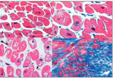
Fig. 3 - Microphotograph of endomyocardial biopsy from specimens obtained during surgery, showing important histopathological alterations. A) size variation of the myocyte fiber; B) hypertrophy of myocytes; C) loss of myofibrils (arrows); D) extensive interstitial fibrosis (hematoxylin and eosin: A and C, 200x, and B, 400x; Masson’s trichrome: D, 100x). The ejection fraction was 25% in the preoperative period and passed to 17% in the postoperative period.
A B
383
reversion of remodeling after revascularization. In fact, 2 of the 3 histopathological characteristics worsened after surgery: a greater number of cells had myocytolysis, and fibrosis increased. Hyper-trophy did not change when comparing the pre- and postoperative specimens. Based on these observations, some speculative hypo-theses may be elaborated: 1) the time elapsed after surgery for histological analysis was not sufficient for showing the reversion of remodeling at the tissue level; 2) restoration of blood flow was not sufficient to promote the beneficial effect at the tissue level; and 3) as no previous publication had focused on studying ventricular re-modeling before and after coronary artery bypass grafting, our ob-servations may have simply reflected the progressive natural his-tory of remodeling, independently of reversion of the ischemic injury. Considering that histological evidence of improvement was not observed after surgery, we tried to study the morphological charac-teristics that could predict which patients would benefit with the procedure. Other authors identified an improvement in myocardial function after revascularization, when only a small amount of fibrosis was observed in biopsies performed during surgery 25,26. In this study,
the degree of fibrosis alone was not sufficient to predict functional recovery of the myocardium. Other authors have not systematically studied other histopathological characteristics, such as myocytolysis and hypertrophy, but we observed no association between these variables and postoperative functional changes. However, because remodeling is a dynamic and multifactorial process, we developed a score with a combination of different histological characteristics, and observed that a smaller degree of fibrosis, myocytolysis, and hypertrophy in the preoperative period was associated with a signi-ficant increase in left ventricular ejection fraction after surgery. Thus, the simultaneous analysis of 3 histopathological characteristics, all indicating adverse remodeling, may constitute a better and more reliable tool to qualify the degree of structural alterations, and, therefore, better represent the amount of potentially recoverable myocardium.
Our results should be interpreted considering the specific li-mitations of the study. First, the specimens were collected from the right interventricular septum, which may account, at least
partially, for the lack of correlation between the histological variables and the functional clinical results. On the other hand, as a second biopsy was planned in the postoperative period, this approach was considered ethically more appropriate than biopsying the left ventricle in patients followed up on an outpatient care basis. In addition, the site of biopsy could not represent the specific segment of abnormal motility, although left ventricular remodeling is assu-med to be an overall and diffuse process, especially considering the severity of the cases studied. This study did not aim at assessing the association of histological variables with the presence or ab-sence of viable myocardium. This information should be explored in the future, as myocardial viability has been used for selecting patients with a greater potential of postoperative recovery. Finally, our data should be seen as generators of hypotheses to be tested in larger studies.
Our study demonstrated that: 1) patients with severe ischemic left ventricular dysfunction undergoing coronary artery bypass surgery had an improvement in the functional class of heart failure, although this is a subjective parameter, but without an increase in left ventricular ejection fraction; 2) no improvement in the histopa-thological characteristics was observed; on the contrary, the degree of fibrosis and myocytolysis worsened; 3) the individual analysis of the preoperative histological characteristics was not able to predict which patients might improve their postoperative ejection fraction. However, the composition of a histological score showed that a lower degree of remodeling was present in the preoperative biopsies of patients, who had an increase in the ejection fraction after surgery. Thus, our results suggest that, despite the improvement in the functional class of heart failure in the whole group and in the left ventricular ejection fraction in a subgroup of patients, evidence of histological improvement, at least in the short run (6 months), after coronary artery bypass grafting in patients with advanced left ventricular ischemic dysfunction should not be ex-pected. Finally, although evidence of moderate histological ab-normalities may indicate a potential for ventricular function im-provement, the characteristics of remodeling may require more time to evidence signs of reversion.
1. Pfeffer JM, Pfeffer MA, Fletcher PJ, et al. Progressive ventricular remodeling in rat with myocardial infarction. Am J Physiol 1991; 260(5Pt. 2)H1406-14. 2. Weber KT, Brilla CG. Pathological hypertrophy and the cardiac interstitium:
fibro-sis and the renin-angiotensin-aldosterone system. Circulation 1991;83:1849-65. 3. Weber KT, Anversa P, Armstrong PWL et al. Remodeling and reparation of the
car-diovascular system.J Am Coll Cardiol 1992;20:3-16.
4. Klappacher G, Franzen P, Haab D et al. Measuring extracellular matrix turnover in the serum of patients with idiopathic or ischemic dilated cardiomyopathy and im-pact on diagnosis and prognosis. Am J Cardiol 1995; 75:913-18.
5. Marijianowsky MM, Teeling P, Mann J, et al. Dilated cardiomyopathy is associa-ted with an increase in type I/typeIII collagen ratio: quantitative assessment. J Am Coll Cardiol 1995; 25: 1263-72.
6. SOLVD Investigators. Effect of enalapril on survival in patients with reduced left ventricular ejection fraction and congestive heart failure. N Engl J Med 1991; 325:293-302.
7. MERIT-HF Study Group. Effect of metoprolol CR/XL in chronic heart failure: Me-toprolol CR/XL Randomized Intervention Trial in Congestive Heart Failure (MERIT-HF). Lancet 1999; 353:2001-07.
8. Elefteriades J, Tolis Jr G, Levi E, et al. Coronary artery grafting in severe left ventri-cular dysfunction: excellent survival, improved ejection fraction and functional state. J Am Coll Cardiol 1993:22:1411-17.
9. Lee KS, Marwick TH, Cook SA et al. Prognostic of patients with left ventricular
dys-References
function with and without viable myocardium after myocardial infarction: relative efficacy of medical therapy and revascularization. Circulation 1994;90(6):2687-94. 10. Udelson JE, Coleman PS, Metherall J, et al. Predicting recovery of severe regional ventricular dysfunction: comparison of rest scintigraphy with 201-thallium and 99mTc-sestamibi. Circulation 1994;89:2552-61.
11. Allman KC,. Shaw LJ, Hachamovitch R, et al. Myocardial Viability Testing and Im-pact of Revascularization on Prognosis inPatients With Coronary Artery Disease and Left Ventricular Dysfunction: A Meta-Analysis. J Am Coll Cardiol 2002;39:1151-8.
12. Loop FD. Coronary artery bypass surgery. In: Topol EJ. Textbook of Cardiovascular Medicine. Lippincott-Raven Publishers, Philadelphia. 1998;2011-31. 13. Mason JW. Techniques for right and left ventricular biopsy. Am J Cardiol
1978;41:887-92.
14. Buckberg GD. Update on current techniques of myocardial protection. Ann Thorac Surg1995; 60: 805-14.
15. Luciani GB, Faggian G, Razzolini R, et al. Severe ischemic left ventricular failure: coronary operation or heart transplantation? Ann Thorac Surg 1993;55:719-23 16. Vanoverschelde JL, Melin JA, Depré C, et al. Time-course of functional recovery of hibernating myocardium after coronary revascularization (Abstract). Circulation 1994;90(suppl): I-378.
384
18. Perrone-Filardi P, Pace L, Prastaro M et al. Assessment of myocardial viability in patients with chronic coronary artery disease: Rest-4-hour-24-hour 201Thallium tomography vs Dobutamine Echocardiography. Circulation 1996;94:2712-19. 19. Narula J, Haider N, Virmani R et al. Apoptosis in myocytes in end-stage heart
fai-lure. N Engl J Med 1996;335:1182-89.
20. Reiss K, Capasso JM, Huang HE, et al. ANG II receptors, c-myc and c-jun in myo-cytes after myocardial infarction and ventricular failure. Am J Physiol 1993;264:H760-9.
21. Sadoshima J, Izumo S. Molecular characterization of angiotensin II induced hy-pertrophy of cardiac myocytes and hyperplasia of cardiac fibroblasts. Critical role of the AT1 receptor subtype. Circ Res 1993;73:413-23.
22. Everett AD, Tufro-McReddie A, Fisher A, et al. Angiotensin receptor regulates
cardiac hypertrophy and transforming growth factor-beta 1 expression. Hyperten-sion 1994;23:587-92.
23. Mitchell GF, Lamas GA, Vaughan DE, et al. Left ventricular remodeling in the year after first anterior myocardial infarction: a quantitative analysis of contractile seg-ment lengths and ventricular shape. J Am Coll Cardiol 1992;19:1136-44. 24. Grossman W, Jones D, McLaurin LP. Wall stress and patterns of hypertrophy in
the human left ventricle. J Clin Invest 1975; 56:56-64.
25. Depré C, Vanoversehelde JLJ, Melin JA et al. Structural and metabolic correlates of the reversibility of chronic left ventricular ischemic dysfunction in humans. Am J Physiol 1995;268:H1265-H1275.