Exercise Stress Test: Prognostic Value for Elderly
Patients with Stable Coronary Atherosclerosis
Marcelo Eidi Ochiai, Otávio C. E. Gebara, João Batista Serro-Azul, Lígia B. Pinto,
Amit Nussbacher, Humberto Pierri, Mauricio Wajngarten
Instituto do Coração do Hospital das Clínicas – FMUSP - São Paulo, SP - Brazil
M a i l i n g A d d r e s s : M a r c e l o E i d i O c h i a i • R u a C a r a í b a s , 1 3 4 2 – 0 5 0 2 0 - 0 0 0 – S ã o P a u l o , S P - B r a z i l
E-mail: mochiai@osite.com.br Received on 03/10/05 • Accepted on 06/14/05
O
BJECTIVETo study the prognostic value of exercise stress test variables in elderly patients with coronary atherosclerosis and exercise-induced ischemia.
M
ETHODSSixty-four elderly patients (61 men, 73 ± 5 years old) with coronary atherosclerosis, verifi ed by cardiac catheterization, that were clinically stable, had a left ventricle ejection fraction greater than or equal to 0.40 and developed myocardial ischemia during the exercise stress test were studied. The patients were evaluated every six months for cardiac events (death, myocardial infarction, unstable angina, angioplasty and myocardial revascularization).
R
ESULTSAfter a mean follow-up period of 48 months, 23 (36%) patients suffered cardiac events. There was no clinical or angiographical differences among the patients that suffered cardiac events and those that did not. Using multivariate analysis, the presence of chest pain during the exercise stress test (relative risk 2.668, p = 0.031) and the heart rate at the onset of ischemia (relative risk 0.966, p = 0.009) were associated with cardiac events.
C
ONCLUSIONIn this elderly population, the presence of chest pain during the exercise stress test and the heart rate at the onset of ischemia were associated with cardiac events. These variables could be useful for risk evaluation in patients with stable coronary atherosclerosis
K
EY WORDSThe growth of the elderly population and the high prevalence of coronary atherosclerosis in this group represents a challenge for cardiologists. The guidelines of the American Heart Association recommend that sedentary elderly people have an exercise stress test before beginning a rigorous physical activity program in order to identify coronary atherosclerosis1. However, Gill
et al 2 imply that the guidelines are not applicable for the
majority of elderly individuals and do not recommend an exercise stress test for asymptomatic individuals. Nevertheless, there is no clear reason to contraindicate the exercise stress test for the elderly3. Aging in itself is
associated with alterations of the cardiovascular system in response to exercise, reduction of physical capacity and a lower frequency of angina pain in patients with coronary atherosclerosis4,5. Clinical evaluation in order
to begin or continue a physical exercise program should be accurate enough to identify high risk patients and the possible benefi t of intervention procedures. Surprisingly, according to the National Medical Care Survey study, it is less likely that patients over 75 will be indicated to take an exercise stress test6. Additionally, if the exercise
stress test is requested, it is generally for diagnostic purposes and not for risk stratifi cation. There is limited information regarding risk stratifi cation in the elderly with stable coronary atherosclerosis. Therefore, the present study was conducted in order to evaluate the prognostic value of exercise stress test variables in the elderly with stable coronary atherosclerosis and exercise-induced myocardial ischemia.
M
ETHODSSixty-four patients were consecutively selected from the outpatient clinic of the Geriatric Unit of the Heart Institute (InCor), University of São Paulo Medical School. The average age of the patients was 73 years (standard deviation 5 years), and all had coronary atherosclerosis that had been verified by cardiac catheterization. All patients were clinically stable, had a left ventricular ejection fraction greater than or equal to 40% and had developed myocardial ischemia during the exercise stress test. Exclusion criteria included artifi cial pacemakers, left bundle branch block, atrial fi brillation, a high atrioventricular block classifi cation or ventricular hypertrophy.
The study was approved by the institution’s Research Ethics Committee and after being informed regarding the study, all the patients agreed to participate.
Cardiac catheterization and left ventriculography - Cardiac catheterization was performed using the Sones e Shirey technique7. A reduction in the coronary artery
luminal diameter greater than or equal to 70% was considered signifi cant. In accordance with the number of main coronary arteries with signifi cant obstruction, the patients were classifi ed as single artery, double artery and triple artery. The left ventricular ejection fraction was
calculated using the Dodge-Kennedy method8,9.
Exercise stress test - The use of cardiovascular medication was discontinued fi fteen days before the test was conducted. The equipment used was a computerized system (Fukuda-Denshi, model 8000, Japan) with twelve traditional leads and three bipolar leads (CC5, CM5 e ML) and an inclinable treadmill. A modifi ed Naughton protocol was used that consists of fi ve stages of progressive inclination (3.5%, 7%, 10.5%, 14% and 17.5%) each with a three minute duration and a speed of 3.6 km/h. For the next fi ve stages, the speed was increased to 4.8 km/h and the inclination reduced to 12.5%, and then progressively increased by 2.5%. Functional capacity was estimated in metabolic equivalents (METs) according to the duration of the test.
Interruption criteria for the exercise stress test were fatigue or intense angina, sloping of the ST segment greater than or equal to 0.3 mV and a systolic blood pressure drop of more than 20 mmHg during the exercise. The presence of myocardial ischemia was defi ned as horizontal or down sloping of the ST segment greater than 0.1 mV to 80 msec. after the J-point. Heart rate, systolic and diastolic blood pressure were recorded at rest, after each stage, at the onset of ischemia and at the peak of exercise. The time of the ischemia onset and the total duration of the test were recorded. The double product was calculated by multiplying the heart rate by the systolic blood pressure, at rest, at the onset of ischemia and at the peak of exercise. The patients were asked during the test if they were experiencing angina chest pain.
Clinical follow-up - Follow-up of the patients was conducted every six months at outpatient clinics. Medicinal therapy was given in accordance with the criteria of each patient’s doctor. Conclusion of the program was defi ned as the following cardiac events: cardiac related death, nonfatal myocardial infarction, unstable angina that led to hospitalization, percutaneous coronary angioplasty and surgical myocardial revascularization. Cardiac related death and a nonfatal myocardial infarction were considered major cardiac events. The patients that suffered cardiac events were classifi ed as group A and the others as group B.
Statistical analysis - Continuous variables were expressed as an average and standard deviation and were compared between the groups A and B using the Student’s t-test. The categorical variables were expressed by frequency and proportion and were compared using the chi-square test or Fisher exact test.
Variables that were signifi cantly different between the groups A and B were entered in the multivariate model using the Cox method, and the computer program SAS (SAS Institute, Cary, North Carolina, USA).10
R
ESULTSAfter a mean follow-up period of 48 months, 23 patients suffered cardiac events (group A, 36%). Ten patients suffered cardiac related deaths, one patient suffered a non-fatal myocardial infarction, fi ve patients presented unstable angina, five patients underwent surgical myocardial revascularization and two patients underwent percutaneous angioplasty. Four patients suffered non-cardiac related deaths of which two died from neoplastic diseases and two from strokes.
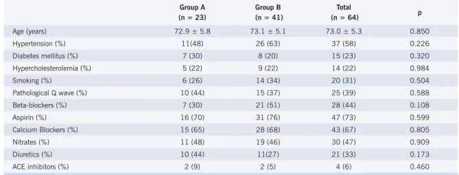
Thirty-seven (58%) patients were hypertensive, fifteen (23%) were diabetics, fourteen (22%) had hypercholesterolemia, twenty (33%) were smokers, and twenty-five (39%) presented a pathological Q wave on the EKG. These variables did not present any signifi cant statistical differences between the Groups A and B (tab.1).
The medications used during the follow-up were beta-blockers for 28 (44%) patients, aspirin for 47 (73%) patients, calcium channel blockers for 43 (67%) patients, nitrates for 34 (53%) patients, diuretics for 21 (33%) patients and angiotensin-converting enzyme inhibitors for 4 (6%) patients. The groups A and B did not present any signifi cant differences in relation to medication (tab.1).
In relation to the coronary disease pattern, 19 (30%) patients were single artery, 18 (28%) double artery and 27 (42%) were triple artery. The left ventricular ejection fraction was 0.69 ± 0.14. The coronary pattern and ventricular function were no different between the groups A and B (tab.2).
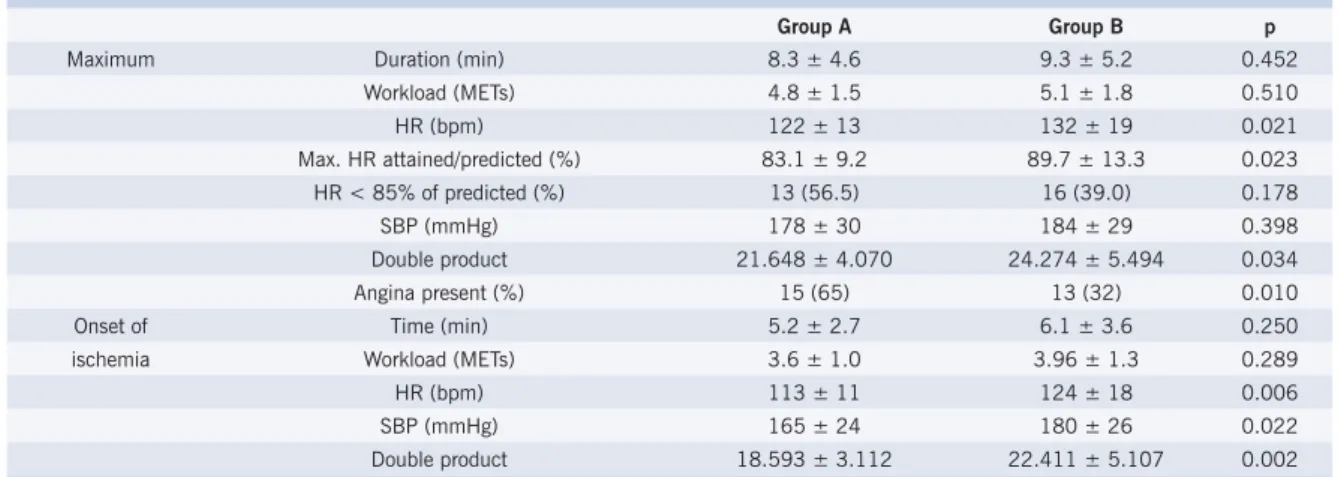
Considering all patients, the average duration of the exercise stress test was 8.9 ± 5.6 minutes, and the average time for the onset of ischemia was 5.8 ± 3.3 minutes. At peak exertion, heart rate, systolic blood pressure and double product were, respectively, 128 ± 17 beats per minute, 182 ± 29 mmHg and 23.300 ± 5.154. At the onset of ischemia, these variables had the following values: 120 ± 18 beats per minute, 174 ± 26 mmHg and 21.038 ± 4.833. Twenty-eight (44%) patients reported chest pain during the test. In these patients, the maximum heart rate was lower than the group that did not have chest pain (122 ± 14 vs. 133 ± 18, p = 0.008), however the duration of the test (7.8 ± 4.6 vs. 9.8 ± 5.2 minutes; p = 0.095) and number of METs (4.6 ± 1.6 vs. 5.3 ± 7.8; p = 0.105) were the same.
Student’s t-test showed that the heart rate for the patients in group A was lower than those in group B (122 ± 13 vs. 132 ± 19; p = 0.021). The chi-square test
Table 1– Clinical characteristics according to cardiac event occurrence (group A) or no occurrence (group B)
Group A (n = 23)
Group B (n = 41)
Total
(n = 64) p
Age (years) 72.9 ± 5.8 73.1 ± 5.1 73.0 ± 5.3 0.850
Hypertension (%) 11(48) 26 (63) 37 (58) 0.226
Diabetes mellitus (%) 7 (30) 8 (20) 15 (23) 0.320
Hypercholesterolemia (%) 5 (22) 9 (22) 14 (22) 0.984
Smoking (%) 6 (26) 14 (34) 20 (31) 0.504
Pathological Q wave (%) 10 (44) 15 (37) 25 (39) 0.588
Beta-blockers (%) 7 (30) 21 (51) 28 (44) 0.108
Aspirin (%) 16 (70) 31 (76) 47 (73) 0.599
Calcium Blockers (%) 15 (65) 28 (68) 43 (67) 0.805
Nitrates (%) 11 (48) 19 (46) 30 (47) 0.909
Diuretics (%) 10 (44) 11(27) 21 (33) 0.173
ACE inhibitors (%) 2 (9) 2 (5) 4 (6) 0.460
ACE = angiotensin-converting enzyme
Table 2 – Angiographic characteristics according to cardiac event occurrence (group A) or no occurrence (group B)
Group A (n = 23)
Group B (n = 41)
Total
(n = 64) p
LVEF 0.66 ± 0.14 0.71 ± 0.14 0.69 ± 0.14 0.226
Single artery (%) 7 (30) 12 (29) 19 (30)
Double artery (%) 5 (22) 13 (32) 18 (28) 0.670
Triple artery (%) 11(48) 16 (39) 27 (42)
showed that the proportion of patients that reached the predicted heart rate was lower in group A (83 + 9 vs. 90 ± 13; p = 0.023). However, the proportion of patients that did not attain 85% of the predicted heart rate for their age was the same for both groups. The duration of the test and the systolic blood pressure at the peak of the exercise were the same for both groups (tab. 3).
At the onset of ischemia, the heart rate (113 ± 11 vs. 124 ± 18 bpm), systolic blood pressure (165 ± 24 vs. 180 ± 26 mmHg) and double product (18.593 ± 3.112 vs. 22.411 ± 5.107) were lower in group A. However, the time for the onset of ischemia was similar (5.2 ± 2.7 vs. 6.1 ± 3.6 min, group A and B respectively; p = 0.250).
Fifteen (65%) patients in group A presented angina during the test compared to thirteen (32%) in group B.
Multivariate analysis using the Cox method demonstrated that the heart rate at the onset of ischemia was associated with the occurrence of cardiac events, with a relative risk of 0.966 (confi dence interval 95%: 0.942 to 0.992; p = 0.009). This signifi es that a reduction of the heart rate by 1 bpm at the onset of ischemia increases the risk for cardiac events by 3.4%. The presence of angina during the exercise stress test revealed an association with the occurrence of cardiac events, with a relative risk of 2.668 (confi dence interval 95%: 1.091 to 6.523; p = 0.031). The frequency of the 23 cardiac events, according to time of occurrence, in the patients with and without pain during the exercise stress test was, respectively, up to six months: 13% and 0%, from six to twelve months: 4% and 0%, and more than twelve months: 48% and 35%. This indicates that the majority of events, including revascularization procedures, occurred long after the test and probably had no relation with the presence of angina during the exercise stress test.
When only the major events are considered, that is,
cardiac related death and non-fatal myocardial infarction, there was no signifi cant association with heart rate at the onset of ischemia or the presence of angina during the test.
Figure 1 shows the probability, estimated using the Cox model, of a given heart rate at the onset of ischemia (60, 90 and 120 bpm) with or without the presence of angina during the test. The lower the heart rate at the onset of ischemia the higher the infl uence of the presence of angina during the test.
D
ISCUSSIONThis study demonstrated that in elderly patients with stable coronary atherosclerosis, exercise-induced myocardial ischemia and minimal ventricular dysfunction, the heart rate at the onset of ischemia and the presence of angina during the exercise stress test were associated with the occurrence of cardiac events. In patients with a lower heart rate at the onset of ischemia, the infl uence of the presence of angina was more evident.
The patients in this sample were older than those in other studies11-19 that evaluated the prognostic value of
the exercise stress test. Only Goraya et al 20 analyzed the
prognostic value of the exercise stress test specifi cally in the elderly. The follow-up timeframe in this study was longer than in most of the studies with the exception of the studies conducted by Dagenais et al14 and Goraya et
al20. Additionally, only the ACIP study, Dagenais et al and
Mark et al also performed cardiac catheterization. Similar to most of the studies, the majority of our patients had coronary disease with a multi-artery pattern. Dagenais et al14 and the researchers of the ACIP study11 also
objectively evaluated the left ventricular function while the other studies did not. Just as in this study, the ACIP study evaluated patients with minimal ventricular dysfunction while the Degenais et al sample included patients with accentuated dysfunction.
Table 3 –Exercise stress test variables according to cardiac event occurrence (group A) or no occurrence (group B)
Group A Group B p
Maximum Duration (min) 8.3 ± 4.6 9.3 ± 5.2 0.452
Workload (METs) 4.8 ± 1.5 5.1 ± 1.8 0.510
HR (bpm) 122 ± 13 132 ± 19 0.021
Max. HR attained/predicted (%) 83.1 ± 9.2 89.7 ± 13.3 0.023
HR < 85% of predicted (%) 13 (56.5) 16 (39.0) 0.178
SBP (mmHg) 178 ± 30 184 ± 29 0.398
Double product 21.648 ± 4.070 24.274 ± 5.494 0.034
Angina present (%) 15 (65) 13 (32) 0.010
Onset of Time (min) 5.2 ± 2.7 6.1 ± 3.6 0.250
ischemia Workload (METs) 3.6 ± 1.0 3.96 ± 1.3 0.289
HR (bpm) 113 ± 11 124 ± 18 0.006
SBP (mmHg) 165 ± 24 180 ± 26 0.022
Double product 18.593 ± 3.112 22.411 ± 5.107 0.002
Cox Regression Analysis
0,0 0,1 0,2 0,3 0,4 0,5 0,6 0,7 0,8 0,9 1,0
0 10 20 30 40 50 60 70 80
Months
Probability of event-free survival
60 no chest pain 60 chest pain 90 no chest pain 90 chest pain 160 no chest pain 160 chest pain
Although the incidence of cardiac events in this sample (36%) was higher than in other studies, the coronary pattern and ventricular function suggest that the condition of these patients was compatible with the patients in the other studies. Additionally, the prevalence of other risk factors for coronary atherosclerosis was not higher than expected for this population. Considering the medicinal treatment, beta-blockers, ASA and angiotensin-converting enzyme inhibitors were used less often than expected. Since risk factors, coronary patterns, ventricular function, and medicinal treatment were similar for groups A and B, the high incidence of cardiac events cannot be attributed to these factors alone. The advanced age of this population could have contributed to the high incidence of events, however, the ages for the two groups were similar.
Exercise stress test variables associated with cardiac events -Unlike other studies11,13-15,17,18,20, that show an
inverse relationship between functional capacity and the incidence of cardiac events, no association was found between the exercise stress test duration and incidence of events for this sample. This difference can be attributed to the great variations in the duration of the exercise stress test in this study, a fi nding expected for the elderly, and also by the fact that the other studies included patients with and without exercise-induced ischemia. Unlike the fi nding of Lauer et al19, the maximum heart rate in this
study was not associated with cardiac events. It is also important to note that 45% of these patients did not attain the submaximum heart rate predicted for their age. These fi ndings could be the result of other non-cardiac related causes such as fatigue, low motivation or arthropathies, as well as myocardial ischemia. Angina during the exercise
Fig.1 – Event free curve, based on estimated probability using the Cox regression model, according to heart rate at the onset of ischemia and the presence of chest pain during the exercise stress test
stress test did not limit the duration since total exercise time and the number of METs were similar for both groups, with and without angina.
The heart rate at the onset of ischemia, that is less dependent on non-cardiac factors, was the predictor for cardiac events. In the ACIP study11, this variable
was used to predict the occurrence of ischemia in everyday activities, however, it was not associated with a greater incidence of cardiac events during the one year follow-up. The onset of ischemia during a lower cardiac response phase could indicate more extensive coronary atherosclerosis, however, the coronary pattern was not associated with the group that had cardiac events. The reasons why the heart rate at the onset of the ischemia was associated with a worse evolution cannot be clarifi ed in this study and should be investigated in elderly patients with stable coronary atherosclerosis.
In this study, the presence of angina during the exercise stress test was associated with a worse evolution which agrees with other previous studies12,15,17; but there are
also other studies that do not confi rm this fi nding14,16.
Therefore, both the heart rate at the onset of ischemia and the presence of angina during the exercise stress test had prognostic value in this study and could be clinically relevant. It is important to note that cardiac events, including revascularization procedures, did not occur immediately following the exercise stress test which indicates that the presence of angina is an indication of mid and long term clinical deterioration. Therefore, the presence of angina during the exercise stress test suggests the need for more attentive preventative actions in order to avoid invasive procedures, risks and discomfort.
Heart rate at the onset of ischemia and angina during the exercise stress test were not indicative of more serious cardiac events (death and myocardial infarction). There are two possible explanations for this fi nding. The fi rst is that the small number of events (eleven patients) in the sample was not signifi cant enough to detect an eventual association. Another possibility is that angina during an exercise stress test is simply a determining factor for other cardiac events such as revascularization procedures.
Study limitations -The study population consisted of a sample of elderly patients with similar characteristics such as extensive coronary disease without diminished
ventricular function or functional capacity. The fi ndings of this study can not be generalized for all the elderly with coronary disease. However, this population is very similar to most elderly patients who want to participate in physical exercise programs.
This study did not have a control group of younger patients for the comparison of the clinical and exercise stress test variables, however, external controls from other studies were used for comparison purposes. Therefore, it is not possible to conclude that our fi ndings are caused only by aging.
C
ONCLUSIONSIn elderly patients with stable coronary atherosclerosis with minimal ventricular dysfunction and exercise-induced ischemia, there is a high incidence of cardiac events. Heart rate at the onset of ischemia and angina during the exercise stress test were indicators of cardiac events. Therefore, exercise stress tests for elderly patients with stable coronary atherosclerosis should be used to improve risk evaluation.
No potential confl ict of interest relevant to this article was reported.
R
EFERENCES1. Gibbons RJ, Balady GJ, Beasley JW, Bricker JT, Duvernoy WFC, Froelicher VF et al. ACC/AHA Guidelines for exercise testing. A report of the American College of Cardiology/American Heart Association Task Force on Practice Guidelines (Committee on Exercise Testing). J Am Coll Cardiol. 1997;30: 260-315.
2. Gill TM, DiPietro L, Krumholz HM. Role of exercise stress testing and safety monitoring for older persons starting an exercise program. JAMA. 2000; 284: 342-9.
3. Ashley EA, Myers J, Froelicher V. Exercise testing in clinical medicine. Lancet. 2000; 356:1592-7.
4. Wajngarten M, Wajngarten M, Negrão CE, Kalil LMP, Ramires PR, Rondon E et al. Infl uence of aging and exercise training on the increase in oxygen uptake as a function of the increase in work rate. Cardiol in the Elderly. 1994;2: 421-6.
5. Callaham PR, Frolicher VF, Klein J, Rish M, Dubach P, Friis R et al. Exercise-induced silent ischemia: age, diabetes mellitus, previous myocardial infarction and prognosis. J Am Coll Cardiol. 1989; 14:1175-80.
6. Cohen MC, Stafford RS, Misra B. Stress testing: National patterns and predictors of test ordering. Am Heart J.1999;138: 1019-24.
7. Sones FM, Shirey EK. Cinecoronary arteriography. Mod Concepts Cardiovasc Dis. 1962; 31:735-8.
8. Dodge HT, Sandler H, Ballew DW, Lord JD. The use of biplane angiocardiography for the measurement of left ventricular volume in man. Am Heart J. 1960; 60:762-76.
9. Kennedy JW, Trenholme SE, Kasser IS. Left ventricular volume and mass from single-plane cineangiocardiogram. A comparison of anteroposterior and right anterior oblique methods. Am Heart J. 1970; 80:343-52.
10. Lee ET. Statistical Methods for Survival Data Analysis. 1st ed. Belmont
(CA): Lifetime Learning Publications, 1980.
11. Stone PH, Chaitman BR, Forman S, Andrews TC, Bitter V, Bourassa
MG et al. Prognostic signifi cance of myocardial ischemia detected by ambulatory electrocardiography, exercise treadmill testing, and electrocardiogram at rest to predict cardiac events by one year (The Asymptomatic Cardiac Ischemia Pilot [ACIP] Study). Am J Cardiol. 1997;80: 1395-401.
12. Villella M, Villella A, Barlera S, Franzosi MG, Maggioni AP et al. Prognostic signifi cance of double product and inadequate double product response to maximal symptom-limited exercise stress testing after myocardial infarction in 6296 patients treated with thrombolytic agents. Am Heart J.1999;137: 443-52.
13. Forslund L, Hjemdahl P, Held C, Björkander I, Eriksson SV, Brodin U et al. Prognostic implications of results from exercise testing in patients with chronic stable angina pectoris treated with metoprolol or verapamil. A report from The Angina Prognosis Study in Stockholm (APSIS). Eur Heart J. 2000;21: 901-10.
14. Dagenais GR, Hjemdahl P, Held C, Björkander I, Eriksson SV, Brodin U et al. Survival with painless strongly positive exercise electrocardiogram. Am J Cardiol. 1988; 62:892-5.
15. Narins CR, Zareba W, Moss AJ, Goldstein RE, Hall WJ et al. Clinical implications of silent versus symptomatic exercise-induced myocardial ischemia in patients with stable coronary disease. J Am Coll Cardiol. 1997; 29:756-63.
16. Weiner DA, Ryan TJ, McCabe CH, Ng G, Chaitman BR, Sheffi eld LT et al. Risk of developing an acute myocardial infarction or sudden coronary death in patients with exercise-induced silent myocardial ischemia. A report from the Coronary Artery Surgery Study (CASS) Registry. Am J Cardiol. 1988; 62:1155-8.
17. Mark DB, Shaw L, Harrell FE Jr et al. Prognostic value of a treadmill exercise score in outpatients with suspected coronary artery disease. N Engl J Med. 1991; 325:849-53.
19. Lauer MS, Francis GS, Okin PM, Pashkow FJ, Snader CE, Marwick TH et al. Impaired chronotropic response to exercise stress testing as a predictor of mortality. JAMA. 1999; 281:524-9.