Brief Comments
Characterization of the Apical Aneurysm of Chronic Chagas’ Heart
Disease by Scintigraphic Image Co-Registration
Marcus Vinicius Simões, Lucas Ferrari de Oliveira, Flavio Cantarelli Hiss, Alexandre Baldini de Figueiredo, Antonio Osvaldo Pintya, Benedito Carlos Maciel, José Antonio Marin-Neto
Divisão de Cardiologia - Hospital das Clínicas da Faculdade de Medicina de Ribeirão Preto - USP - Ribeirão Preto, SP - Brazil
Key words
Heart aneurysm; Chagas’ cardiomyopathy; radionuclide imaging; Chagas’ disease.
Mailing address: Marcus Vinicius Simões •
Rua Marechal Deodoro, 1085/101 - 14010-190 - Ribeirão Preto, SP - Brazil E-mail: simoesmv@yahoo.com
Manuscript received February 6, 2007; revised manuscript received March 26, 2007; accepted April 23, 2007.
Methods
Thirteen patients (mean aged 48 ± 3 years, nine male) with chronic Chagas’ heart disease, defined as positive serology and epidemiological history, were investigated in a clinical trial designed to analyze the correlation between cardiac sympathetic denervation and myocardial perfusion changes8. They had normal global systolic ventricular function
(LV ejection fraction was 55 ± 2%) and no ventricular dilation, yet showed segmental wall motion abnormalities on MUGA-SPECT. Of these patients, four had apical aneurysm, confirmed by two-dimensional echocardiography and contrast ventriculography during cardiac catheterization. All of them had angiographically normal coronaries. The study was approved by the Institutional Ethics Committee, and all patients signed an informed consent form.
Image acquisition and processing - Myocardial perfusion studies were performed at rest after an injection of thallium-201 (148 MBq) and, on the second day, MUGA-SPECT, after
in vivo blood-pool labeling with injection of stannous chloride, followed by technetium-99m (740 MBq). Images were acquired using a dual-head digital gamma camera DST-SMV (Sopha Medical Vision - Twinsburg, Ohio, USA) equipped with a low-energy, high-resolution (LEHR) collimator with a 20% energy window centered at 70 KeV for thallium-201 images and at 140 KeV for technetium-99m images. ECG-gated SPECT studies were performed by acquiring 32 projections (60 seconds per projection) over a 180° semicircular orbit with the patient supine. Images were processed on a dedicated computer (Vision PowerStation – IBM – SMV), and tomographic slices were reconstructed and exported in a DICOM 3.0 format to a dedicated workstation with specific software for co-registration calculation.
Image co-registration - The free software package VTK CISG Registration ToolKit (vtKCisg)9, which relies on registration
algorithms based on similarity of voxel cross-correlation, was used. After image alignment, the insertion of the right ventricular myocardium into the interventricular septum was used as an anatomical landmark to check alignment accuracy. Whenever necessary, manual corrections were performed to improve alignment.
Results
In the four patients with apical aneurysms, scintigraphic features were quite consistent. The summed images of myocardial perfusion studies showed severe perfusion defects, yet relatively small when compared to the size of
Introduction
In its early phases, chronic Chagas’ cardiomyopathy may distinctively present segmental wall motion abnormalities, ranging from mild hypokinesis to regional aneurysm formation1. Apical aneurysm is the most notable segmental
disorder in chronic Chagas’ heart disease2.
Recent clinical trials have shown that the presence of this cardiac lesion is an independent risk factor for cardioembolic stroke in patients with chronic Chagas’ heart disease3-5.
Therefore, early detection of the aneurysm using several diagnostic imaging methods is clinically relevant because of its therapeutic implications.
ECG-gated SPECT myocardial perfusion imaging has been recently validated for assessing left ventricular function, and is now widely used6. Visual analysis of tomographic slices
displayed in cine mode allows the identification of dyskinetic areas, including left ventricle aneurysms, associated with ischemic heart disease7. Nevertheless, while many Chagasic
patients undergo myocardial perfusion imaging due to symptoms suggestive of myocardial ischemia, there is no report on characterization of the apical aneurysm of chronic Chagas’ disease by gated-SPECT.
Multi-gated blood pool SPECT (MUGA-SPECT), which produces images of the ventricular cavity, and gated-SPECT (which produces images of the ventricular walls) provide complementary information for better characterization of this form of ventricular lesion in chronic Chagas’ heart disease. Recent computational techniques allow these images to be aligned and co-registered, with simultaneous visualization of these structures.
This report describes the anatomical and functional characteristics of apical aneurysms in chronic Chagas’ heart disease patients using different modalities of three-dimensional cardiac image co-registration, most specifically, myocardial perfusion and labeled blood pool images.
Brief Comments
Simões et al Apical aneurysm of Chagas heart disease
Arq Bras Cardiol 2007; 89(2) : 119-121
the aneurysms identified by radionuclide ventriculography (Figures 1 and 2). This was associated with virtually preserved
segmental wall motion in the region surrounding the aneurysm, resulting in an aneurysm with a narrow neck.
Fig. 1 -Tomographic slices of myocardial perfusion imaging (upper panels), blood pool imaging (middle panels) and fused images of myocardial perfusion (in blue) and blood pool (in red), shown in the lower panels. The left panels are the diastolic frames, and right panels are the systolic frames. A severe perfusion defect involves a moderate portion of the cardiac apex, with preserved motion of the segments surrounding it (short arrows). Blood pool images revealing an apical aneurysm extending beyond the left ventricular walls (long arrows). The fused images clearly demonstrate the topographic correlation between the aneurysm cavity, the myocardial segments that form its “neck”, and the perfusion defects (wide arrows).
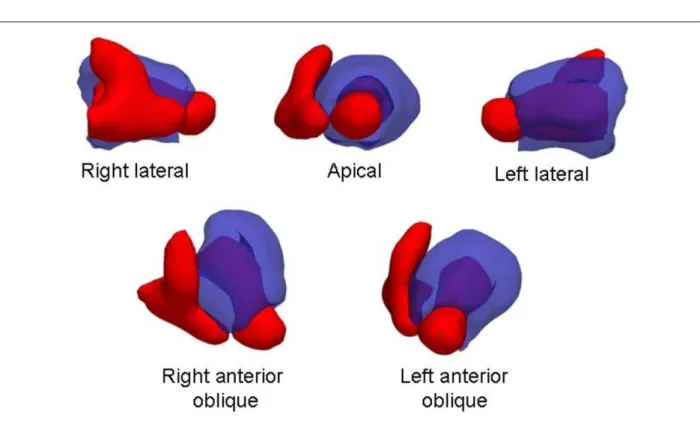
Fig. 2 -The fusion of myocardial perfusion (in semitransparent blue) and blood pool (solid red) images is represented in three-dimensional volumes with view angles. An aneurysmal projection beyond the left ventricular wall at the apex is noted.
Brief Comments
Simões et al
Apical aneurysm of Chagas heart disease
Arq Bras Cardiol 2007; 89(2) : 119-121
References
1. Carrasco HA, Barboza JS, Inglessis G, Fuenmayor A, Molina C. Left ventricular cineangiography in Chagas’ disease: detection of early myocardial damage. Am Heart J. 1982; 104: 595-602.
2. Oliveira JSM, Oliveira JAM, Frederigue Junior U, Lima-Filho EC. Apical aneurysm in Chagas’ heart disease. Br Heart J. 1981; 46: 432-7.
3. Oliveira JSM, Araújo RRC, Navarro MA, Muccillo G. Cardiac thrombosis and thromboembolism in Chronic Chagas’ heart disease. Am J Cardiol. 1983; 52: 147-51.
4. Carod-Artal FJ, Vargas PA, Horan TA, Nunes LGN. Chagasic cardiomyopathy is independently associated with ischemic stroke in Chagas disease. Stroke. 2005; 36: 965-70.
5. Oliveira-Filho J, Viana LC, Vieira-de-Melo RM, Faical F, Torreao JA, Villar FA, et al. Chagas disease is an independent risk factor for stroke: baseline characteristics of a Chagas disease cohort. Stroke. 2005; 36
(9): 2015-7.
6. Berman DS, Germano G. Evaluation of ventricular ejection fraction, wall motion, wall thickening, and other parameters with gated myocardial perfusion single-photon emission computed tomography. J Nucl Cardiol. 1997; 4: S169-S171.
7. Koszegi K, Kolozsvari R, Varga J, Galuska L, Szuk T, Csapo K, et al. 99mTc-MIBI-SPECT assessment of the effects of aneurysm resection on the left ventricular morfology. Acta Cardiol. 2004; 59: 541-6.
8. Simões MV, Pintya AO, Bromberg-Marin G, Sarabanda AV, Pazin-Filho A, Maciel BC, et al. Relation of regional sympathetic denervation and myocardial perfusion disturbance to wall motion impairment in Chagas’ cardiomyopathy. Am J Cardiol. 2000; 86: 975-81.
9. Hartkens T. Measuring, analyzing, and visualizing brain deformation using non-rigid registration. [thesis]. London: , University of London; 2003.
This aspect contrasts with that classically found in the apical aneurysm secondary to ischemic heart disease, in which a large perfusion defect with dyskinetic motion of the segments surrounding the aneurysm is noted. In chronic Chagas’ heart disease aneurysms, left ventricular walls show a convergent spatial orientation toward the apex, unlike the divergent orientation classically identified in aneurysms associated with ischemic heart disease. Therefore, in Chagas’ heart disease, the ventricular chamber becomes protruded, forming the aneurysm cavity, and extends beyond the ventricular wall segments identifiable by perfusion images, which form the neck of the aneurysm, making it resemble a diverticulum.
Discussion
The digital alignment technique, or co-registration, of myocardial perfusion images with those of radionuclide ventriculography used in this study allowed a perfect visualization of the topographic correlation between the myocardium, which constitutes the aneurysm neck, and the saccular formation at the apex of the heart, typical of chronic Chagas’ heart disease.
These aspects may be better understood from the rather distinctive pathologic features of this type of lesion2. Typically,
this form of ventricular aneurysm has a saccular morphology, with a narrow neck. The aneurysm walls, which are formed by the apposition of the epicardial and endocardial layers, are quite thin, resembling a diverticulum2. Consequently, they do
not have enough myofibers to take up significant amounts of perfusion tracer. However, as demonstrated in this study, the wall motion in the myocardial segments adjacent to the apical aneurysm is preserved.
Conversely, given the virtual lack of dyskinesis in the wall segments and also the relatively small perfusion defect, gated-SPECT myocardial perfusion imaging alone may not be able to identify the presence of an aneurysm clearly.
Together, these morphological and functional characteristics enable an accurate distinction to be made between the apical aneurysm of chronic Chagas’ heart disease and that secondary to ischemic heart disease, the latter showing a divergent orientation of the ventricular walls and abnormal motion of the segments adjacent o the aneurysm, forming a large neck.
Considering that many Chagasic patients experience precordial pain, requiring nuclear studies to explain this symptom, these results suggest that it may be appropriate to expand this investigation using radionuclide ventriculography for early detection and characterization of any possible apical aneurysm. This diagnostic approach may have important clinical implications, such as the need for anticoagulation, since this lesion is often associated with thromboembolic events.
Sources of Funding
This study was funded by FAPESP.
